Download presentation
Presentation is loading. Please wait.

1
The Foot Bones Joint Muscles Artery & Nerves

2
Superficial veins

3
Great saphenous vein

4
Small saphenous vein

5
Arteries

6
Ant. Tibial Dorsalis pedis artery

7
Ant. Tibial

8
Post. Tibial Medial plantar Lateral plantar
Crosses over the two tendons long flexor Lateral plantar Deep to flexor digitorum brevis

9
Nerves

10
Fibular n.

11
Common plantar digital n.
Medial plantar n. Flexor digitorum brevis Flexor hallucis brevis Abductor hallucis

12
Lateral plantar n. Flexor accessories Abductor digiti minimi
Interosseous muscles (deep brunch) Adductor hallucis (deep brunch) Flexor digiti minimi brevis (sup. brunch)
 Adductor hallucis (deep brunch) Flexor digiti minimi brevis (sup. brunch)")
13
Surface anatomy of lower limb

15
Ankle and foot medial and lateral malleolus

16
Tarsal tunnel

17
Tendons

18
Dorsalis pedis a.

19
Plantar arch

20
Normal Knee – Anterior, Extended

21
Surface Anatomy - Anterior, Extended
Patella Indented Hollow Appears hollow on either side of patella There is a slight indentation above the patella A small amount of fluid will make these hollow-appearing areas disappear. Larger effusions are most conspicuous as a fullness proximal to the patella.

22
Normal Knee – Anterior, Flexed

23
Surface Anatomy - Anterior, Flexed
Patella Tibial Tuberosity Head Of Fibula

24
Lateral and Medial Patellar Facets
Palpation – Anterior* Patella: Lateral and Medial Patellar Facets Superior And Inferior Patellar Facets *Assess for tenderness, edema, warmth **Palpate the insertion of the patellar tendon on tibial tubercle in adolescents (location of pain in Osgood-Schlatter syndrome in adolescents) Medial Fat Pat Lateral Fat Pad Patellar Tendon**
 Medial Fat. Pat. Lateral Fat Pad. Patellar Tendon**")
25
Surface Anatomy - Medial
Patella Tibial Tuberosity Medial Femoral Condyle Joint Line Medial Tibial Condyle

26
Palpation - Medial Medial Collateral Ligament (MCL)* Pes anserine
bursa** Medial joint line *Assess for tenderness along entire course of ligament from origin on medial femoral condyle to insertion on proximal tibia. **Pes anserine bursa is about 3 finger widths inferior to the medial joint line and contains the insertion site for the sartorius, gracilis, and semitendinosis muscles

27
Surface Anatomy – Lateral
Patella Quadriceps Tibial Tuberosity Head Of Fibula

28
Common fibular nerve

29
Injury to the common peroneal nerve
The common fibular nerve may be severed during fracture of the fibula neck. Results in paralysis of all muscles in the anterior and lateral compartments of the leg. The loss of eversion of the foot and dorsiflexion of the ankle causes foot-drop. Foot-drop: the foot drops and the toes drag of the floor when walking.

30
Popliteal fossa

31
PNS Throughout Life Dermatome – an area of skin
Innervated by cutaneous branches of a single spinal nerve Embryonic muscles migrate to new locations Some skin dermatomes become displaced Muscles and skin always retain their original nerve supply

32
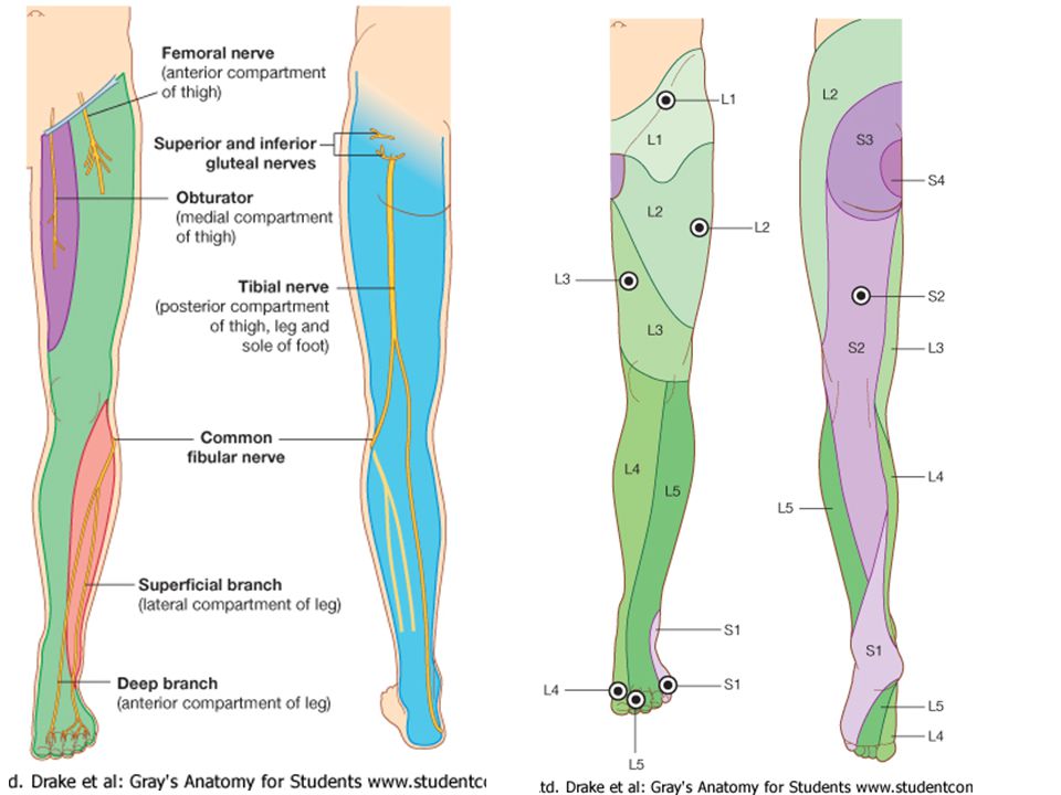
Posterior Anterior Map of Dermatomes

33
Innervation of the Skin: Dermatomes
Upper limb – skin is supplied by nerves of the brachial plexus Lower limb: Lumbar nerves – anterior surface Sacral nerves – posterior surface

34
Elsie (L.3 ) is trying to rescue her clumsy man Slim (SI) from a septic tank (SciaTIC nerve), using a rope and a balloon. He has some GLUe (nerves to GLUteus muscles) on his leg. She is pregnant· is FEMale (FEMoral nerve) and has an OBstetric condition (OBturator nerve).
 on his leg. She is pregnant· is FEMale (FEMoral nerve) and has an OBstetric condition (OBturator nerve)..")
36
lumbar disc herniation

37
lumbar disc herniation
Disc Level Root Comp. Weakness Reflex Involvement Sensory Loss Pain Distribution L3-L4 L4 quadriceps, tibialis anterior knee jerk medial knee and shin anterior thigh L4-L5 L5 extension of big toe no significant big toe back of thigh, lateral calf L5-S1 S1 gastrocnemius (ankle plantar flexion) Achilles lateral foot and heel back of thigh and calf
 Achilles. lateral foot and heel. back of thigh and calf.")
38
Lower limb dermatomes L1 Dermatome: over trochanter and groin
L2 Dermatome: front of thigh to knee L3 Dermatome: upper buttock, anterior thigh and knee, medial lower leg L4 Dermatome: lateral Buttock, lateral thigh, medial leg, dorsum of foot, big toe L5 Dermatome: Buttock, posterior and lateral thigh, lateral aspect of leg, dorsum of foot, medial half of sole, first, second, and third toes S1 Dermatome: Buttock, thigh, and posterior leg S2 Dermatome: Buttock, thigh, and posterior leg S3 Dermatome: Groin, medial thigh to knee S4 Dermatome: Perineum, genitals, lower sacrum

Similar presentations

>")


