Download presentation
Presentation is loading. Please wait.

2
Lower Limb CONTENTS OF THE ANTERIOR FASCIAL COMPARTMENT OF THE LEG
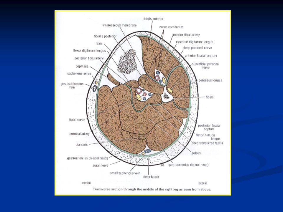
Muscles: The tibialis anterior, extensor digitorum longus, peroneus tertius and extensor hallucis longus. Blood supply: Anterior tibial artery. Nerve supply: Deep peroneal nerve.

4
RETINACULA OF THE ANKLE
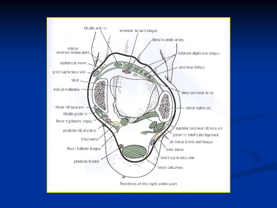
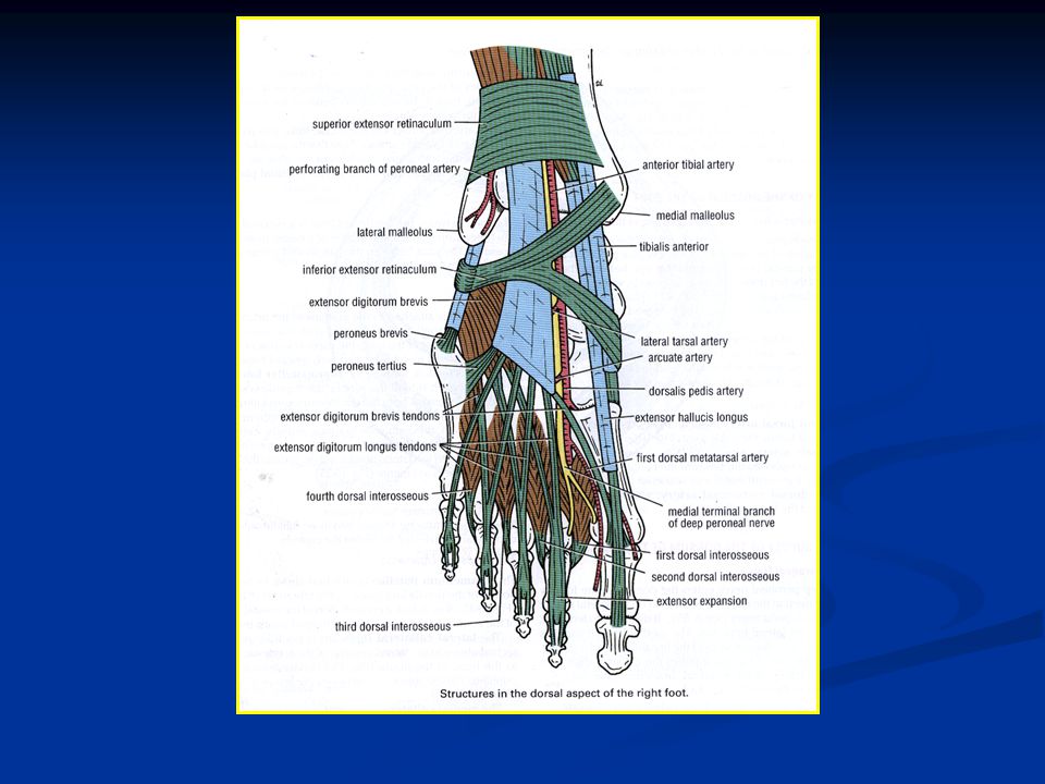
In the region of the ankle joint, the deep fascia is thickened to form a series of retinacula. They serve to keep the long tendons in position and act as modified pulleys. The superior extensor retinaculum It is attached to the distal ends of the anterior borders of the fibula and tibia. Near its medial end, it splits to enclose the tendon of the tibialis anterior muscle.

5
RETINACULA OF THE ANKLE
The inferior extensor retinaculum It is a Y-shaped band of deep fascia that is attached by its stem to the upper surface of the calcaneum. The upper limb of the Y is attached to the medial malleolus and the lower limb is continuous with the plantar fascia on the medial border of the foot. The muscles of the anterior group of the leg pass through the retinaculum.

6
ANTERIOR ASPECT OF THE ANKLE
Structures That Pass Anterior to the Extensor Retinacula from Medial to Lateral: Saphenous nerve and great saphenous vein (in front of the medial malleolus). Superficial peroneal nerve (medial and lateral branches). Structures That Pass Beneath or Through the Extensor Retinacula from Medial to Lateral: Tibialis anterior tendon. Extensor hallucis longus tendon. Anterior tibial artery with venae comitantes. V Deep peroneal nerve. Extensor digitorum longus tendons. Peroneus tertius.
. Superficial peroneal nerve (medial and lateral branches). Structures That Pass Beneath or Through the Extensor Retinacula from Medial to Lateral: Tibialis anterior tendon. Extensor hallucis longus tendon. Anterior tibial artery with venae comitantes. V. Deep peroneal nerve. Extensor digitorum longus tendons. Peroneus tertius.")
12
Anterior Compartment of Leg
Tibialis Anterior Muscle Extensor Digitorum Longus Extensor Hallucis Peroneus Tertius Origin of the Muscles Ext. Hallucis (Insertion) Ext. Digitorum Longus (Insertion) Peroneus Tertius (Insertion)
 Ext. Digitorum Longus (Insertion) Peroneus Tertius (Insertion)")
13
Front of Leg Tibialis Anterior
Origin: From the upper half of the lateral surface of the tibia and from the interosseous membrane. Insertion: The tendon passes through both extensor retinacula and is attached to the medial cuneiform bone and the adjoining base of the first metatarsal bone.

14
Front of Leg Tibialis Anterior Nerve supply: Deep peroneal nerve.
Action: Extends (dorsiflexes) the foot at the ankle joint. Inverts the foot. It assists in holding up the medial longitudinal arch of the foot.
 the foot at the ankle joint. Inverts the foot. It assists in holding up the medial longitudinal arch of the foot.")
15
Front of Leg Extensor Digitorum Longus
Origin: From the upper two thirds of the anterior surface of the fibula and from the interosseous membrane Insertion: The tendons pass behind the superior and through the inferior extensor retinacula. Each tendon forms an extensor expansion with the other tendons of the same toe and is inserted onto: The middle and distal phalanges of the lateral four toes.

16
Front of Leg Extensor Digitorum Longus
Nerve supply: Deep peroneal nerve. Action: Extends the toes and extends the foot at the ankle joint.

17
Front of Leg Peroneus Tertius Origin: Insertion:
It arises from the lower third of the anterior surface of the fibula and the interosseous membrane. Insertion: Its tendon follows the tendons of extensor digitorum longus behind the superior and through the inferior extensor retinacula. It is inserted into the medial side of the dorsal aspect of the base of the fifth metatarsal bone.

18
Front of Leg Peroneus Tertius Nerve supply: Deep peroneal nerve.
Action: Extends the foot at the ankle joint. Everts the foot.

19
Front of Leg Extensor Hallucis Longus
Origin: From the middle half of the anterior surface of the fibula and from the interosseous membrane. Insertion: The tendon passes behind the superior and through the inferior extensor retinacula. It is inserted into the base of the distal phalanx of the great toe.

20
Front of Leg Extensor Hallucis Longus
Nerve supply: Deep peroneal nerve. Action: Extends the big toe and extends the foot at the ankle joint. It also assists in inversion of the foot.

23
Blood Supply of the anterior Compartment of the Leg
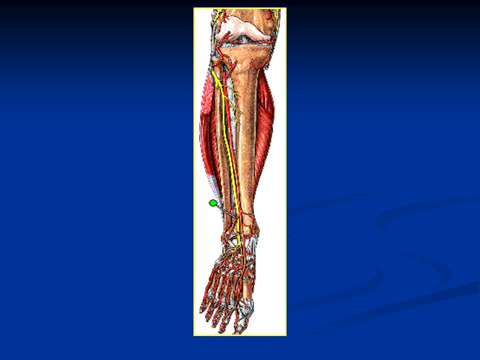
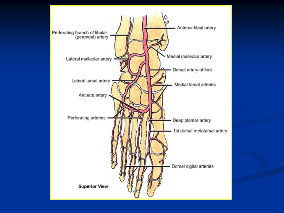
Anterior Tibial Artery The anterior tibial artery is the smaller of the terminal branches of the popliteal artery. It arises at the level of the lower border of the popliteus muscle and passes forward into the anterior compartment of the leg through an opening in the upper part of the interosseous membrane. It descends on the anterior surface of the interosseous membrane, accompanied by the deep peroneal nerve.

26
Anterior Tibial Artery
The nerve first lies lateral to the artery, then anterior to it and finally lateral again. In the upper part of its course, it lies deep beneath the muscles of the compartment. In the lower part of its course, it lies superficial in front of the lower end of the tibia. Having passed behind the superior extensor retinaculum, it has the tendon of the extensor hallucis longus on its medial side and the deep peroneal nerve and the tendons of extensor digitorum longus on its lateral side. In front of the ankle joint, the artery becomes the dorsalis pedis artery.

27
Anterior tibial artery & nerve

28
Anterior Tibial Artery
Branches Muscular branches to neighboring muscles. Anastomotic branches that anastomose with branches of other arteries around the knee and ankle joints. Venae comitantes of the anterior tibial artery join those of the posterior tibial artery in the popliteal fossa to form the popliteal vein.

29
Nerve Supply Deep Peroneal Nerve
The deep peroneal nerve is one of the terminal branches of the common peroneal nerve. It arises in the substance of the peroneus longus muscle on the lateral side of the neck of the fibula. The nerve enters the anterior compartment by piercing the anterior fascial septum. It then descends deep to the extensor digitorum longus muscle, first lying lateral, then anterior and finally lateral to the anterior tibial artery.

31
Nerve Supply Deep Peroneal Nerve
The nerve passes behind the extensor retinacula. Branches: Muscular branches to the tibialis anterior, the extensor digitorum longus, the peroneus tertius and the extensor hallucis longus. Articular branch to the ankle joint.

32
Lower Limb CONTENTS OF THE LATERAL FASCIAL COMPARTMENT OF THE LEG
Muscles: Peroneus longus and peroneus brevis. Blood supply: Branches from the peroneal artery. Nerve supply: Superficial peroneal nerve.

34
RETINACULA OF THE ANKLE
The superior peroneal retinaculum It is a thickened band of deep fascia that extends from the lateral malleolus to the lateral surface of the calcaneum. The tendons of the peroneus longus and brevis pass deep to the retinaculum.

35
RETINACULA OF THE ANKLE
The inferior peroneal retinaculum It is a thickened band of deep fascia that is attached to the peroneal tubercle and to the calcaneum above and below the peroneal tendons. The tendons of the peroneus longus and brevis pass deep to the retinaculum.

37
Lateral ASPECT OF THE ANKLE
Structures That Pass behind the Lateral Malleolus Superficial to the Superior Peroneal Retinaculum: The sural nerve. Small saphenous vein. Structures That Pass behind the Lateral Malleolus beneath the Superior Peroneal Retinaculum: The peroneus longus and brevis tendons. The two muscles also pass beneath the inferior peroneal retinaculum.

38
Lateral side of Leg Peroneus Longus
Origin: From the upper two thirds of the lateral surface of the fibula. Insertion: It is inserted into the medial cuneiform and the base of the first metatarsal. Nerve supply: Superficial peroneal nerve.

40
Lateral side of Leg Peroneus Longus Action:
Plantar flexes the foot at the ankle joint. Everts the foot. It plays an important part in holding up the lateral longitudinal arch in the foot and helps to maintain the transverse arch of the foot.

41
Lateral side of Leg Peroneus Brevis
Origin: From the lower two thirds of the lateral surface of the fibula. Insertion: It is inserted into the tubercle on the base of the fifth metatarsal bone. Nerve supply: Superficial peroneal nerve. Action: Plantar flexes the foot at the ankle joint. Everts the foot. It assists in holding up the lateral longitudinal arch of the foot.

43
Artery of the Lateral Fascial Compartment of the Leg
Numerous branches from the peroneal artery which lies in the posterior compartment of the leg, pierce the posterior fascial septum and supply the peroneal muscles.

44
Nerve of the Lateral Fascial Compartment of the Leg
Superficial Peroneal Nerve The superficial peroneal nerve is one of the terminal branches of the common peroneal nerve. It arises in the substance of the peroneus longus muscle on the lateral side of the neck of the fibula. It descends between the peroneus longus and brevis muscles and in the lower part of the leg it becomes cutaneous.

45
Nerve of the Lateral Fascial Compartment of the Leg
Superficial Peroneal Nerve Branches: Muscular branches: to the peroneus longus and brevis. Cutaneous: Medial and lateral branches are distributed to the skin on the lower part of the front of the leg and the dorsum of the foot. In addition, branches supply the dorsal surfaces of the skin of all the toes, except the adjacent sides of the first and second toes and the lateral side of the little toe.

46
The Dorsum of the Foot Extensor Digitorum Brevis
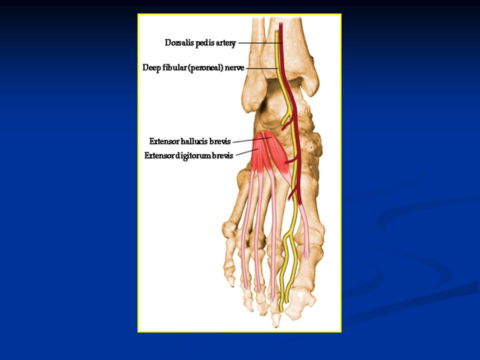
Origin: From the anterior part of the upper surface of the calcaneum and from the inferior extensor retinaculum. Insertion: The muscle gives rise to four tendons that pass forward and medially. The most medial tendon (sometimes called tendon of extensor hallucis brevis) is inserted into the base of the proximal phalanx of the big toe. Dr. Iman Abdel Aal
 is inserted into the base of the proximal phalanx of the big toe. Dr. Iman Abdel Aal.")
47
Dr. Iman Abdel Aal

48
The Dorsum of the Foot Extensor Digitorum Brevis
The lateral three tendons join the long extensor tendons passing to the second, third and fourth toes. Nerve supply: Deep peroneal nerve. Action: Extends the first, second, third and fourth toes at the interphalangeal and metatarsophalangeal joints.

52
ARTERY OF THE DORSUM OF THE FOOT
Dorsalis Pedis Artery (the Dorsal Artery of the Foot) The dorsalis pedis artery begins in front of the ankle joint as a continuation of the anterior tibial artery. It terminates by passing downward into the sole between the two heads of the first dorsal interosseous muscle, where it joins the lateral plantar artery and completes the plantar arch.
 The dorsalis pedis artery begins in front of the ankle joint as a continuation of the anterior tibial artery. It terminates by passing downward into the sole between the two heads of the first dorsal interosseous muscle, where it joins the lateral plantar artery and completes the plantar arch.")
54
ARTERY OF THE DORSUM OF THE FOOT
Dorsalis Pedis Artery It is superficial in position and is crossed by the inferior extensor retinaculum and the first tendon of extensor digitorum brevis. On its lateral side lie the terminal part of the deep peroneal nerve and the extensor digitorum longus tendons. On the medial side lies the tendon of extensor hallucis longus.

55
ARTERY OF THE DORSUM OF THE FOOT
Dorsalis Pedis Artery Branches Lateral tarsal artery: which crosses the dorsum of the foot just below the ankle joint. Arcuate artery: which runs laterally under the extensor tendons opposite the bases of the metatarsal bones. It gives off metatarsal branches to the toes. First dorsal metatarsal artery: which supplies both sides of the big toe.

Similar presentations


.>")


