Download presentation
Presentation is loading. Please wait.

1
Blood supply of the leg and foot
WINDSOR UNIVERISITY SCHOOL OF MEDICINE

2
ARTERIES IN THE POSTERIOR COMPARTMENT
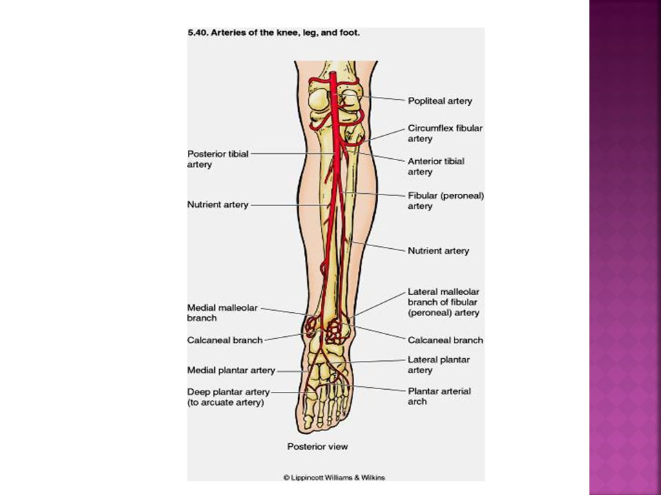
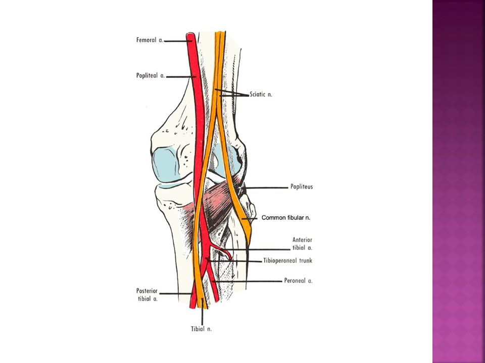
The posterior tibial artery, the larger and more direct terminal branch of the popliteal artery, provides the blood supply to the posterior compartment of the leg and to the foot It begins at the distal border of the popliteus Branches largest branch, the fibular artery . During its descent, the posterior tibial artery is accompanied by the tibial nerve and veins. The artery runs posterior to the medial malleolus, from which it is separated by the tendons of the tibialis posterior and flexor digitorum longus(tarsal tunnel) Inferior to the medial malleolus, it runs between the tendons of the flexor hallucis longus and flexor digitorum longus. Deep to the flexor retinaculum the posterior tibial artery divides into medial and lateral plantar arteries
 Inferior to the medial malleolus, it runs between the tendons of the flexor hallucis longus and flexor digitorum longus. Deep to the flexor retinaculum the posterior tibial artery divides into medial and lateral plantar arteries.")
4
Posterior Tibial Pulse
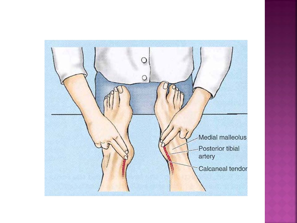
The posterior tibial pulse can usually be palpated between the posterior surface of the medial malleolus and the medial border of the calcaneal tendon Palpation of the posterior tibial pulses is essential for examining patients with occlusive peripheral arterial disease. intermittent claudication, characterized by leg pain and cramps, develops during walking and disappears after rest. These conditions result from ischemia of the leg muscles caused by narrowing or occlusion of the leg arteries

6
ARTERIES IN THE ANTERIOR COMPARTMENT
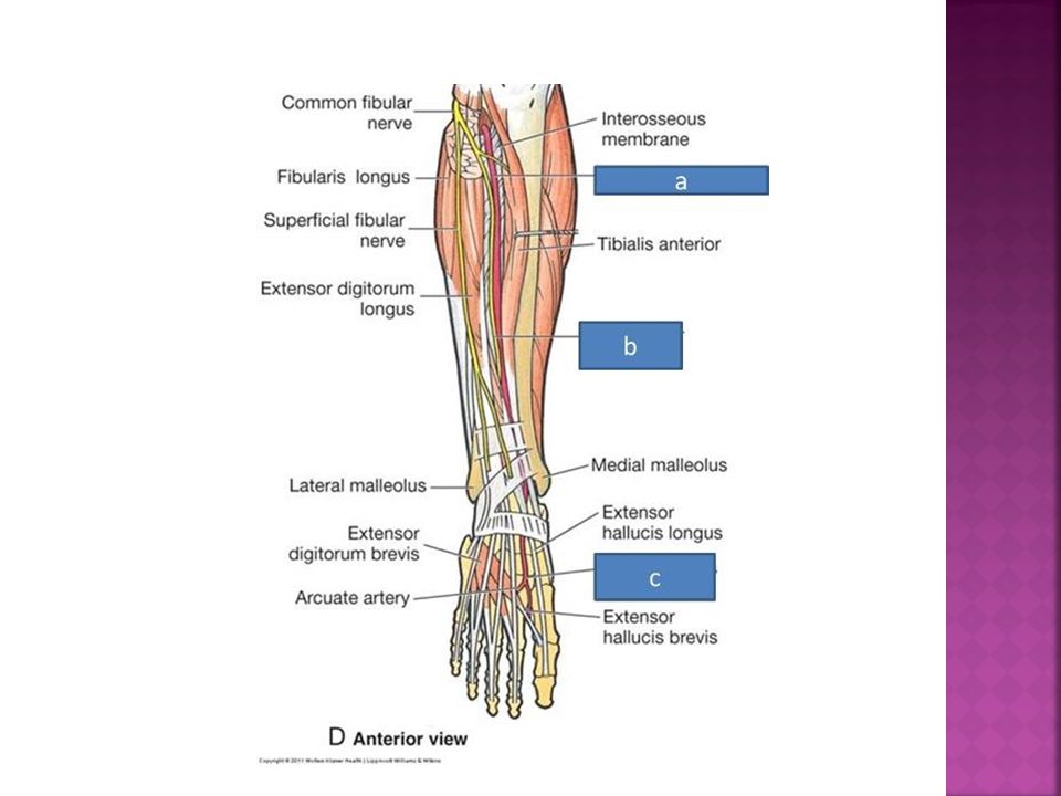
Anterior tibial artery supplies structures in the anterior compartment Begins at the inferior border of popliteus muscle Passes anterior through the gap in the superior part of the interroseous membrane to descend between the tendons of tibialis anterior and extensor digitorum longus muscles Continues in the foot as the dorsalis pedis artery

9
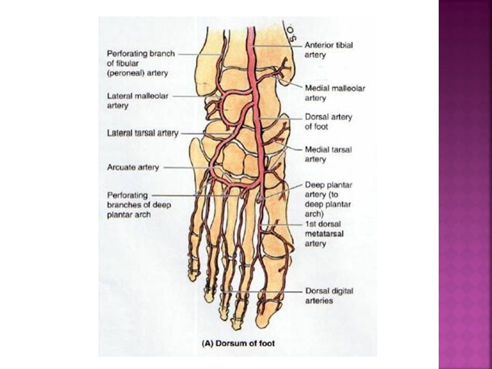
Dorsal Artery of the Foot
The dorsal artery of the foot (L. arteria dorsalis ped is the direct continuation of the anterior tibial artery The dorsal artery begins midway between the malleoli and runs anteromedially, deep to the inferior extensor retinaculum between the extensor hallucis longus and the extensor digitorum longus tendons on the dorsum of the foot. it divides into the 1st dorsal metatarsal artery and a deep plantar artery. The deep plantar artery enters the sole of the foot, where it joins the lateral plantar artery to form the deep plantar arch

11
Other arteries on the dorsum of the foot
The lateral tarsal artery The 1st dorsal metatarsal artery The arcuate artery perforating branches dorsal digital arteries

12
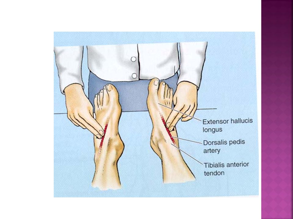
Palpation of the Dorsalis Pedis Pulse
Is evaluated during a physical examination of the peripheral vascular system. The pulses are usually easy to palpate because the dorsal arteries of the foot are subcutaneous and pass along a line from the extensor retinaculum to a point just lateral to the Extensor Hallucis Longus tendons absent dorsalis pedis pulse usually suggests vascular insufficiency resulting from arterial disease. The five P signs of acute arterial occlusion are pain, pallor, paresthesia, paralysis, and pulselessness.

14
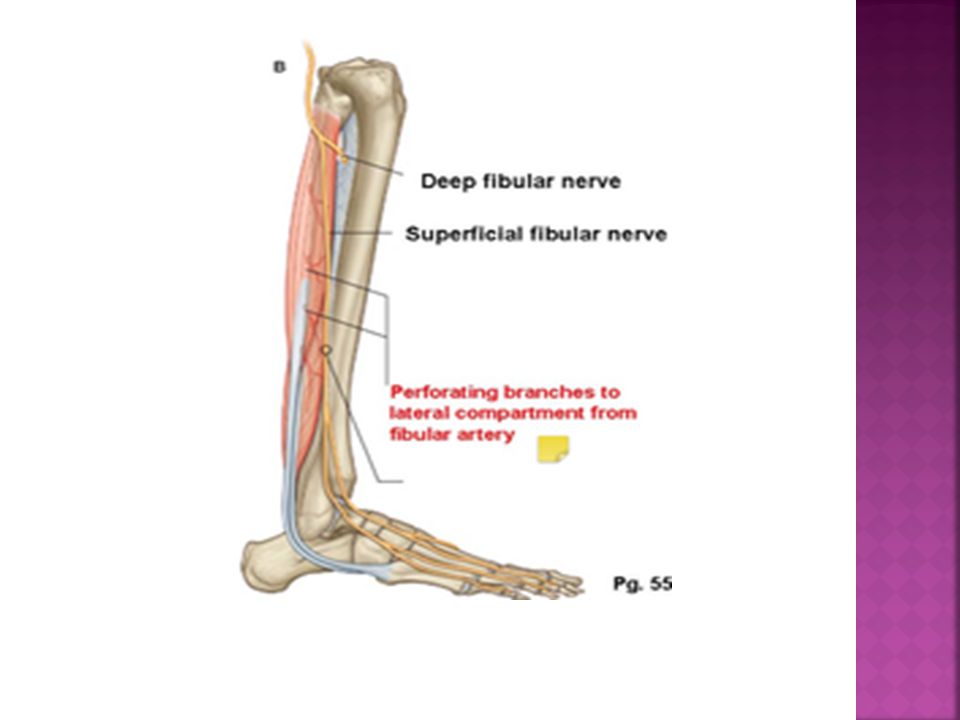
ARTERIES IN THE LATERAL COMPARTMENT
By perforating branches of anterior tibial artery proximally Distally by perforating bracnches of fibular artery

16
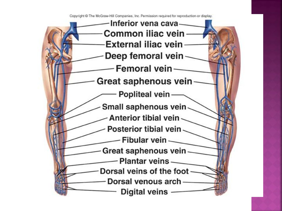
Venous drainage of the lower limb.
Significance of venous return from the lower limb- antigravity. The are subdivided superficial and deep veins the superficial veins are between the two layers of superficial fascia while the deep veins accompany the arteries. Both sets of veins are provided with valves more numerous in the deep than in the superficial set.

17
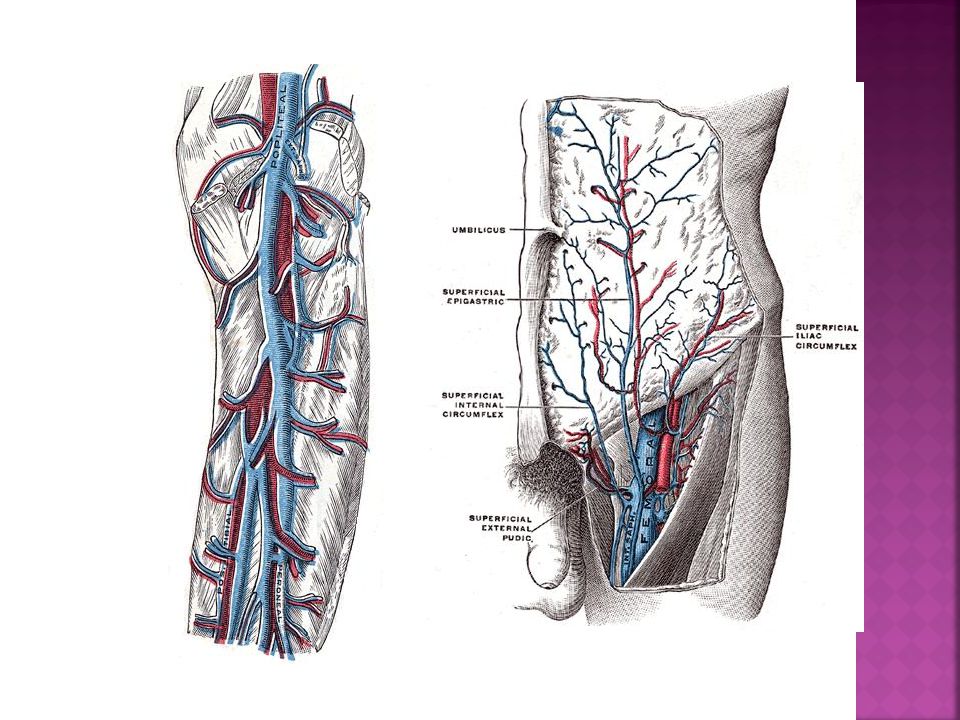
The great saphenous vein (long saphenous vein)
is the longest vein in the body. Begins in the medial marginal vein of the dorsum of the foot and ends in the femoral vein about 3 cm below the inguinal ligament. It passes in front of the medial malleolus and along the medial side of the leg in relation with the saphenous nerve behind the medial condyles of the tibia and femur and along the medial side of the thigh to end in the femoral vein. Main Tributaries: -superficial circumflex iliac -superficial external pudendal

18
Small saphenous vein ( short saphenous vein)
- Small saphenous vein ( short saphenous vein) begins behind the lateral malleolus as a continuation of the lateral marginal vein,ascends and then crosses it to reach the middle of the back of the leg. It perforates the deep fascia at the popliteal fossa and ends in the popliteal vein Tributaries -Branch from great saphenous vein -Lateral marginal vein - Numerous tributaries from the back of the leg.
 begins behind the lateral malleolus as a continuation of the lateral marginal vein,ascends and then crosses it to reach the middle of the back of the leg. It perforates the deep fascia at the popliteal fossa and ends in the popliteal vein. Tributaries. -Branch from great saphenous vein. -Lateral marginal vein. - Numerous tributaries from the back of the leg.")
19
The great and small saphenous veins and their tributaries
Greys Atlas The great and small saphenous veins and their tributaries

20
Deep veins They accompany the arteries and their branches; they possess numerous valves. The deep plantar venous arch lies alongside the plantar arterial arch drains into the medial and lateral plantar veins The posterior tibial vein accompanies the posterior tibial artery and joined by the peroneal vein. The anterior tibial vein is the upward continuation of the venæ comitantes of the dorsalis pedis artery. They leave the front of the leg by passing between the tibia and fibula, over the interosseous membrane, and unite with the posterior tibial vein to form the popliteal vein.

21
The Popliteal Vein ascends through the popliteal fossa to the opening in the Adductor magnus, where it becomes the femoral vein. It receives tributaries corresponding to the branches of the popliteal artery, and the small saphenous vein. The femoral vein accompanies the femoral artery through the upper two-thirds of the thigh. It receives many muscular tributaries, and about 4 cm below the inguinal ligament is joined by the deep femoral vein and is joined by the great saphenous vein before it terminates. The deep femoral vein receives tributaries corresponding to the perforating branches of the profunda artery, and through these establishes communications with the popliteal vein below and the inferior gluteal vein above. It also receives the medial and lateral femoral circumflex veins.

22
Popliteal vein Formed by union of venae comitantes of ant, post tibial arteries. At the lower border of popliteus. Continues as femoral vein at adductor opening. TRIBUTARIES: 1- Veins accompany arteries. 2- Small saphenous v.

25
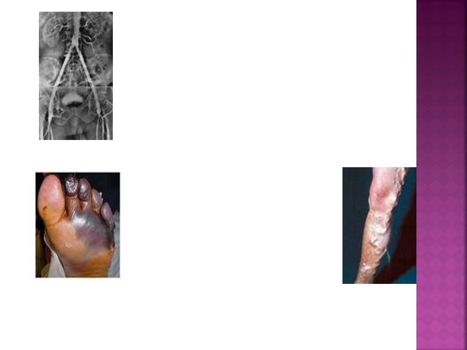
Clinical application clinical study- angiography palpation for pulses gangrene varicose veins

Similar presentations

.>")

>")


