Download presentation
Presentation is loading. Please wait.

1
Hypothalamus & Limbic System Chapter 12 Excluding pages pg263-278

2
Hypothalamus Regulates Homeostasis Hunger Thirst Body Temp, Blood Pressure Sex Drives & Behavior Emotions Via Limbic System Pituitary Gland Circadian Rhythms

3
Neuroanatomy of Hypothalamus Know the names of the nuclei on both sections –Periventricular, medial and lateral –Preoptic anterior, middle and posterior

4
Neural Basis of Emotion Fear, Anxiety, & Envy& Love, Joy Role of Cingulate Gyrus, Amygdala, Hypothalamus, Hippocampus

6
Emotions Emotional Experience Input from senses Processed by cerebral cortex Emotional Expression Behavioral output from somatic motor, autonomic and hypothalamus

8
Theories of Emotion James Lange Theory 1884 Experience emotions IN RESPONSE to physiological changes in our body Feel sad because we cry NOT cry because we feel sad The emotion is the physiology

9
Cannon-Bard Theory 1927: Emotional experience can occur independently of emotion expression Transect animal spinal cord and emotional expression observed. Removal or damage to somatic sensory system does not diminish emotion experience.

11
Discrepency for James-Lange The same physiological characteristics can occur without the emotion such as in illness fever etc. Difference according to Cannon is the activation of the thalamus (hypothalamus) for the emotional response
 for the emotional response.")
12
Limbic Lobe 1878 Paul Broca identified medial surface of cerebrum that are different from the rest of cortex—called it border=limbic lobe Cortex surrounding corpus callosum Thought to be involved in olfaction

13
Papez Circuit James Papez 1930s identified limbic structures involved in emotion (added the thalamic structures to the limbic lobe) Cingulate cortex to hippocampus to hypothalamus via the fornix and from hypothalamus to anterior nuclei of thalamus Neocortex connects to cingulate cortex Allows one to experience emotion
 Cingulate cortex to hippocampus to hypothalamus via the fornix and from hypothalamus to anterior nuclei of thalamus Neocortex connects to cingulate cortex Allows one to experience emotion")
16
Limbic System Limbic Lobe and Papez Circuit together This distinguishes human emotions and responses to situations from the stereotypical response of animals due to reflexive systems involving brainstem

17
Frontal Lobes of Cortex Provides Rationale Control of emotional disposition & involved in personality Injury to frontal lobes causes change in personality Control of emotions and impulse control Example of Phineas Gage

19
Pathologies Tumors and injury to areas of the brain lead to emotional changes. Damage to cingulate cortex lead to emotional disturbances: fear, depression, irritability

20
Fear, Agression & Anxiety Learned Fear, Anxiety & Temporal Lobes and AMYGDALA

22
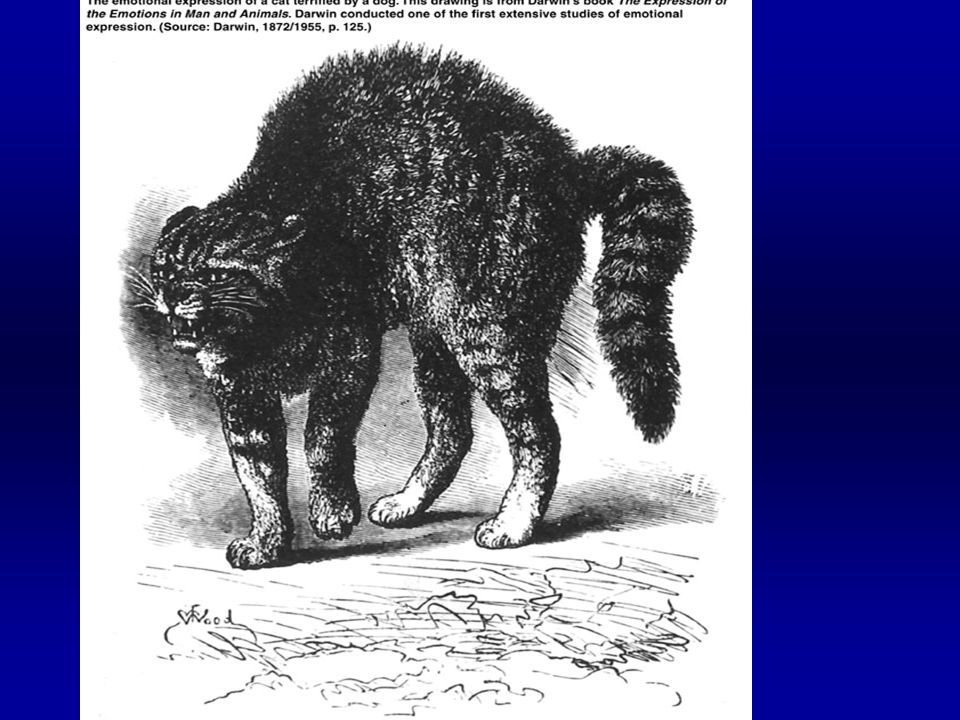
Kluver & Bucy Neuroscientist Remove bilateral temporal lobes and monkeys cannot experience fear, approach humans other monkeys and dangerous situtations Cannot recognize objects by vision; called psychic blindness-use mouth to identify objects seen striking increase in sexual activity

23
Kluver-Bucy Syndrome Humans with temporal lobe lesions show similar behavior as monkeys with temporal lobectomy Have flattened emotions, don’t feel happy, sad etc

24
Amygdala Neurons at the pole of the temporal lobe below the cortex on the medial side Greek name for almond shape Has 3 nuclei, basolateral, corticomedial and central Afferents from all lobes of neocortex & hippocampus and cingulate gyrus

26
Input to Amygdala Basolateral nuclei receive sensory input (visual, gustatory, auditory and tactile); also projects to cortex for perception of emotion Corticomedial nuclei receive olfactory inputs Central nuclei contain output neurons to hypothalamus and periaqueductal grey in brainstem for physiological responses
; also projects to cortex for perception of emotion Corticomedial nuclei receive olfactory inputs Central nuclei contain output neurons to hypothalamus and periaqueductal grey in brainstem for physiological responses")
28
Damage to Amygdala Decreases emotional response Kluver-Bucy Syndrome=reduced emotionality Fearlessness SM human cannot recognize emotional expressions on faces that are fearful, anxious & angry but recognize happy & disgust Bilateral amygdala removal reduces memory

29
Electrical Stimulation of Amygdala Cause affective rage when basalateral nuclei is stimulated Corticomedial stimulation reduces aggression

30
Learned Behaviors Require the amygdala and work through 2 pathways. Integrate information from all sensory systems and orchestrate the physiological and physchological response –Ventral amygdofugal pathway –Stria terminalis

31
Do Not learn Pathway Names

32
Hypothalamus-brainstem Autonomic nuclei in the brainstem receive synaptic input from hypothalamus via –Medial forebrain bundle –Dorsal longitudinal fasciculus

33
Aggressive Behaviors Androgen levels in males can alter aggressive behaviors Predatory aggression: purpose is getting food, little sympathetic NS activity –Medial hypothalamus Affective aggresion: purpose is scare off enemies/protection –Lateral hypothalamus

34
Hypothalamus and Rabies Rabies causes excess rage and aggression Rabies virus damages hypothalamic neurons Led identification of hypothalamus as critical brain area involved in anger

35
Electrical Stimulation of Hypothalamus Depending on area, animal shows different behaviors Associated with eating, sniff & eat Associated with fear or anger Demonstrates 2 functions of hypothalamus –Metabolic regulation; homeostasis –Coordinated somatic & visceral responses

36
Serotonin Serotonin containing neurons located in Raphe nucleus in brainstem that project via medial forebrain bundle to hypothalamus & other limbic structures Aggressive mice have decreased serotonin turnover Drugs that block serotonin release or synthesis cause increase in aggression

37
Serotonin Receptors 14 5HT receptor subtypes Mice with no (knock-out) gene for 1A and 1B isoform, the type found in Raphe Nucleus are more aggressive & anxious when stressed otherwise act normally Specific agonist of 1A and 1B reduce anxiety
 gene for 1A and 1B isoform, the type found in Raphe Nucleus are more aggressive & anxious when stressed otherwise act normally Specific agonist of 1A and 1B reduce anxiety")
38
Memory Systems Hippocampus

41
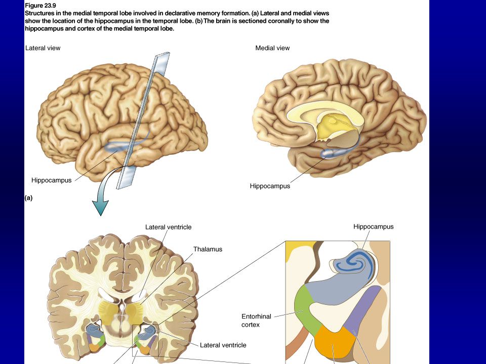
Hippocampus & Relational Memory Highly processed information from association cortex areas enter hippocampus Hippocampus integrates them—ties them together and then output is stored in other cortical areas Allows you to retrieve all the information about an event

42
Patients & Syndromes HM-mediotemporal lobe NA--thalamus Korsakoffs-thalamus & hypothalamus

43
Amnesia Anterograde –Cannot form any new types of memories so always live at time of injury Retrograde –Cannot recall stored memories for a specific time period

44
Memory Declarative: Explicit –Facts & Events Easy to form, easy to lose Medial Temporal Lobe & Thalamus Non-Declarative: Implicit Takes repetition, hard to lose –Procedural Skills & Habits –Striatum –Classical Conditioning Skeletal Muscles –Cerebellum Emotional Responses –Amygdala

46
Conscious Recollection Only declarative memories & not non- declarative memories

47
Declarative Memory Essential Anatomy –Medial Temporal Lobe –Entorhinal and Perirhinal, Parahippocampal Cx –Hippocampus –Fornix to Mammilary Body of Hypothalamus –Anterior & Dorsomedial Thalamus that project to cingulate cx (limbic system)
")
48
HM Had bilateral mediotemporal lobes removed due to epilepsy Removed amygdala, anterior 2/3 of hippocampus, temporal cortex Had anterograde amnesia Studied by Brenda Milner Could learn by procedural memory but had no recollection of having learned task

50
Squire & Mishkin Neuroscientists create an animal model for HM symptoms Lesioned amygdala, hippocampus and perirhinal cortex in temporal lobe of monkeys and found that they could no longer perform in recognition memory tests Later showed that perirhinal cortex is most important for new memory; temporary storage? Memory consolidation?

52
Diencephalon & Memory Processing Anterior thalamic nucleus Dorsal Medial Thalamic nucleus Mammillary bodies in hypothalamus

54
Dorsal medial thalamic nucleus Receives input from temporal lobe structures including amygdala & inferiortemporal cortex Projects to all frontal cortex areas

55
NA Air Force technician injured by fencing foil –penetrated the dorsalmedial thalamus Developed retrograde amnesia of previous 2 years and severe anterograde amnesia Supports role of thalamus in memory

57
Lashley Lashley: 1920s studied rats in maze after cortical lesions Found that all cortical areas are involved in memory

59
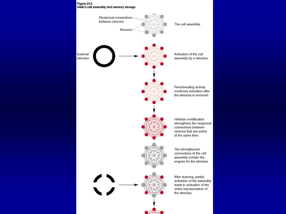
Hebb, Lashley student suggested CELL ASSEMBLY = all cells that respond to an external stimulus & are reciprocally interconnected Neurons that fire together, wire together 1949 Organization of Behavior Sensory cortex also stores memory Led to neural networks computer modeling

61
Circuit using limbic structures Hippocampal output axons travel as a bundle, the fornix, to the mammillary bodies of the hypothalamus Mammillary body axons project to anterior thalamic nucleus

62
Definitions Declarative & NonDeclarative Long term & Short Term Procedural & Working Experience Dependent Brain Development Anterograde and Retrograde Amnesia

63
Learning & Memory Adaptations of brain circuitry to life experience Learning = acquisition of new information or knowledge Memory = retention of learning

64
Long Term/Short Term Memory Long Term: last years but is selective Short term: last seconds to hours

66
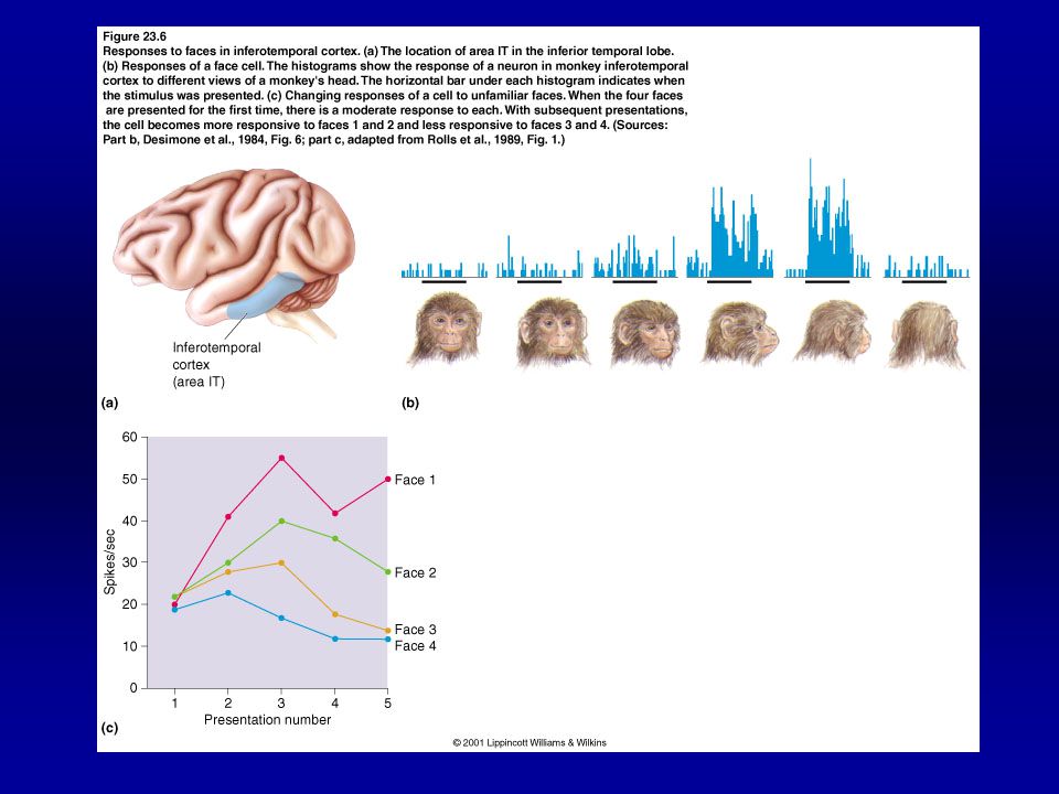
Memory based on Vision Should be found in cortical area involved in vision processing inferiortemporal cortex: higher order processing of visual information—stores memory of previously seen objects Allows recognition of visual objects –Remember Kluver-Bucy pyschic blind monkeys

68
Penfield Neurosurgeon in the 1950’s removed epileptic foci after stimulation Found that stimulation of temporal lobe in awake patients caused halucinations or memory retrieval

Similar presentations