Download presentation
Presentation is loading. Please wait.

1
BLOOD

2
Learning Objectives Describe the primary functions' of blood
Describe the characteristics of blood Discuss the blood types including Rh factor Describe common disorders of blood

4
Hematophobia = fear of blood
Blood transports substances and maintains homeostasis in the body Hematophobia = fear of blood

5
Blood and Blood Cells Blood is a type of CONNECTIVE TISSUE
It has two basic components: CELLS (rbc, wbc, platelets) = 45% Plasma (water, proteins, amino acids..etc) = 55% What is hematology? (study of blood) Is blood a fluid tissue (yes)
 = 45% Plasma (water, proteins, amino acids..etc) = 55% What is hematology (study of blood) Is blood a fluid tissue (yes)")
6
Hematocrit - volume of blood cells in a sample, should be 45%
Hematocrit - volume of blood cells in a sample, should be 45%. The remaining fluid is plasma (55%). To determine the percentages, blood is placed in a centrifuge What is the difference between blood plasma and blood serum? (Plasma contains clotting factors , serum does not)
. To determine the percentages, blood is placed in a centrifuge. What is the difference between blood plasma and blood serum (Plasma contains clotting factors , serum does not)")
7
Three Types of Blood Cells
RBC – million WBC – 5-10,000 Platelets – 250,000 Three Types of Blood Cells red blood cells (erythrocytes) white blood cells (leukocytes) platelets (thrombocytes)
 white blood cells (leukocytes) platelets (thrombocytes)")
8
BLOOD RBC live about 4 months before going to the liver Granular leukocytes live only a few days Non-granular leukocyte 6 months or more

10
HEMATOPOEISIS – formation of blood cells (bone marrow)
Biconcave discs 5 million per cubic millimeter Lack nuclei What is myeloid tissue better known as : Red bone marrow – sternum, ribs, and hip bones HEMATOPOEISIS – formation of blood cells (bone marrow) Liver & Spleen - phagocytosis
 Liver & Spleen - phagocytosis.")
11
Main Functions of RBCs Transports oxygen, picks up carbon dioxide
HEMOGLOBIN - molecule that combines with O2 IRON is critical to synthesize hemoglobin Disk shape without nuclei Because of their large number and their unique shape their total surface area is enormous , if laid out their total surface area is bigger than a football field

12
Oxygen Levels Oxyhemoglobin = plenty of oxygen; bright red
Deoxyhemoglobin = low in O2, “bluish red”

13
Elements Critical to RBC Production
Folic Acid Vitamin B12 Iron Too few RBC = anemia

14
WHITE BLOOD CELLS (Leukocytes)
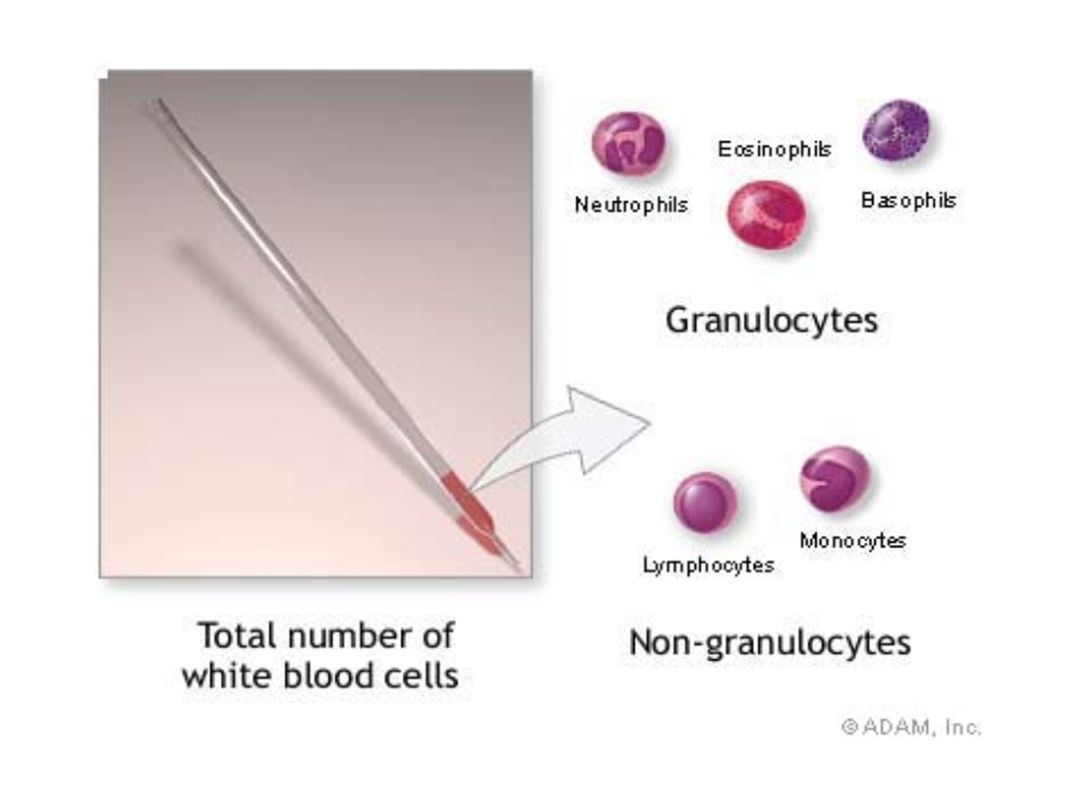
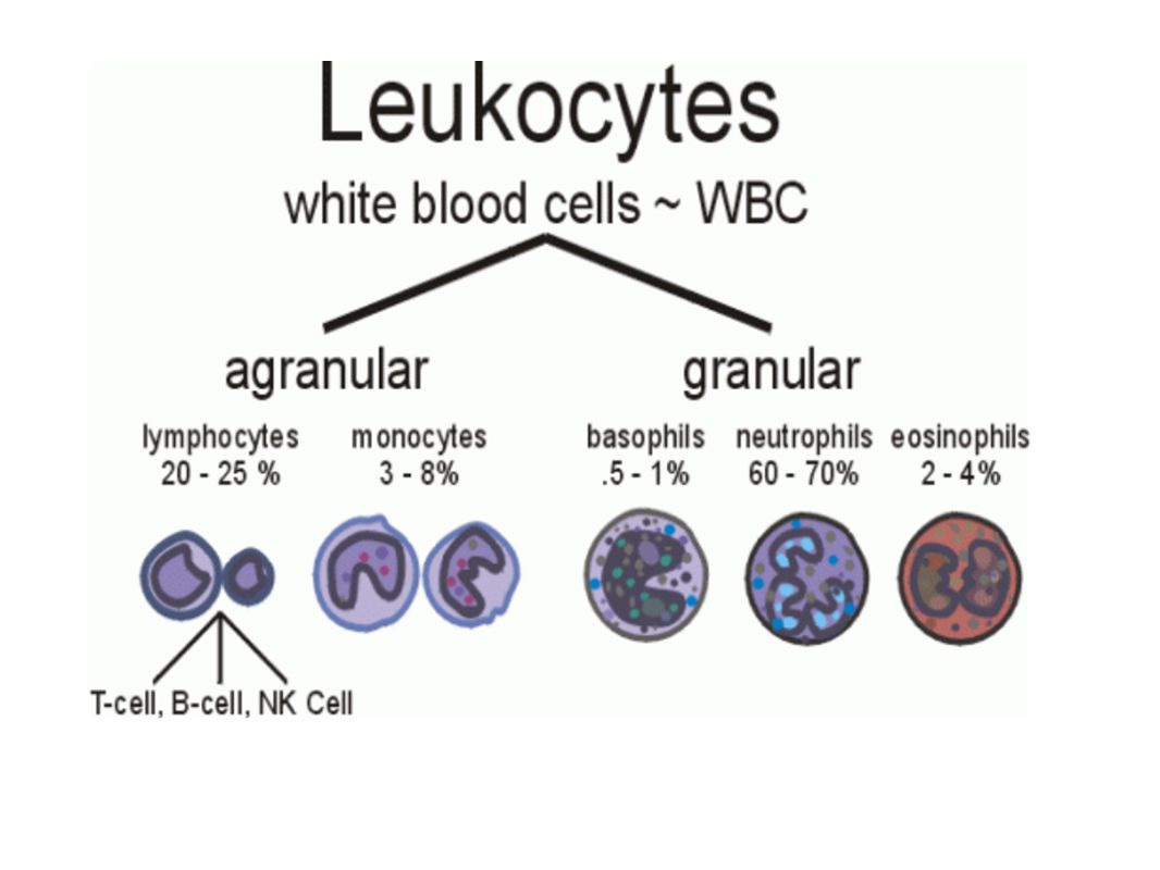
General function is to protect the body against disease There are FIVE different kinds of WBCs Granulocytes (granular cytoplasm) Neutrophils, Eosinophils, Basophils Agranulocytes (lacking granular cytoplasm) Monocytes, Lymphocytes
 Neutrophils, Eosinophils, Basophils. Agranulocytes (lacking granular cytoplasm) Monocytes, Lymphocytes.")
19
Neutrophil (nucleus has several lobes)
Active phagocytes 60% of WBC Present in the pus of wounds Carry out phagocytosis (condition in which neruophils and monocytes digest microbes

20
Basophil Produces Heparin and Histamines
Important in Inflammatory Reaction 1% WBC

21
Eosinophil Mainly attack parasites 2% WBC
Are granulocytic are capable of phagocytosis Attack parasitic irritants - allergies

22
Monocyte (larger cell, horseshoe shaped nucleus)
Become macro-phages

23
Lymphocyte (nucleus is dark and takes up almost whole cell; almost no cytoplasm seen)
Defense against invaders Yield Antibodies 30% WBC Produce antibodies or attack foreign cells – T lymphocytes

24
Left: Lymphoctye | Right: Neutrophil

26
Platelets (thrombocytes)
Blood clots and vessel repair

27
Platelets Platelets and blood clotting
Platelets play an essential role in blood clotting Blood clot formation Clotting factors released at the injury site produce prothrombin activator Prothrombin activator and calcium convert prothrombin to thrombin Thrombin triggers formation of fibrin, which traps RBC to form a clot What does thrombin combine with to form fibrin? (fibrinogen) When does clotting become dangerous? (Clots are dangerous when they occlude normal blood flow.) What is an embolism? (This is a clot that has moved from the site of formation.) What is a thrombus? (This is a stationary clot.)
 When does clotting become dangerous (Clots are dangerous when they occlude normal blood flow.) What is an embolism (This is a clot that has moved from the site of formation.) What is a thrombus (This is a stationary clot.)")
29
PLASMA The liquid portion of blood is 92% water
Also contains nutrients, gases, vitamins (etc) and plasma proteins Plasma – blood minus its cells Composition – water containing many dissolved substances (food, salts, and hormones) Amount of blood varies with person’s size – 4-6 liters (7-9% of body weight)
 and plasma proteins. Plasma – blood minus its cells. Composition – water containing many dissolved substances (food, salts, and hormones) Amount of blood varies with person’s size – 4-6 liters (7-9% of body weight)")
30
Fibrinogen converted to FIBRIN
Plasma Proteins Albumins – blood pressure Globulins (alpha, beta, gamma) – transport lipids and antibodies for immunity Fibrinogen – important for blood clotting MAJOR EVENT IN BLOOD CLOTTING = Fibrinogen converted to FIBRIN
 – transport lipids and antibodies for immunity. Fibrinogen – important for blood clotting. MAJOR EVENT IN BLOOD CLOTTING = Fibrinogen converted to FIBRIN.")
32
This machine removes the plasma from the blood and returns the RBC’s to the donor.

34
HEMOSTASIS The process of stopping bleeding
Involves the coagulation and clotting of the blood to seal the site of damage

35
THREE EVENTS IN HEMOSTASIS
1. Blood Vessel Spasm Seratonin = vasoconstrictor 2. Platelet plug formation 3. Blood coagulation conversion of fibrinogen to fibrin *thrombin is an enzyme that causes the conversion

36
Hemostasis Blood Clot Formation Animated(Video)
2D animation Medivisual

37
COAGULATION - the thickening of blood to form a clot (hematoma)
")
38
THROMBUS – blood clot (abnormal)
EMBOLUS – when the clot moves to another place.

39
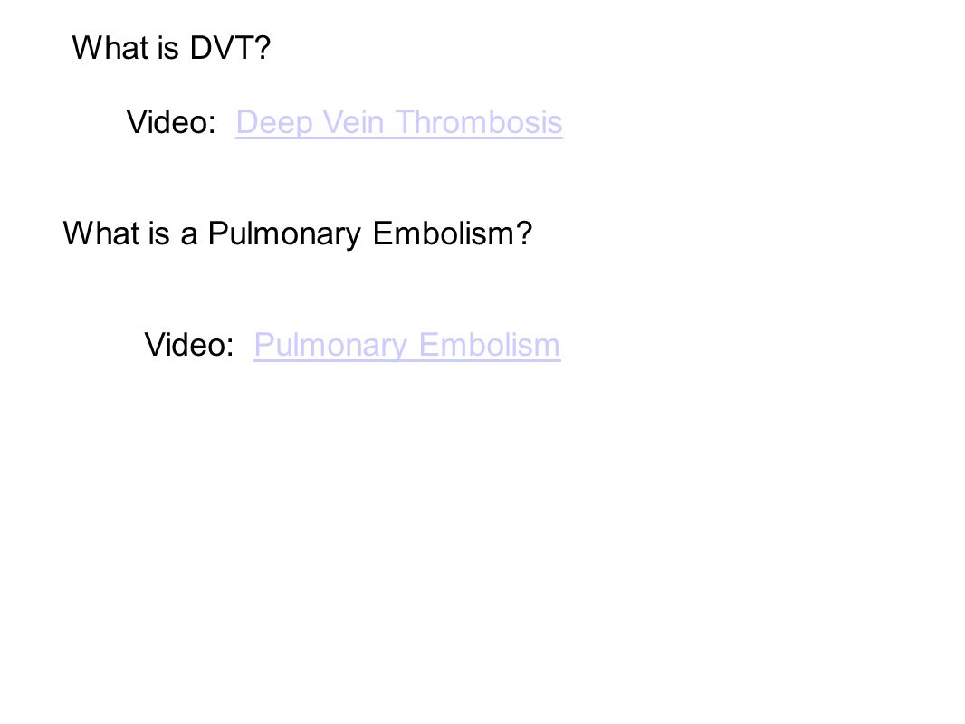
What is DVT? Video: Deep Vein Thrombosis What is a Pulmonary Embolism? Video: Pulmonary Embolism
40
BLOOD TYPES

41
On a cold day in 1667, a renegade physician named Jean Denis transfused calf's blood into one of Paris's most notorious madmen. In doing so, Denis angered not only the elite scientists who had hoped to perform the first animal-to-human transfusions themselves, but also a host of powerful conservatives who believed that the doctor was toying with forces of nature that he did not understand. Just days after the experiment, the madman was dead, and Denis was framed for murder. From: Blood Work: A Tale of Medicine and Murder in the Scientific Revolution

42
Even animals have blood types
Austrian Karl Landsteiner discovered human blood groups Even animals have blood types

43
Blood Type is Controlled by 3 Alleles
4 Possible Blood Types Alleles: A, B, O A & B are codominant O is recessive

44
Rh factor antigen present in RBCs Rh-negative blood
Rh system Rh-positive blood Rh factor antigen present in RBCs Rh-negative blood No Rh factor present in RBCs No anti-Rh antibodies present naturally in plasma Anti-Rh antibodies appear in the plasma of Rh-negative people if Rh-positive RBCs have been introduced into their bodies Where did the term “Rh” come from? (Rh was discovered in a rhesus monkey, thus its name.) What is an antigen in blood typing? What is an antibody? (An antigen is a substance that can activate the immune system. An antibody is the substance made in response to the stimulation of an antigen.) What happens when many antibodies react with their antigens? (Agglutination can occur and lead to death. Cross matching of blood is essential to avoid agglutination.)
 What is an antigen in blood typing What is an antibody (An antigen is a substance that can activate the immune system. An antibody is the substance made in response to the stimulation of an antigen.) What happens when many antibodies react with their antigens (Agglutination can occur and lead to death. Cross matching of blood is essential to avoid agglutination.)")
45
Genotypes What are antigens and antibodies, and how do they relate to each other? (Antigens are substances that can react with antibodies. Antibodies are substances which can react with antigens which are labeled as “foreign” to the body.) What can happen if a patient receives a blood transfusion with a blood type not compatible with her own? (illness and/or death) Which type is known as the universal donor? (type O blood) What is the universal recipient? (type AB blood)
 What can happen if a patient receives a blood transfusion with a blood type not compatible with her own (illness and/or death) Which type is known as the universal donor (type O blood) What is the universal recipient (type AB blood)")
46
Consider Both Parents Type A (genotype AA) x Type O (genotype OO)
 x Type O (genotype OO)")
47
Blood Type Antigens

48
Blood that has antibodies on it that is not recognized by the body will be attacked by your immune system AB is the Universal Acceptor O is the Universal Donor

49
Rh Factor A person can either be Rh + or Rh – (positive is dominant)
")
50
Rh Factor and Pregnancy
*Problem: When a fetus is Rh+ and the mother is Rh-, this can cause the mother’s immune system to attack the fetus. There are drugs that will suppress this reaction. Rh system Erythroblastosis fetalis: May occur when Rh-negative mother carries a second Rh-positive fetus; caused by mother’s Rh antibodies reacting with baby’s Rh-positive cells

51
What is the trade name of the protein usually given to all Rh-negative mothers who carry an Rh-positive baby? (RhoGAM) What does this protein do? (It stops the mother’s body from forming anti-Rh antibodies.) Discuss Research, Issues & Trends: Artificial Blood.
 Discuss Research, Issues & Trends: Artificial Blood.")
52
Blood Type Test

53
Blood Safety EXAMPLES OF BLOODBORNE PATHOGENS HEPATITIS B (HBV)
HEPATITIS C (HCV) Other NON A, NON B HEPATITIS HUMAN IMMUNODEFICIENCY VIRUS (HIV) MALARIA OTHER POTENTIALLY INFECTIOUS MATERIALS
 Other NON A, NON B HEPATITIS. HUMAN IMMUNODEFICIENCY VIRUS (HIV) MALARIA. OTHER POTENTIALLY INFECTIOUS MATERIALS.")
54
TRANSMISSION HIV, hepatitis B virus, and hepatitis C virus are the viruses most likely to be transmitted via the following routes in an occupational setting: needle stick / sharps injuries skin or eye contact mucous membrane and non-intact skin exposure to contaminated blood or other potentially infectious materials ( scratches, cuts, bites, or wounds )
")
55
Avoid Contact With Blood
Wear gloves Dispose of items that have been contaminated (tissues, needles, bandaids) in biohazard containers Do not “horse around” Treat every person as if they may be carrying an infectious disease
 in biohazard containers. Do not horse around Treat every person as if they may be carrying an infectious disease.")
56
BLOOD DISORDERS

57
Carbon Monoxide Poisoning
CO binds to your hemoglobin, prevents oxygen from binding. Can be fatal. It is a "silent killer" as people often die in their sleep when a heater fails. Carbon monoxide deaths are more likely to occur in winter Article from 2010, St Clair County

59
ANEMIA Iron-Deficiency Anemia (most common)
Aplastic Anemia – bone marrow does not produce enough RBC Hemorrhagic anemia – due to extreme blood loss Pernicious anemia – B12 deficiency Sickle Cell Anemia (genetic) - blood cells abnormally shaped Polycythemia – too many RBC
 - blood cells abnormally shaped. Polycythemia – too many RBC.")
60
SICKLE CELL ANEMIA Genetic Disorder Abnormally shaped blood cells
Parents can be carriers (asymptomatic)
")
61
Complications Pain Lethargy Lifelong anemia (low red blood count) Organ failure Stroke
 Organ failure Stroke")
62
Leukemia Type of cancer Overproduction of immature white blood cells
They take the place of RBCs Treatable with bone marrow transplants, chemothemotherapy, radiation

63
Leukemia Leukopenia - Abnormally low WBC
Leukocytosis – Abnormally high WBC count Leukemia – Elevated WBC, but they do not function properly – immature

64
Blood Smear of a patient with Leukemia

65
Blood Smear; Leukemia

66
St. Jude Hospital Leukemia is one of the most common childhood cancers. It occurs when large numbers of abnormal white blood cells fill the bone marrow and sometimes enter the bloodstream. Because these abnormal blood cells are defective, they don't help protect the body against infection the way normal white blood cells do. And because they grow uncontrollably, they take over the bone marrow and interfere with the body's production of other important types of cells in the bloodstream, like red blood cells (which carry oxygen) and platelets (which help blood to clot).
 and platelets (which help blood to clot).")
67
Infectious mononucleosis
sometimes called "mono" or "the kissing disease," is an infection usually caused by the Epstein-Barr virus (EBV). The designation "mononucleosis" refers to an increase in one type of white blood cells (lymphocytes) in the bloodstream relative to the other blood components as a result of the EBV infection. EBV is very common, and many people have been exposed to the virus at some time in childhood. Article at Medicinenet
. The designation mononucleosis refers to an increase in one type of white blood cells (lymphocytes) in the bloodstream relative to the other blood components as a result of the EBV infection. EBV is very common, and many people have been exposed to the virus at some time in childhood. Article at Medicinenet.")
68
Blood poisoning - Septicemia
An infection enters the blood stream Can be deadly Treated with antibiotics

69
Thrombocytopenia Low production of Platelets
Causing bleeding or bruising A bruise is caused when tiny blood vessels are damaged or broken as the result of a blow to the skin (be it bumping against something or hitting yourself with a hammer). The raised area of a bump or bruise results from blood leaking from these injured blood vessels into the tissues as well as from the body's response to the injury.
. The raised area of a bump or bruise results from blood leaking from these injured blood vessels into the tissues as well as from the body s response to the injury.")
70
Hemophilia - inability or reduced ability of the blood to clot; genetic disorder (more on this later) von Willebrand Disease - also a clotting disorder, but not as severe, excessive bruising occurs

71
HEMOPHILIA This disorder causes a failure of the blood to clot
Patients can be treated with blood transfusions that include clotting agents.

72
Queen Victoria Carrier for Hemophilia

73
Jaundice In newborns, caused by the liver not functioning fully
Secretes bilirubin into the blood causing the yellow color Exposure to flourescent lights (bili lights) will break down the substance
 will break down the substance.")
75
Quick Genetics Review A gene consists of 2 alleles (represented by letters) One allele is usually dominant over the other Example: Genotype Phenotype PP widow’s peak Pp widow’s peak pp straight hairline

76
A person with a widow's peak (Pp) is married to a person with a straight hairline (pp), what percentage of their children will have a straight hairline?
 is married to a person with a straight hairline (pp), what percentage of their children will have a straight hairline")
77
Two people who are both heterozygous for the widow's peak trait are married. What percentage of their children will have a straight hairline?

78
Sickle Cell Anemia is actually codominant
AA = normal Aa = sickle cell trait (few symptoms) aa = sickle cell anemia
 aa = sickle cell anemia.")
79
If both parents are carriers, child has a ¼ chance of having the disease

80
A female has sickle cell anemia and is married to a man who appears normal. A doctor tests the man and determines that he does NOT have sickle cell trait. What is the chance that this couple will have a child with sickle cell anemia?

81
What happens when a female who is a carrier marries man with sickle cell anemia?

82
Hemophilia is carried on the X chromosome
Females X H X H normal X H X h carrier X h X h hemophiliac Males X H Y normal X h Y hemophiliac

83
What happens when a female who is a carrier marries a normal man?

84
What happens when a female who is normal marries a man who has hemophilia?

Similar presentations


 Aplastic Anemia – bone marrow does not produce enough RBC Hemorrhagic anemia – due to extreme.>")


 –Formed elements 45%– rbc’s, wbc’s, platelets –Buffy coat – wbc and platelets.>")


. What does blood do? Transports substances around the body to maintain homeostasis Transports substances around the body to maintain.>")