Download presentation
Presentation is loading. Please wait.

1
Copyright © 2004 Pearson Education, Inc., publishing as Benjamin Cummings Fundamentals of Anatomy & Physiology Frederic H. Martini Lecture 4: Chapter 4 The Tissue Level of Organization Pages: 106 - 152 Lecturer: Dr. Barjis Room: P313 Phone: (718) 260-5285 E-Mail: ibarjis@citytech.cuny.edu
")
2
Learning Objectives Identify the four major tissue types and describe their functions. Describe the relationship between form and function for each tissue type. Discuss the types and functions of epithelial tissues. Compare the structure and function of connective tissues.

3
Learning Objectives Explain the structure and function of the four types of membrane. Describe the three types of muscle tissue and the structural features of each. Discuss the basic structure and role of neural tissue.

4
Tissues are: Collections of specialized cells and cell products organized to perform a limited number of functions Histology = study of tissues The four tissue types are: Epithelial Connective Muscular Nervous Tissues of the Body: An Introduction Tissues and tissue types

5
Includes glands and epithelium Glands are secretory Is avascular Forms a protective barrier that regulates permeability Cells may show polarity Tissues and tissue types Epithelial tissue

6
Physical protection Control permeability Provide sensation Produce specialized secretions Functions of epithelium

7
Perform secretory functions Perform transport functions Maintain physical integrity Ciliated epithelia move materials across their surface Specializations of epithelium

8
The Polarity of Epithelial Cells

9
Cells attach via cell adhesion molecules (CAM) Cells attach at specialized cell junctions Tight junctions Desmosomes Gap junctions Maintaining the integrity of epithelium
 Cells attach at specialized cell junctions Tight junctions Desmosomes Gap junctions Maintaining the integrity of epithelium")
10
Intercellular connections Animation: check tutorials

11
Basal lamina attaches to underlying surface Lamina lucida Lamina densa Germinative cells replace short-lived epithelial cells Structure of typical epithelium

12
Number of cell layers Simple Stratified Shape of apical surface cells Squamous Cuboidal Columnar Classification of epithelia

13
Squamous Epithelia

14
Cuboidal Epithelia

16
Transitional Epithelium

17
Columnar Epithelia

20
Exocrine glands Secrete through ducts onto the surface of the gland Endocrine glands Release hormones into surrounding fluid Glandular epithelia

21
Merocrine (product released through exocytosis) Apocrine (involves the loss of both product and cytoplasm) Holocrine (destroys the cell) Glandular secretions can be:
 Apocrine (involves the loss of both product and cytoplasm) Holocrine (destroys the cell) Glandular secretions can be:")
22
Mechanisms of Glandular Secretion Animation: Mechanisms of glandular secretion (check tutorial)
")
23
Unicellular Individual secretory cells Multicellular Organs containing glandular epithelium Classified according to structure Glands

24
A Structural Classification of Exocrine Glands

25
Establishing a structural framework Transporting fluids and dissolved materials Protecting delicate organs Supporting, surrounding and interconnecting tissues Storing energy reserves Defending the body from microorganisms Connective Tissues Connective tissue functions:

26
A Classification of Connective Tissues

27
Specialized cells Matrix Composed of extracellular protein fibers and a ground substance Connective tissues contain

28
Contains varied cell populations Contains various fiber types A syrupy ground substance Connective tissue proper

29
Fluid connective tissue Contains a distinctive cell population Watery ground substance with dissolved proteins Two types Blood Lymph

30
Less diverse cell population Dense ground substance Closely packed fibers Two types Cartilage Bone Supporting connective tissues

31
Contains fibers, a viscous ground substance, and a varied cell population Fibroblasts Macrophage Adipocytes Mesenchymal cells Melanocytes Mast cells Lymphocytes Microphages Connective tissue proper

32
Three types of fiber Collagen fibers Reticular fibers Elastic fibers Connective tissue proper

33
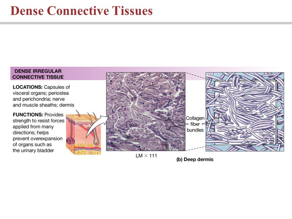
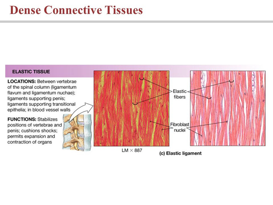
Classified as loose or dense Loose Embryonic mesenchyme, mucous connective tissues Areolar tissue Adipose tissue Reticular tissue Dense Dense regular CT Dense irregular CT Connective tissue proper

34
The Cells and Fibers of Connective Tissue Proper

35
Connective Tissue in Embryos

36
Adipose and Reticular Tissues

37
Dense Connective Tissues

40
Distinctive collections of cells in a fluid matrix Blood Formed elements and plasma Red blood cells, white blood cells and platelets Arteries carry blood away, veins carry to the heart Capillaries allow diffusion into the interstitial fluid Lymph Interstitial fluid entering the lymphatic vessels Fluid connective tissues

41
Formed Elements of the Blood

42
Cartilage and bone support the rest of the body Cartilage Grows via interstitial and appositional growth Matrix is a firm gel containing chondroitin sulfate Cells called chondrocytes Cells found in lacunae Perichondrium separates cartilage from surrounding tissues Three types: hyaline, elastic and fibrocartilage Supporting connective tissues

43
The Perichondrium and Types of Cartilage

46
Has osteocytes Depend on diffusion through canaliculi for nutrients Little ground substance Dense mineralized matrix Surrounded by periosteum Bone, or osseus tissue

47
Bone

48
Form a barrier Composed of epithelium and connective tissue Four types Cutaneous Synovial Serous Mucous Membranes Membranes are simple organs

49
Membranes

50
Line cavities that communicate with the exterior Contain lamina propria Mucous membranes

51
Line sealed internal cavities Form transudate Serous membranes

52
Cutaneous membrane Covers the body surface Synovial membrane Incomplete lining within joint cavities Membranes continue

53
Network of connective tissue proper consisting of Superficial fascia Deep fascia Subserous fascia The Connective Tissue Framework of the Body Organs and systems are interconnected

54
The Fasciae

55
Specialized for contraction Three types Skeletal Cardiac Smooth Muscle tissue

56
Muscle Tissue

59
Cells are multinucleate Striated voluntary muscle Divides via satellite cells Skeletal muscle

60
Cardiocytes occur only in the heart Striated involuntary muscle Relies on pacemaker cells for regular contraction Cardiac muscle

61
Non-striated involuntary muscle Can divide and regenerate Smooth muscle tissue

62
Conducts electrical impulses Conveys information from one area to another Neural tissue

63
Neurons Transmit information Neuroglia Support neural tissue Help supply nutrients to neurons Neural tissue cells

64
Neural Tissue

65
Cell body Dendrites Axon (nerve fiber) Carries information to other neurons Neural anatomy
 Carries information to other neurons Neural anatomy")
66
Injured tissues respond in coordinated fashion Homeostasis restored by inflammation and regeneration Tissue Injuries and Aging Inflammation and regeneration

67
Isolates injured area Damaged cells, tissue components and dangerous microorganisms removed Infection avoided Regeneration restores normal function Tissue Injuries and Aging Inflammatory response

68
An Introduction to Inflammation

69
Change with age Repair and maintenance less efficient Structure altered Chemical composition altered Aging and tissue repair

70
Aging and cancer incidence Incidence of cancer increases with age 70-80% of all cases due to exposure to chemicals or environmental factors

71
Changes in a Tissue under Stress

72
You should now be familiar with: The four major tissue types and their functions. The relationship between form and function for each tissue type. The types and functions of epithelial tissues. The structure and function of connective tissues. The structure and function of the four types of membrane. The three types of muscle tissue and the structural features of each. The basic structure and role of neural tissue.

Similar presentations