Download presentation
Presentation is loading. Please wait.

2
Integumentary Anatomy Laura E. Edsberg, Ph.D.

5
The Skin Epidermis Dermis Subcutaneous Fat

6
Largest Organ System 1/3 of cardiac output is directed to skin Surface area 18-20 Sq Ft 7% of body weight Thickness 1.5-4mm

7
Functions of The Skin Large & Complex Sensory Functions Barrier –Harmful Chemicals –Ultraviolet Radiation –Infection Temperature Regulation & Sweating Required for Vitamin D Production

9
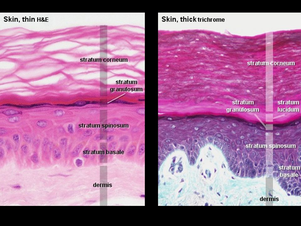
Epidermis Tough, leathery outer surface Composed mainly of Keratinocytes.06-.6mm, thickest portion palms and soles Epidermis avascular, receives nutrients by diffusion through semi-permeable Basement Membrane (BM)
")
10
Epidermal Cellular Layers Five Layers –Represent different stages of cellular differentiation, gradual loss of nuclear material & accumulation of keratin proteins –New cells form –Older cells elongate, membranes thicken as they are pushed up

12
Basal Layer Stratum Basale, deepest layer, attached to dermis by thin, acellular (BM) Single row of keratinocytes Typically, mitotic activity of keratinocytes is confined to this layer Epidermal turnover time Approx. 4 weeks Keratinocytes produce Keratin (protective protein)
.")
13
Stratum Spinosum Above Stratum Basale Several rows of more mature keratinocytes Appear spiny due to keratin filaments

14
Stratum Granulosum Above Stratum Spinosum Three to five flattened cell rows Increased concentration of Keratin

15
Stratum Lucidum Above Stratum Granulosum As keratinocytes migrate up away from their dermal blood supply, they slowly die Stratum Lucidum contains layers of flattened dead keratinocytes

16
Stratum Corneum “Horney” Layer Consists of dead keratinocytes 20-30 cells thick 75% of thickness of the epidermis Cells are continuously abraded & replaced by cells below A cells journey from the basale layer up through the corneum takes 14-21 days Friction or pressure will increase the thickness of the stratum corneum (Callus)
")
18
Epidermal Cells Keratinocytes - Keratin (protective protein) Merkel Cells – Specialized mechanoreceptors (light touch) Langerhan’s Cells – deeper layers of epidermis assist in fighting infection (attack & engulf foreign material) Melanocytes
 Merkel Cells – Specialized mechanoreceptors (light touch) Langerhan’s Cells – deeper layers of epidermis assist in fighting infection (attack & engulf foreign material) Melanocytes")
19
Melanocytes Produce pigment Melanin –Protects skin from UV –Gives the skin its color More = Darker Less = Lighter Grey Hair - loss of melanocytes in hair bulb –Faster turnover in scalp vs body

21
Epidermal Appendages Specialized epidermal structures, extend down into the dermis - Hair, Glands, and Nails Hair Follicles (soft keratin) –everywhere except palms and soles –Helps regulate body temp by trapping air between hair and skin surface Sebaceous Gland –Each hair follicle contains a sebaceous gland –Secretes Sebum – oily substance that lubricates skin & hair –May slow bacterial growth, reducing colonization
 –everywhere except palms and soles –Helps regulate body temp by trapping air between hair and skin surface Sebaceous Gland –Each hair follicle contains a sebaceous gland –Secretes Sebum – oily substance that lubricates skin & hair –May slow bacterial growth, reducing colonization")
22
Epidermal Appendages Sudoriferous Glands –Present everywhere except lips and ears –Secrete sweat into ducts that lead to skin’s surface –Evaporation of sweat helps cool the body Nails – dorsal tips of digits –Hard Keratin –Protect terminal digit & assist with function

23
Functions of the Epidermis Provides a physical & chemical barrier Regulates fluid Provides light touch sensation Assists with thermoregulation Assists with excretion Assists with vitamin D production Contributes to appearance

24
Basement Membrane Zone Dermo-Epidermal Junction Where epidermis and dermis join Contains many proteins and structures Site of inflammation in many diseases Congenital defects Important in skin neoplasia

25
Aging and the Dermo- Epidermal Junction Flattening with age –Dermal papillae –Epidermal rete pegs Flattening = Less Surface Area –Less communication –Less resistance to shearing

26
Dermis 2 to 4mm thick, fibrous part of skin Contains collagen and elastic fibers contained in an amorphous ground substance, nerve fibers, and nerve-end sensory organs Highly vascular –Capillaries provide color (pale pink to rosy red) –Superficial Lymphatics Assist in returning H 2 O, proteins, and other substances from tissue to blood stream Dermis should appear shiny or moist due to high H 2 0 content
 –Superficial Lymphatics Assist in returning H 2 O, proteins, and other substances from tissue to blood stream Dermis should appear shiny or moist due to high H 2 0 content")
27
Dermal Layers Two layers –Papillary Dermis (thin superficial) Loosely woven fibers embedded in gelatinous matrix (ground substance) Blisters occur here if friction between epidermis & dermis –Reticular Dermis Dense irregular arranged connective tissue Provide increased structural support to the skin
 Loosely woven fibers embedded in gelatinous matrix (ground substance) Blisters occur here if friction between epidermis & dermis –Reticular Dermis Dense irregular arranged connective tissue Provide increased structural support to the skin")
28
Aging and the Dermis Decreased thickness More Avascular Decrease in elastin content

29
Dermal Cell Types Fibroblasts – main cells found in dermis, produce collagen & elastin fibers, ground substance –Give dermis strength & flexibility Macrophages & Polymorphonuclear Leukocytes (neutrophils) –Help fight infection by engulfing harmful substances & releasing destructive enzymes
 –Help fight infection by engulfing harmful substances & releasing destructive enzymes")
30
Dermal Cell Types Mast Cells –Specialized secretory cells Produce chemical mediators of inflammation such as histamine –Attract other cells and cause vasodilation to fight infection or repair injury Dermis also contains sensory receptors for: –Touch –Vibration –Temperature –Pressure

31
Functions of the Dermis Support & nourish epidermis House epidermal appendages Assists with infection control Assists with thermoregulation Provides sensation

32
Subcutaneous Tissue Hypodermis Supports the skin Adipose Tissue –Highly Vascular, loose connective tissue, stores fat for energy, insulation, protection (cushion over structures such as bony prominences) –Healthy adipose is glisteny white to pale yellow (darker if dehydrated) Fascia –Highly fibrous connective tissue –Separates and surrounds structures, facilitates movement between adjacent structures (muscle, tendon, bone) Deeper lymphatic vessels are located in subcutaneous tissue
 –Healthy adipose is glisteny white to pale yellow (darker if dehydrated) Fascia –Highly fibrous connective tissue –Separates and surrounds structures, facilitates movement between adjacent structures (muscle, tendon, bone) Deeper lymphatic vessels are located in subcutaneous tissue")
33
Deeper Tissues Wounds can extend beyond subcutaneous tissue Muscle –Regularly arranged fibers surrounded by fascia –Rich vascular supply – red in color – bleeds easily –Non-viable muscle will appear gray or black in color Tendons –Regularly arranged fibers, may be enclosed in fibrous sheath

34
Deeper Tissues Ligaments & Joint Capsules –consist of dense connective tissue –Ligaments – regularly arranged fibers –Joint Capsule – direction of fibers vary –When Healthy, glisteny (silky) white appearance –Non-viable, dry, leathery, dark, and may be disconnected Bone –Shiny, smooth, milky white appearance, hard when probed –Unhealthy – moth-eaten, irregular surface, dark discoloration Pressure ulcers, Diabetic lesions, or burns may involve these deep tissues
 white appearance –Non-viable, dry, leathery, dark, and may be disconnected Bone –Shiny, smooth, milky white appearance, hard when probed –Unhealthy – moth-eaten, irregular surface, dark discoloration Pressure ulcers, Diabetic lesions, or burns may involve these deep tissues")
35
Depth of Tissue Involvement Extent of tissue involvement is characterized as: –Superficial –Partial-thickness –Full-thickness

36
Superficial Wounds Affect only the epidermis –Ex. Abrasion – top layer of integument is removed Dermis may be exposed

37
Partial-thickness Wounds Involve epidermis & part of the dermis –Ex. Second degree burn (sunburn), deep with blistering & peeling
, deep with blistering & peeling.")
38
Full-thickness Wounds Extends through epidermis & dermis to the subcutaneous tissue layer May be further categorized as –Subcutaneous –Sub-dermal ** if tissues such as: tendon, muscle, or bone are involved

39
Pressure Ulcer Stages National Pressure Ulcer Advisory Panel (NPUAP) classification system Stage I: Nonblanchable erythema of intact skin Stage II: Partial-thickness skin loss involving epidermis or dermis or both. The ulcer is superficial and presents clinically as an abrasion, blister, or shallow crater.

40
Pressure Ulcer Stages Stage III: Full-thickness skin loss involving damage or necrosis of subcutaneous tissue, which may extend down to, but not through, underlying fascia. The ulcer presents clinically as a deep crater with or without undermining of adjacent tissue. Stage IV: Full-thickness skin loss with extensive destruction, tissue necrosis or damage to muscle, bone, or supporting structures (e.g., tendon, joint capsule, etc.).
..")
41
Questions ????

Similar presentations


 Largest organ of the body (15% of body weight) Skin thickness variable, normally 1-2 mm Protection –chemical barrier (waterproof)>")
 Barrier to keep water and solutes in Barrier to keep bacterial,>")

