Download presentation
Presentation is loading. Please wait.

1
Degenerative Disease Dr. Sharifa AL-Duraibi

2
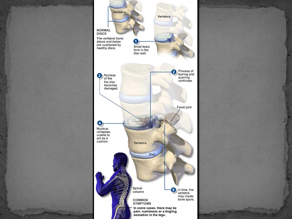
Degenerative disc disease
Degeneration of one or more intervertebral disc(s) of the spine. Disc degeneration is a disease of aging, and though for most people is not a problem, in certain individuals a degenerated disc can cause severe chronic pain if left untreated.
 of the spine. Disc degeneration is a disease of aging, and though for most people is not a problem, in certain individuals a degenerated disc can cause severe chronic pain if left untreated.")
3
low back pain world wide
common complaint among adults. lifetime prevalence in working population up to 80%. 60% experience functional limitation or disability. second most common reason for work disability. despite advances in imaging and surgical techniques LBP prevalence and its cost are relatively unchanged. It is one of the most common complaints by adults to their primary care doctor. Of the working population 80% will experience low back pain. 60% will have some form of functional limitation or disability. Second most common reason for work disability. Economically it is estimated that low back pain cost 100 to 200 billion dollars annually in health care cost and lost wages. And despite advances in medicine low back pain prevalence and cost are relatively unchanged. In fact according to medicare cost are rising.

4
Pathologic changes Fibrocartilage replaces the gelatinous mucoid material of the nucleus pulposus as the disc changes with age. There may be splits in the annulus fibrosis, permitting herniation of elements of nucleus pulposus.

5
Pathologic changes Shrinkage of the nucleus pulposus that produces prolapse or folding of the annulus with secondary osteophyte formation at the margins of the adjacent vertebral body.

7
Disc pathology vs Pain degree of disc injury (size of tear / herniation), nor the degree of nerve root compression correlate with subjective pain or functional disability. Karppinen J. et al. “Severity of Symptoms and Signs in Relation to MRI Findings Among Sciatica Patients.” Spine 2001; 26(7):E149-E154
, nor the degree of nerve root compression correlate with subjective pain or functional disability. Karppinen J. et al. Severity of Symptoms and Signs in Relation to MRI Findings Among Sciatica Patients. Spine 2001; 26(7):E149-E154.")
8
Cervical Radiculopathy

9
Lumbosacral Radiculopathy (Sciatica)
Important: A herniated disc at (e.g.) L4-5 may impinge either the L4 or L5 nerve roots!
 L4-5 may impinge either the L4 or L5 nerve roots!")
10
Degenerative Disc (and Facet Joint) Disease
Foraminal stenosis Thickening/Buckling of Ligamentum Flavum

12
MRI - Degenerative Disc Disease
Age: 36% have degenerated disc. 85-95% have degenerated disc. 98% have degenerated disc. ** <60 20% have asymptomatic disc herniation. Conclusion: Abnormal findings on MRI frequently DO NOT relate to symptoms (and vice versa) !!
 !!")
13
MRI – Herniated Disc Levels
85-95% at L4-L5/ L5-S1. 5-8% at L3-L4. 2% at L2-L3. 1% at L1-L2/ T12-L1. ** Cervical: most common C4-C7. **Thoracic: 15% in asymptomatic pts. at multiple levels, not often symptomatic.

14
Anular tear Separations between anular fibers, avulsion of fibers from their vertebral body insertions, or breaks through fibers involving one or many layers of the anular lamellae. The terms 'tear' or 'fissure' does not imply that the lesion is consequent to trauma. In case of a traumatic event the term 'rupture' is appropriate.a

15
Disc herniation Displacement of disc material beyond the limits of the intervertebral disc space. A herniated disc can be contained (covered by outer anulus fibrosus) or uncontained.
 or uncontained.")
16
Disc herniation Focal Herniation Broad based hernia
Is a herniated disc less than 90? of the disc circumference. Is a herniated disc in between 90?-180? of the disc circumference.

17
Disc herniation Bulging Disc
Is the presence of disc tissue 'circumferentially' (180?-360?) beyond the edges of the ring apophyses and is NOT considered a form of herniation.
 beyond the edges of the ring apophyses and is NOT considered a form of herniation.")
18
Focal disc herniation Disc Protrusion Disc Extrusion
Indicates that the distance between the edges of the disc herniation is less than the distance between the edges of the base. present when the distance between the edges of the disc material is greater the distance at the base.

19
Disc herniation Migration Sequestration
indicates displacement of disc material away from the site of extrusion, regardless of whether sequestrated or not. used to indicate that the displaced disc material has lost completely any continuity with the parent disc

20
Axial localisation of herniated discs

21
Symmetrical presence (or apparent presence) of disc tissue "circumferentially" (50-100%) beyond the edges of the ring apophyses may be described as a "bulging disc" or "bulging appearance", and is not considered a form of herniation. Furthermore, “bulging” is a descriptive term for the shape of the disc contour and not a diagnostic category. Adapted from: “Nomenclature and Classification of Lumbar Disc Pathology: Recommendations of the Combined Task Forces of the North American Spine Society, American Society of Spine Radiology, and American Society of Neuroradiology,” 2001.

22
Asymmetrical bulging of the disc margin (50%-100%), such as is found in severe scoliosis, is also not considered a form of herniation. Adapted from: “Nomenclature and Classification of Lumbar Disc Pathology: Recommendations of the Combined Task Forces of the North American Spine Society, American Society of Spine Radiology, and American Society of Neuroradiology,” 2001.

23
By convention, a "broad-based" herniation involves between 25% and 50% (90-180) of the disc circumference. Adapted from: “Nomenclature and Classification of Lumbar Disc Pathology: Recommendations of the Combined Task Forces of the North American Spine Society, American Society of Spine Radiology, and American Society of Neuroradiology,” 2001.

24
By convention, a "focal herniation" involves less than 25% (90) of the disc circumference.
Adapted from: “Nomenclature and Classification of Lumbar Disc Pathology: Recommendations of the Combined Task Forces of the North American Spine Society, American Society of Spine Radiology, and American Society of Neuroradiology,” 2001.

25
Herniated discs may take the form of protrusion or extrusion, based on the shape of the displaced material (see definitions in text). Adapted from: “Nomenclature and Classification of Lumbar Disc Pathology: Recommendations of the Combined Task Forces of the North American Spine Society, American Society of Spine Radiology, and American Society of Neuroradiology,” 2001.

26
Protrusion Extrusion Extrusion
When a relatively large amount of disc material is displaced, distinction between protrusion (A) and extrusion (B or C) will generally only be possible on sagittal MR sections or sagittal CT reconstructions. In Figure C, although the shape of the displaced material is similar to that of a protrusion, the greatest cranio-caudal diameter of the fragment is greater than the cranio-caudal diameter of its base at the level of the parent disc, and the lesion therefore qualifies as an extrusion. In any situation, the distance between the edges of the base, which serves as reference for the definition of protrusion and extrusion, may differ from the distance between the edges of the aperture in the anulus, which cannot be assessed on CT images and is seldom appreciated on MR images. In the cranio-caudal direction, the length of the base cannot exceed, by definition, the height of the intervertebral space (Adapted from Milette PC. Classification, diagnostic imaging and imaging characterization of a lumbar herniated disc. Radiol Clin North Am 2000; 38: ) Protrusion Extrusion Extrusion Adapted from: “Nomenclature and Classification of Lumbar Disc Pathology: Recommendations of the Combined Task Forces of the North American Spine Society, American Society of Spine Radiology, and American Society of Neuroradiology,” 2001.
 and extrusion (B or C) will generally only be possible on sagittal MR sections or sagittal CT reconstructions. In Figure C, although the shape of the displaced material is similar to that of a protrusion, the greatest cranio-caudal diameter of the fragment is greater than the cranio-caudal diameter of its base at the level of the parent disc, and the lesion therefore qualifies as an extrusion. In any situation, the distance between the edges of the base, which serves as reference for the definition of protrusion and extrusion, may differ from the distance between the edges of the aperture in the anulus, which cannot be assessed on CT images and is seldom appreciated on MR images. In the cranio-caudal direction, the length of the base cannot exceed, by definition, the height of the intervertebral space (Adapted from Milette PC. Classification, diagnostic imaging and imaging characterization of a lumbar herniated disc. Radiol Clin North Am 2000; 38: ) Protrusion Extrusion Extrusion. Adapted from: Nomenclature and Classification of Lumbar Disc Pathology: Recommendations of the Combined Task Forces of the North American Spine Society, American Society of Spine Radiology, and American Society of Neuroradiology,")
27
Protrusion w/ migration + sequestration Protrusion w/ migration
Schematic representation of various types of posterior central herniations. A, Small sub-ligamentous herniation (or protrusion) without significant disc material migration. B, Sub-ligamentous herniation with downward migration of disc material under the posterior longitudinal ligament (PLL). C, Sub-ligamentous herniation with downward migration of disc material and sequestered fragment (arrow). (From Milette PC. Classification, diagnostic imaging and imaging characterization of a lumbar herniated disc. Radiol Clin North Am 2000; 38: ) Protrusion w/ migration + sequestration Protrusion w/ migration Protrusion Adapted from: “Nomenclature and Classification of Lumbar Disc Pathology: Recommendations of the Combined Task Forces of the North American Spine Society, American Society of Spine Radiology, and American Society of Neuroradiology,” 2001.
 without significant disc material migration. B, Sub-ligamentous herniation with downward migration of disc material under the posterior longitudinal ligament (PLL). C, Sub-ligamentous herniation with downward migration of disc material and sequestered fragment (arrow). (From Milette PC. Classification, diagnostic imaging and imaging characterization of a lumbar herniated disc. Radiol Clin North Am 2000; 38: ) Protrusion w/ migration + sequestration. Protrusion w/ migration. Protrusion. Adapted from: Nomenclature and Classification of Lumbar Disc Pathology: Recommendations of the Combined Task Forces of the North American Spine Society, American Society of Spine Radiology, and American Society of Neuroradiology,")
28
Lumbar Spinal Stenosis

29
Lumbar Spinal Stenosis
Disc bulge, facet hypertrophy and flaval ligament thickening frequently combine to cause central spinal stenosis. Note the trefoil shape of stenotic spinal canal.

30
Lumbar Spinal Stenosis
Disc bulge, facet hypertrophy and ligament flavum thickening frequently combine to cause central spinal stenosis Note the trefoil shape of stenotic spinal canal

31
Foraminal Stenosis Neural foramen

33
Cervical Spinal Stenosis

34
case study - annie 30 y.o. female presents with low back pain.
Pain radiating down right leg. Initial onset approximately 1 year. Referred by orthopedic surgeon. On motrin, previously darvocet, flexeril and valium. Previous treatments: chiropractic and physical therapy. Also large annular L5-S1. Tx’d 5 deg. VAS dropped from 5/10 – 1/10 after first tx. Pain-free after 5th tx. One year F/U – still pain-free. Returned to normal activity including tennis. Only c/o mild left knee pain. 34

35
Diagnostic studies A-P / lateral Plain Film: MRI:
Degenerative disc height loss at L4-5 level. MRI: L4-L5: Large central disc herniation (9mm in AP X 10mm Broad) effacing the ventral thecal sac and impressing upon the central canal. This produces moderate canal stenosis. L5-S1: broad disc bulge with radial tear. mild effacement upon the ventral thecal sac.
 effacing the ventral thecal sac and impressing upon the central canal. This produces moderate canal stenosis. L5-S1: broad disc bulge with radial tear. mild effacement upon the ventral thecal sac.")
36
Imaging

37
Abnormal Disc < 180º > 180º Herniation Tear Bulge 90º–180º
< 90º Broad-based Focal Symmetric Asymmetric Waist* No waist Extrusion Protrusion Sequestered Migrated Neither *(In any plane) Adapted from: “Nomenclature and Classification of Lumbar Disc Pathology: Recommendations of the Combined Task Forces of the North American Spine Society, American Society of Spine Radiology, and American Society of Neuroradiology,” 2001.
 Adapted from: Nomenclature and Classification of Lumbar Disc Pathology: Recommendations of the Combined Task Forces of the North American Spine Society, American Society of Spine Radiology, and American Society of Neuroradiology,")
38
Schmorl’s Nodes protrusions of the cartilage of the intervertebral disc through the vertebral body endplate and into the adjacent vertebra.

39
Confusing “Spondy-” Terminology
Spondylosis = “spondylosis deformans” = degenerative spine. Spondylitis = Inflamed spine (e.g. ankylosing, pyogenic, etc.). Spondylolysis = Chronic fracture of pars interarticularis with nonunion (“pars defect”). Spondylolisthesis = anterior slippage of vertebra typically resulting from bilateral pars defects. Pseudospondylolisthesis = “degenerative spondylolisthesis” (spondylolisthesis resulting from degenerative disease rather than pars defects)
. Spondylolysis = Chronic fracture of pars interarticularis with nonunion ( pars defect ). Spondylolisthesis = anterior slippage of vertebra typically resulting from bilateral pars defects. Pseudospondylolisthesis = degenerative spondylolisthesis (spondylolisthesis resulting from degenerative disease rather than pars defects)")
40
Spondylolysis / Spondylolisthesis

41
current therapies for discogenic pain or disc pathology
Medication and limited activity Spinal rehabilitation. Interventional pain management. Surgery is the last thing I recommend to my patients if possible. I recommend surgery if there is progressive neurological deterioration or intractable pain that interferes with patients life. Be careful not to offend the audience. Note: Each of these options have their time and place (that’s why they’re on the list) – However, the primary objective of these options is not aimed at directly addressing the underlying disc pathology; except maybe surgery. Spinal surgery.
 – However, the primary objective of these options is not aimed at directly addressing the underlying disc pathology; except maybe surgery. Spinal surgery.")
Similar presentations


>")


 - Lumbar>")


 by bony structures (vertebral body, facets, pedicles) as well as soft tissue structures (ligamentum.>")


