Download presentation
Presentation is loading. Please wait.

1
Introduction to Neuroimaging University of Wisconsin–Madison
SPINE Aaron S. Field, MD, PhD Neuroradiology University of Wisconsin–Madison Updated 6/13/06

2
Anatomy

6
Radiographic Anatomy ML Richardson, Univ. Of Washington

7
Cervical Spine – AP View
ML Richardson, Univ. Of Washington

8
Cervical Spine – Lateral View
ML Richardson, Univ. Of Washington

9
Cervical Spine – Oblique View
ML Richardson, Univ. Of Washington

10
Cervical Spine – Open-Mouth (Dens) View
ML Richardson, Univ. Of Washington

11
Lumbar Spine – AP View ML Richardson, Univ. Of Washington

12
Lumbar Spine – Lateral View
ML Richardson, Univ. Of Washington

13
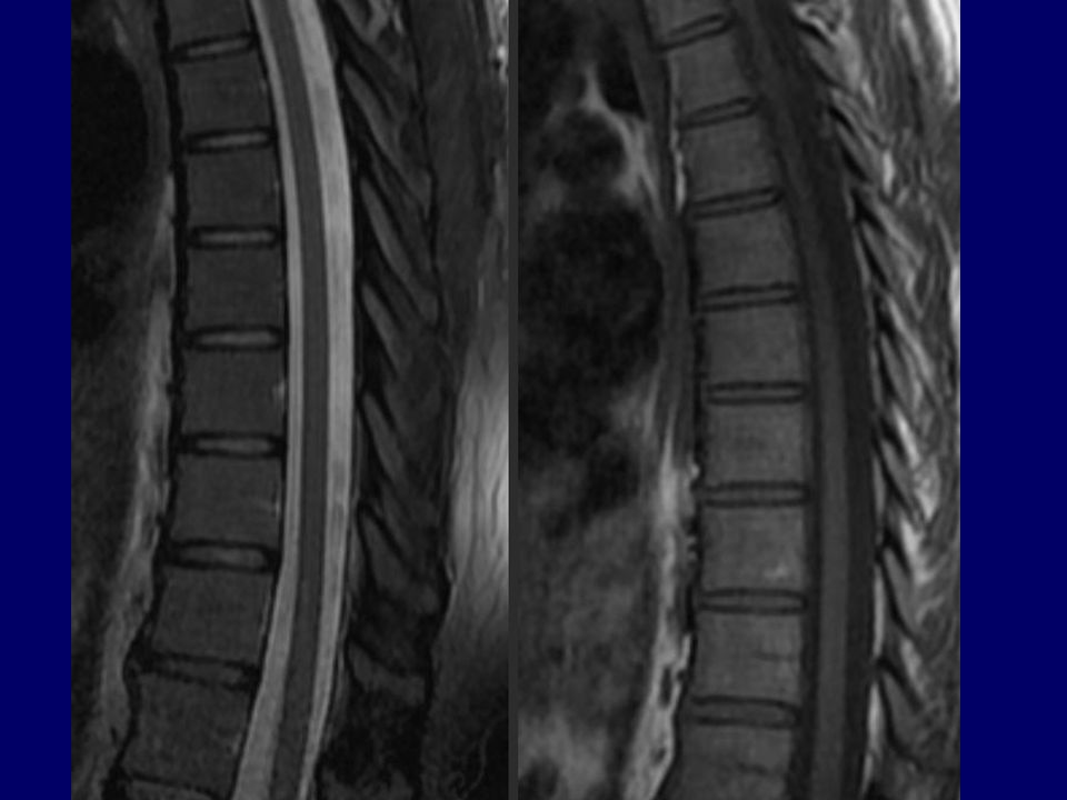
MRI Anatomy Source: CW Kerber and JR Hesselink, Spine Anatomy, UCSD Neuroradiology

19
Source: CW Kerber and JR Hesselink, Spine Anatomy, UCSD Neuroradiology

24
Spine Pathology Trauma Degenerative disease Tumors and other masses
Inflammation and infection Vascular disorders Congenital anomalies

25
Trauma

26
Evaluating Trauma Fracture – plain film / CT
Dislocation – plain film / CT Ligamentous injury – MRI Cord injury – MRI Nerve root avulsion – MRI

27
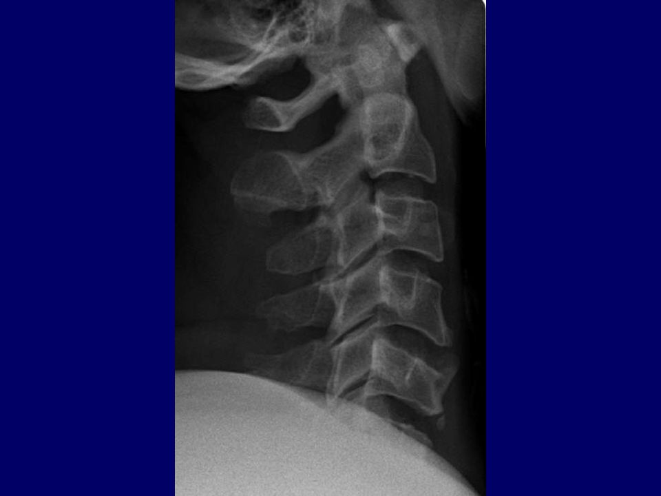
Plain film findings may be very subtle or absent!
Anterolisthesis of C6 on C7 (Why??)
")
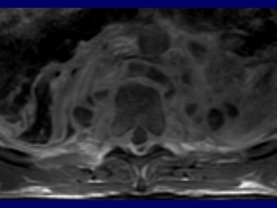
28
Fractures of C6 left pedicle and lamina

29
CT – 2D Reconstructions Acquire images axially…
…reconstruct sagittal / coronal

30
26M MVA

31
Vertebral body burst fx with retropulsion into spinal canal
2D Reformats

34
Vertebral Artery Dissection/Occlusion Secondary to C6 Fracture

35
Hyperflexion fx with ligamentous disruption and cord contusion

36
Nerve root avulsion Axial Coronal Sagittal

37
Degenerative Disease

38
Degenerative Disc (and Facet Joint) Disease
Foraminal stenosis Thickening/Buckling of Ligamentum Flavum

39
Degenerative Disc (and Facet Joint) Disease
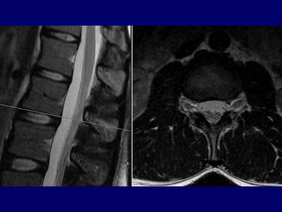
 Disease")
40
Degenerative Disc (and Facet Joint) Disease
 Disease")
42
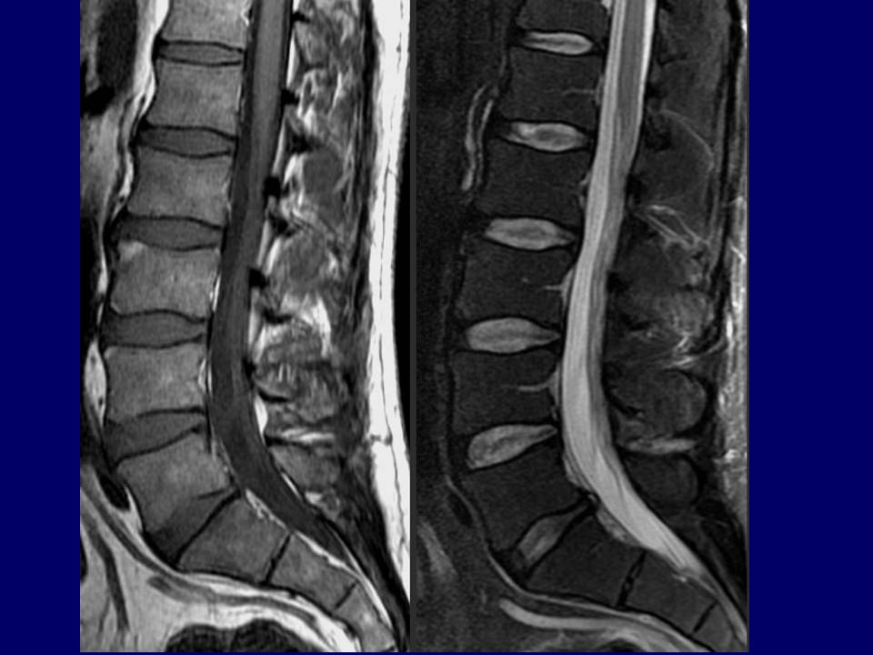
Lumbar Spinal Stenosis

43
Lumbar Spinal Stenosis
MR#

44
Lumbar Spinal Stenosis

45
Lumbar Spinal Stenosis

46
Lumbar Spinal Stenosis

47
Lumbar Spinal Stenosis
Disc bulge, facet hypertrophy and flaval ligament thickening frequently combine to cause central spinal stenosis Note the trefoil shape of stenotic spinal canal

49
Lumbar Spinal Stenosis
Disc bulge, facet hypertrophy and flaval ligament thickening frequently combine to cause central spinal stenosis Note the trefoil shape of stenotic spinal canal

50
Foraminal Stenosis Neural foramen

51
Cervical Spinal Stenosis

53
MRI - Degenerative Disc Disease
Age: % have degenerated disc % have degenerated disc % have degenerated disc <60 20% have asymptomatic disc herniation Conclusion: Abnormal findings on MRI frequently DO NOT relate to symptoms (and vice versa) !!
 !!")
54
MRI – Herniated Disc Levels
85-95% at L4-L5, L5-S1 5-8% at L3-L4 2% at L2-L3 1% at L1-L2, T12-L1 Cervical: most common C4-C7 Thoracic: 15% in asymptomatic pts. at multiple levels, not often symptomatic

55
Schematic sagittal anatomical sections showing the differentiating features of an annular tear (radial tear in this case) and a disc herniation. The term "tear" is used to refer to a localized radial, concentric, or horizontal disruption of the anulus without associated displacement of disc material beyond the limits of the intervertebral disc space. Nuclear material is shown in black, and the annulus (internal and external) corresponds to the white portion of the intervertebral space. The same convention is used in Figures 3, 12, 13, and 14. (Adapted from Milette PC. The proper terminology for reporting lumbar intervertebral disk disorders. AJNR Am J Neurorad 1997;18: ; with permission.) Annular

56
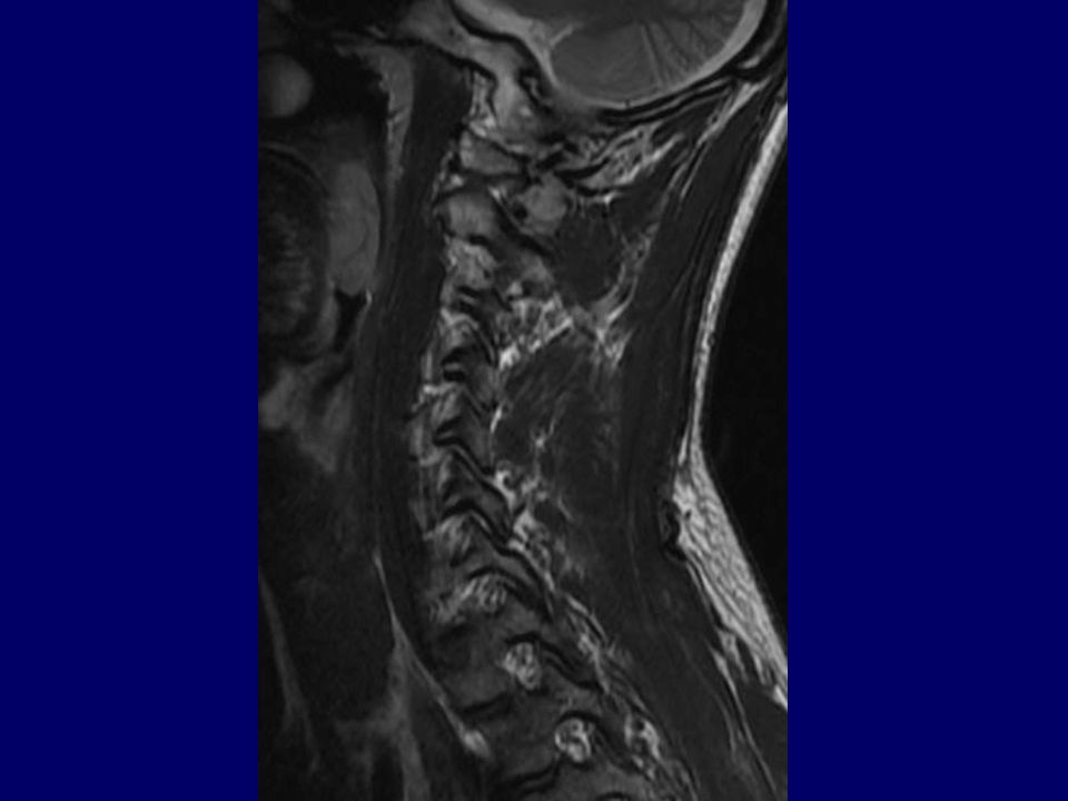
Symmetrical presence (or apparent presence) of disc tissue "circumferentially" (50-100%) beyond the edges of the ring apophyses may be described as a "bulging disc" or "bulging appearance", and is not considered a form of herniation. Furthermore, “bulging” is a descriptive term for the shape of the disc contour and not a diagnostic category. Adapted from: “Nomenclature and Classification of Lumbar Disc Pathology: Recommendations of the Combined Task Forces of the North American Spine Society, American Society of Spine Radiology, and American Society of Neuroradiology,” 2001.

57
Asymmetrical bulging of the disc margin (50%-100%), such as is found in severe scoliosis, is also not considered a form of herniation. Adapted from: “Nomenclature and Classification of Lumbar Disc Pathology: Recommendations of the Combined Task Forces of the North American Spine Society, American Society of Spine Radiology, and American Society of Neuroradiology,” 2001.

58
By convention, a "broad-based" herniation involves between 25% and 50% (90-180) of the disc circumference. Adapted from: “Nomenclature and Classification of Lumbar Disc Pathology: Recommendations of the Combined Task Forces of the North American Spine Society, American Society of Spine Radiology, and American Society of Neuroradiology,” 2001.

59
By convention, a "focal herniation" involves less than 25% (90) of the disc circumference.
Adapted from: “Nomenclature and Classification of Lumbar Disc Pathology: Recommendations of the Combined Task Forces of the North American Spine Society, American Society of Spine Radiology, and American Society of Neuroradiology,” 2001.

60
Herniated discs may take the form of protrusion or extrusion, based on the shape of the displaced material (see definitions in text). Adapted from: “Nomenclature and Classification of Lumbar Disc Pathology: Recommendations of the Combined Task Forces of the North American Spine Society, American Society of Spine Radiology, and American Society of Neuroradiology,” 2001.

61
Protrusion Extrusion Extrusion
When a relatively large amount of disc material is displaced, distinction between protrusion (A) and extrusion (B or C) will generally only be possible on sagittal MR sections or sagittal CT reconstructions. In Figure C, although the shape of the displaced material is similar to that of a protrusion, the greatest cranio-caudal diameter of the fragment is greater than the cranio-caudal diameter of its base at the level of the parent disc, and the lesion therefore qualifies as an extrusion. In any situation, the distance between the edges of the base, which serves as reference for the definition of protrusion and extrusion, may differ from the distance between the edges of the aperture in the anulus, which cannot be assessed on CT images and is seldom appreciated on MR images. In the cranio-caudal direction, the length of the base cannot exceed, by definition, the height of the intervertebral space (Adapted from Milette PC. Classification, diagnostic imaging and imaging characterization of a lumbar herniated disc. Radiol Clin North Am 2000; 38: ) Protrusion Extrusion Extrusion Adapted from: “Nomenclature and Classification of Lumbar Disc Pathology: Recommendations of the Combined Task Forces of the North American Spine Society, American Society of Spine Radiology, and American Society of Neuroradiology,” 2001.
 and extrusion (B or C) will generally only be possible on sagittal MR sections or sagittal CT reconstructions. In Figure C, although the shape of the displaced material is similar to that of a protrusion, the greatest cranio-caudal diameter of the fragment is greater than the cranio-caudal diameter of its base at the level of the parent disc, and the lesion therefore qualifies as an extrusion. In any situation, the distance between the edges of the base, which serves as reference for the definition of protrusion and extrusion, may differ from the distance between the edges of the aperture in the anulus, which cannot be assessed on CT images and is seldom appreciated on MR images. In the cranio-caudal direction, the length of the base cannot exceed, by definition, the height of the intervertebral space (Adapted from Milette PC. Classification, diagnostic imaging and imaging characterization of a lumbar herniated disc. Radiol Clin North Am 2000; 38: ) Protrusion Extrusion Extrusion. Adapted from: Nomenclature and Classification of Lumbar Disc Pathology: Recommendations of the Combined Task Forces of the North American Spine Society, American Society of Spine Radiology, and American Society of Neuroradiology,")
62
Protrusion w/ migration + sequestration Protrusion w/ migration
Schematic representation of various types of posterior central herniations. A, Small sub-ligamentous herniation (or protrusion) without significant disc material migration. B, Sub-ligamentous herniation with downward migration of disc material under the posterior longitudinal ligament (PLL). C, Sub-ligamentous herniation with downward migration of disc material and sequestered fragment (arrow). (From Milette PC. Classification, diagnostic imaging and imaging characterization of a lumbar herniated disc. Radiol Clin North Am 2000; 38: ) Protrusion w/ migration + sequestration Protrusion w/ migration Protrusion Adapted from: “Nomenclature and Classification of Lumbar Disc Pathology: Recommendations of the Combined Task Forces of the North American Spine Society, American Society of Spine Radiology, and American Society of Neuroradiology,” 2001.
 without significant disc material migration. B, Sub-ligamentous herniation with downward migration of disc material under the posterior longitudinal ligament (PLL). C, Sub-ligamentous herniation with downward migration of disc material and sequestered fragment (arrow). (From Milette PC. Classification, diagnostic imaging and imaging characterization of a lumbar herniated disc. Radiol Clin North Am 2000; 38: ) Protrusion w/ migration + sequestration. Protrusion w/ migration. Protrusion. Adapted from: Nomenclature and Classification of Lumbar Disc Pathology: Recommendations of the Combined Task Forces of the North American Spine Society, American Society of Spine Radiology, and American Society of Neuroradiology,")
63
Abnormal Disc Herniation Bulge Broad-based Focal Symmetric Asymmetric
< 180º > 180º Herniation Bulge 90º–180º < 90º Broad-based Focal Symmetric Asymmetric Waist* No waist Extrusion Protrusion Sequestered Migrated Neither *(In any plane) Adapted from: “Nomenclature and Classification of Lumbar Disc Pathology: Recommendations of the Combined Task Forces of the North American Spine Society, American Society of Spine Radiology, and American Society of Neuroradiology,” 2001.
 Adapted from: Nomenclature and Classification of Lumbar Disc Pathology: Recommendations of the Combined Task Forces of the North American Spine Society, American Society of Spine Radiology, and American Society of Neuroradiology,")
64
Central Disc Protrusion

65
L5-S1 Disc Extrusion Into Lateral Recess with Impingement of R S1 Nerve Root
MR#

66
Schmorl’s Nodes

67
Cervical Radiculopathy

68
Lumbosacral Radiculopathy (Sciatica)
Important: A herniated disc at (e.g.) L4-5 may impinge either the L4 or L5 nerve roots!
 L4-5 may impinge either the L4 or L5 nerve roots!")
69
L5-S1 Disc Extrusion Into Lateral Recess with Impingement of R S1 Nerve Root
MR#

70
Spondylolysis / Spondylolisthesis

71
Confusing “Spondy-” Terminology
Spondylosis = “spondylosis deformans” = degenerative spine Spondylitis = inflamed spine (e.g. ankylosing, pyogenic, etc.) Spondylolysis = chronic fracture of pars interarticularis with nonunion (“pars defect”) Spondylolisthesis = anterior slippage of vertebra typically resulting from bilateral pars defects Pseudospondylolisthesis = “degenerative spondylolisthesis” (spondylolisthesis resulting from degenerative disease rather than pars defects)
 Spondylolysis = chronic fracture of pars interarticularis with nonunion ( pars defect ) Spondylolisthesis = anterior slippage of vertebra typically resulting from bilateral pars defects. Pseudospondylolisthesis = degenerative spondylolisthesis (spondylolisthesis resulting from degenerative disease rather than pars defects)")
72
Tumors and Other Masses

73
Classification of Spinal Lesions
Extradural = outside the thecal sac (including vertebral bone lesions) Intradural / extramedullary = within thecal sac but outside cord Intramedullary = within cord
 Intradural / extramedullary = within thecal sac but outside cord. Intramedullary = within cord.")
74
Common Extradural Lesions
Herniated disc Vertebral hemangioma Vertebral metastasis Epidural abscess or hematoma Synovial cyst Nerve sheath tumor (also intradural/extramedullary) Neurofibroma Schwannoma
 Neurofibroma. Schwannoma.")
75
Common Intradural Extramedullary Lesions
Nerve sheath tumor (also extradural) Neurofibroma Schwannoma Meningioma Drop Metastasis
 Neurofibroma. Schwannoma. Meningioma. Drop Metastasis.")
76
Common Intramedullary Lesions
Astrocytoma Ependymoma Hemangioblastoma Cavernoma Syrinx Demyelinating lesion (MS) Myelitis
 Myelitis.")
77
Classification of Spinal Lesions
Dura Cord Intradural Extramedullary Extradural Intramedullary

78
Extradural: Vertebral Body Tumor

79
Extradural: Vertebral Metastases
MR# T2 (Fat Suppressed) T T1+C (fat suppressed)
 T1 T1+C (fat suppressed)")
80
Extradural: Vertebral Metastases
? T2 (Fat Suppressed) T T1+C (fat suppressed)
 T1 T1+C (fat suppressed)")
81
Vertebral Metastases vs. Hemangiomas
Hemangiomas (Benign, usually asymptomatic, commonly incidental): Bright on T1 and T2 (but dark with fat suppression) Enhancement variable Metastases: Dark on T1, Bright on T2 (even with fat suppression) Enhancement
: Bright on T1 and T2 (but dark with fat suppression) Enhancement variable. Metastases: Dark on T1, Bright on T2 (even with fat suppression) Enhancement.")
82
Vertebral Hemangiomas

83
Extradural: Vertebral Metastases
Diffusely T1-hypointense marrow signal may represent widespread vertebral metastases as in this patient with prostate Ca This can also be seen in the setting of anemia, myeloproliferative disease, and various other chronic disease states

84
Extradural: Epidural Abscess

85
Extradural: Nerve Sheath Tumor
(Schwannoma)
")
86
Intradural Extramedullary: Meningioma

87
Intradural Extramedullary: Meningioma

88
Intradural Extramedullary: Nerve Sheath Tumor
(Neurofibroma)
")
89
Intradural Extramedullary: “Drop Mets”
MR# T T T1+C

90
Intradural Extramedullary: “Drop Mets”

91
Intradural Extramedullary: Arachnoid Cyst
T T1

92
Intramedullary: Astrocytoma

93
Intramedullary: Astrocytoma

94
Intramedullary: Cavernoma

95
Intramedullary: Ependymoma

96
Intramedullary: Syringohydromyelia
Seen with: congenital lesions Chiari I & II tethered cord acquired lesions trauma tumors arachnoiditis idiopathic

97
Intramedullary: Syringohydromyelia
Seen with: congenital lesions Chiari I & II tethered cord acquired lesions trauma tumors arachnoiditis idiopathic

98
Confusing “Syrinx” Terminology
Hydromyelia: Fluid accumulation/dilatation within central canal, therefore lined by ependyma Syringomyelia: Cavitary lesion within cord parenchyma, of any cause (there are many). Located adjacent to central canal, therefore not lined by ependyma Syringohydromyelia: Term used for either of the above, since the two may overlap and cannot be discriminated on imaging Hydrosyringomyelia: Same as syringohydromyelia Syrinx: Common term for the cavity in all of the above
. Located adjacent to central canal, therefore not lined by ependyma. Syringohydromyelia: Term used for either of the above, since the two may overlap and cannot be discriminated on imaging. Hydrosyringomyelia: Same as syringohydromyelia. Syrinx: Common term for the cavity in all of the above.")
99
Infection and Inflammation

100
Infectious Spondylitis / Diskitis
Common chain of events (bacterial spondylitis): Hematogenous seeding of subchondral VB Spread to disc and adjacent VB Spread into epidural space epidural abscess Spread into paraspinal tissues psoas abscess May lead to cord abscess
: Hematogenous seeding of subchondral VB. Spread to disc and adjacent VB. Spread into epidural space epidural abscess. Spread into paraspinal tissues psoas abscess. May lead to cord abscess.")
101
Infectious Spondylitis / Diskitis
T T T1+C T1+C

102
Infectious Spondylitis / Diskitis

103
Pyogenic Spondylitis / Diskitis with Epidural Abscess

107
T1 T2

108
Spinal TB (Pott’s Disease)
Prominent bone destruction More indolent onset than pyogenic Gibbus deformity Involvement of several VB’s T1 + C

109
Spinal TB (Pott’s Disease)
Prominent bone destruction More indolent onset than pyogenic Gibbus deformity Involvement of several VB’s

110
Transverse Myelitis Inflamed cord of uncertain cause Viral infections
Immune reactions Idiopathic Myelopathy progressing over hours to weeks DDX: MS, glioma, infarction

111
Multiple Sclerosis Inflammatory demyelination eventually leading to gliosis and axonal loss T2-hyperintense lesion(s) in cord parenchyma Typically no cord expansion (vs. tumor); chronic lesion may show atrophy
; chronic lesion may show atrophy.")
112
Multiple Sclerosis Inflammatory demyelination eventually leading to gliosis and axonal loss T2-hyperintense lesion(s) in cord parenchyma Typically no cord expansion (vs. tumor); chronic lesion may show atrophy
; chronic lesion may show atrophy.")
113
venous hypertension (e.g. AV fistula)
Cord Edema As in the brain, may be secondary to ischemia (e.g. embolus to spinal artery) or venous hypertension (e.g. AV fistula) Dural AVF MR#
 or. venous hypertension (e.g. AV fistula) Dural AVF. MR#")
114
Vascular

115
Spinal AVM / AVF

116
Congenital

117
MR# Lipomyelomeningocele with tethered cord, mistaken for myelomenigocele at birth and partially resected without untethering cord. Now 11 y/o with progressive cavovarus deformity, LE weakness and incontinence.

118
Spine Imaging Guidelines
Uncomplicated LBP usually self-limited, requires no imaging Consider imaging if: Trauma Cancer Immunocompromise / suspected infection Elderly / osteoporosis Significant neurologic signs / symptoms Back pain with signs / symptoms of spinal stenosis or radiculopathy, no trauma: Start with MRI; use CT if: Question regarding bones or surgical (fusion) hardware Resolve questions / solve problems on MRI (typically use CT myelography) MRI contraindicated
 hardware. Resolve questions / solve problems on MRI (typically use CT myelography) MRI contraindicated.")
119
Spine Imaging Guidelines (cont.)
Begin with plain films for trauma; CT to solve problems or to detail known fractures; MRI to evaluate soft-tissue injury (ligament disruption, cord contusion) MRI for sx of radiculopathy, cauda equina syn, cord compression, myelopathy Fusion hardware is safe for MRI but may degrade image quality; still worth a try Indications for IV contrast in MRI: Tumor, infection, inflammation (myelitis), any cord lesion Post-op L-spine (discriminate residual/recurrent disk herniation from scar) Emergent or scheduled? Emergent only if immediate surgical or radiation therapy decision needed (e.g. cord compression, cauda equina syndrome) Difficult to image entire spine in detail; target study to likely level of pathology CT chest/abdomen/pelvis includes T-L spine (no need to rescan trauma pts*) * If image data still on scanner (24-48 hours)
 MRI for sx of radiculopathy, cauda equina syn, cord compression, myelopathy. Fusion hardware is safe for MRI but may degrade image quality; still worth a try. Indications for IV contrast in MRI: Tumor, infection, inflammation (myelitis), any cord lesion. Post-op L-spine (discriminate residual/recurrent disk herniation from scar) Emergent or scheduled Emergent only if immediate surgical or radiation therapy decision needed (e.g. cord compression, cauda equina syndrome) Difficult to image entire spine in detail; target study to likely level of pathology. CT chest/abdomen/pelvis includes T-L spine (no need to rescan trauma pts*) * If image data still on scanner (24-48 hours)")
120
Introduction to Neuroimaging University of Wisconsin–Madison
SPINE Aaron S. Field, MD, PhD Neuroradiology University of Wisconsin–Madison

Similar presentations


 and cervical neurofibroma removal (7/09) MRI studies showed an enhancing.>")


 Seminar by: Dr.>")


 10.1 A 10.1 B 10.1 C Precontrast sagittal T1 wtd. MRI of.>")


