Download presentation
Presentation is loading. Please wait.

1
Chapter 12 The Cardiovascular System

2
* function: circulate blood throughout entire body:
transport O2 and nutrients (glucose) to cells transport CO2 and wastes (urea) away from cells transport immune system cells and antibodies transport hormones to target cells
 to cells. transport CO2 and wastes (urea) away from cells. transport immune system cells and antibodies. transport hormones to target cells.")
3
* consists of 2 components:

4
heart “cardio” - pumps blood

5
blood vessels “ vascular” - carry blood

6
* 2 circuits:

7
pulmonary circuit

8
right side of heart lung capillaries left side of heart

9
systemic circuit

10
body tissue capillaries
left side of heart body tissue capillaries right side of heart

13
Forum The Team Linksystem Clotting Profile Links Groups Awards Pressure Sign Out Disclaimer Donation

14
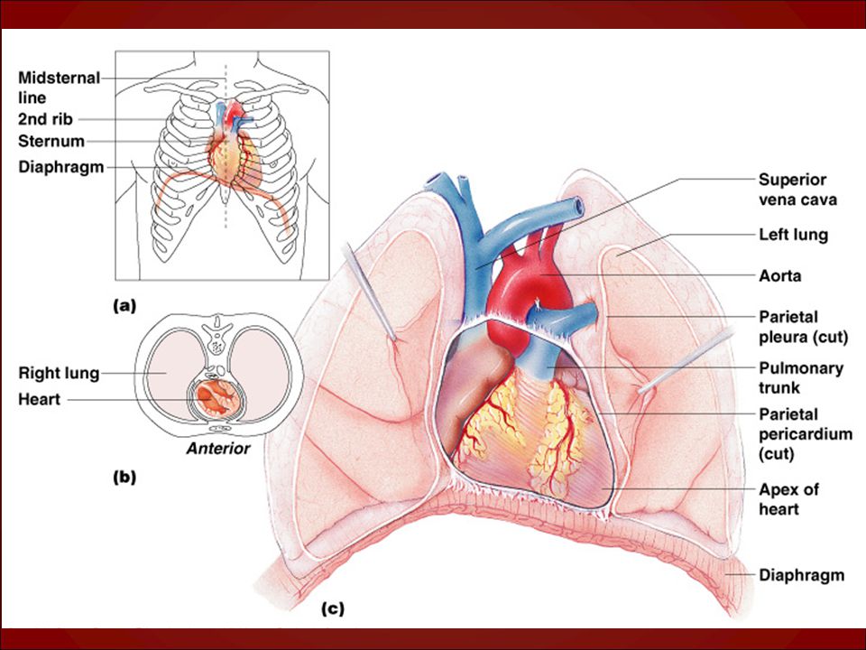
12.1 Anatomy of the Heart

15
* located in thoracic cavity between lungs

17
A. The Wall and Coverings of the Heart

18
1. pericardium- serous membrane, covers the heart

20
2. myocardium – cardiac muscle, wall of heart

21
3. endocardium – inner lining of heart

23
B. Chambers of the Heart

24
1. Right Atrium * right upper chamber of heart
* receives deoxygenated blood from body (venae cavae) tricuspid (R-AV) valve right ventricle
 tricuspid (R-AV) valve right ventricle.")
25
2. Right Ventricle * right lower chamber of heart
* pumps deoxygenated blood pulmonary semilunar valve pulmonary arteries lungs

26
3. Left Atrium * left upper chamber of heart
* receives oxygenated blood from lungs (pulmonary veins) bicuspid (mitral) valve left ventricle
 bicuspid (mitral) valve left ventricle.")
27
4. Left Ventricle * left lower chamber of heart
* pumps oxygenated blood aortic semilunar valve aorta body

28
Interventricular Septum
Heart Chambers Left Atrium Right Atrium Left Ventricle Right Ventricle Interventricular Septum

29
Aortic Semilunar Valve
Heart Valves Aortic Semilunar Valve Pulmonary Semilunar Valve Tricuspid Valve Tricuspid & Bicuspid valves are called atrioventricular valves because they are located between atria & ventricles. These valves snap shut when the ventricles contract. Chordae tendinae are strings of connective tissue that prevent the tricuspid & bicuspid valves from being pushed into the atria when the ventricles contract. Pulmonary & Aortic semilunar valves snap shut when the ventricles relax. Heart murmur - leaky valve(s). Slight leakage is OK, but major leaks must be stopped (replaced with artificial valve). Bicuspid valve is the most common leaky valve. Bicuspid Valve Chordae Tendinae
. Slight leakage is OK, but major leaks must be stopped (replaced with artificial valve). Bicuspid valve is the most common leaky valve. Bicuspid Valve. Chordae Tendinae.")
32
Heart Blood Vessels Aorta Pulmonary Arteries Superior Vena Cava
Right Pulmonary Veins Left Pulmonary Veins Tricuspid & Bicuspid valves are called atrioventricular valves because they are located between atria & ventricles. These valves snap shut when the ventricles contract. Chordae tendinae are strings of connective tissue that prevent the tricuspid & bicuspid valves from being pushed into the atria when the ventricles contract. Pulmonary & Aortic semilunar valves snap shut when the ventricles relax. Heart murmur - leaky valve(s). Slight leakage is OK, but major leaks must be stopped (replaced with artificial valve). Bicuspid valve is the most common leaky valve. Inferior Vena Cava
. Slight leakage is OK, but major leaks must be stopped (replaced with artificial valve). Bicuspid valve is the most common leaky valve. Inferior Vena Cava.")
34
C. Operation of the Heart Valves
1. AV valves are normally open, they close when ventricles contract. 2. Semilunar valves are normally closed, they open when ventricles contract

35
D. Coronary Circuit 1. Heart muscle gets blood supply from the coronary arteries, and drains into coronary veins

36
2. Coronary Circuit Disorders:
a. atherosclerosis / plaque = fatty deposits in coronary arteries b. ischemic heart disease = insufficient blood supply to heart c. thromboembolism = blood clot stuck in coronary artery d. angina pectoris = chest pain left arm e. myocardial infarction = damage to myocardium “heart attack”

37
3. Surgical Procedures a. balloon angioplasty =
balloon inflates to open up a clogged coronary artery b. coronary bypass operation = portion of blood vessel from another part of body is used to bypass blocked coronary arteries

41
12.2 Physiology of the Heart

42
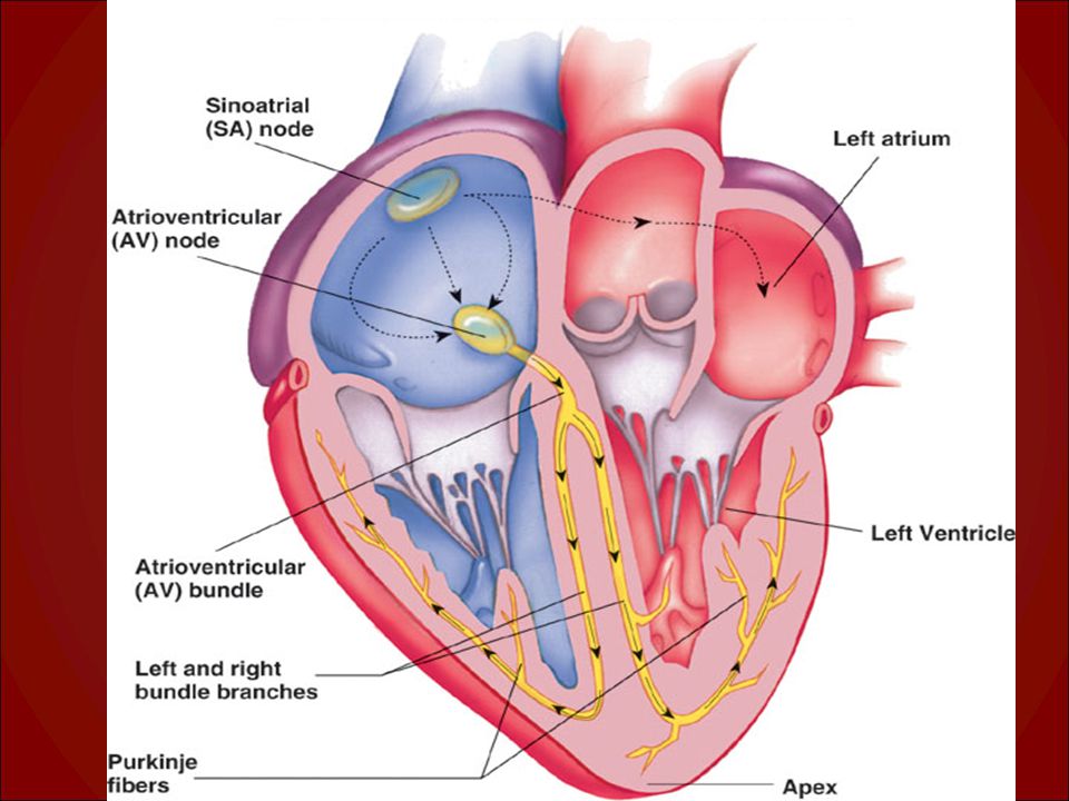
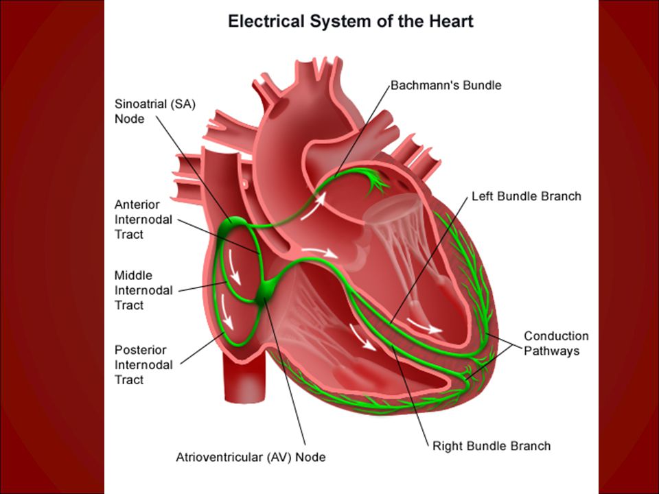
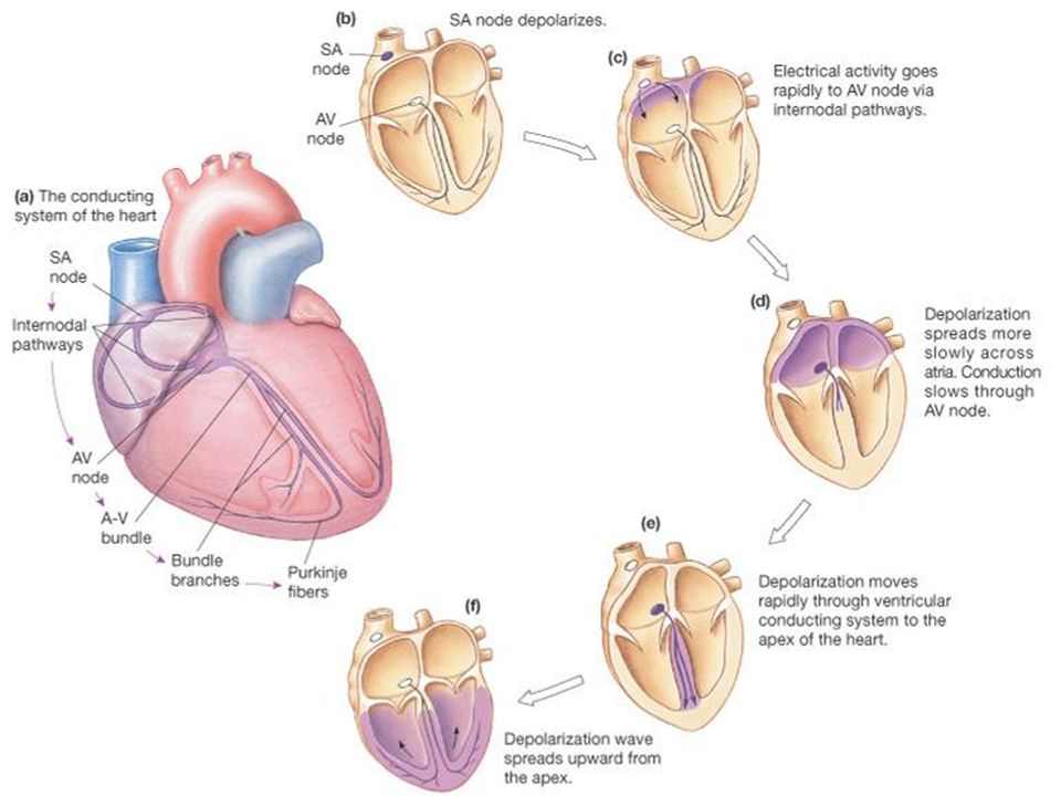
A. Conduction System of the Heart
* electrical system of the heart * causes contraction of the heart muscle * controlled by brain autonomic NS

43
1. Nodal Tissue

44
a. SA (sinoatrial) node * upper posterior wall of right atrium
* initiates heartbeat “pacemaker” * causes atria to contract

45
b. AV (atrioventricular) node * base of right atrium near septum
* “relay station” AV bundle “bundle of HIS” bundle branches Purkinje fibers * causes ventricles to contract

52
2. Electrocardiogram (ECG)
* graph: records electrical activity of heart

54
* consists of a set of waves:

55
P = depolarization (contraction) of atria
 of atria")
56
QRS = depolarization (contraction) of ventricles
 of ventricles")
57
T = ventricles repolarize (recover)
")
58
A - SA node initiates impulse which spreads rapidly to all cardiac muscle cells of atria (via intercalated disks). Atria contract pushing blood into ventricles. Signal is picked up by AV node & transmitted through bundle branches to the Purkinje fibers. Impulse spreads rapidly to all cardiac muscle cells of ventricles (via intercalated disks). Ventricles contract causing bicuspid & tricuspid valves to snap shut (causes “lub” sound). This forces blood in right ventricle into pulmonary arteries & in left ventricle into aorta. When ventricles relax, the pulmonary & aortic semilunar valves snap shut (causes “dup” sound). B - electrocardigram (ECG) showing electrical changes on the body’s surface in response to electrical activities of the heart. P wave corresponds to depolarization of the atria; QRS complex corresponds to ventricular depolarization & atrial repolarization; T wave corresponds to repolarization of ventricles. C - “lub” sound due to closing of tricuspid & bicuspid valves; “dup” sound due to closing of semilunar valves.
 showing electrical changes on the body’s surface in response to electrical activities of the heart. P wave corresponds to depolarization of the atria; QRS complex corresponds to ventricular depolarization & atrial repolarization; T wave corresponds to repolarization of ventricles. C - lub sound due to closing of tricuspid & bicuspid valves; dup sound due to closing of semilunar valves.")
59
Electrocardiogram Intervals show timing of cardiac cycle
P-P = one cardiac cycle P-Q = time for atrial depolarization Q-T = time for ventricular depolarization T-P = time for relaxation

62
B. Cardiac Cycle and Heart Sounds

63
1. cardiac cycle * events that occur during one heartbeat systole = contraction of heart muscle diastole = relaxation of heart muscle

64
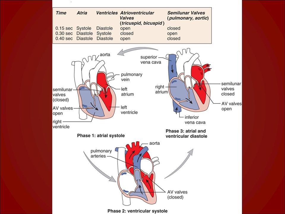
* 3 phases:

65
1. atrial systole atria contract (blood is forced into ventricles)
venrtricles relaxed AV valves open, semilunar valves closed

67
2. ventricular systole ventricles contract (blood forced out of heart)
atria are relaxed AV valves close “lubb”, semilunar valves open

69
3. atrial and ventricular diastole
atria and ventricles are relaxed blood flows from atria to ventricles AV valves open, semilunar valves close “dup”

72
2. Heart Sounds * caused by the closing of the heart valves
* “lubb-dup”

73
C. Cardiac Output (CO)
")
74
* volume of blood pumped out of a ventricle
in one minute * dependent on 2 factors: Heart Rate (HR) - beats per minute Stroke Volume (SV) - amount of blood
 - beats per minute. Stroke Volume (SV) - amount of blood.")
75
A Simple Model of Stroke Volume
Figure 20.19a-d

76
Factors Affecting Cardiac Output
Figure 20.20

77
Cardiac Output: An Example
CO (ml/min) = HR (75 beats/min) x SV (70 ml/beat) CO = 5250 ml/min (5.25 L/min) If HR increases to 150 b/min and SV increases to 120 ml/beat, then CO = 150 b/min x 120 ml/beat CO = 18,000 ml/min or 18 L/min (WOW is right!!)
 = HR (75 beats/min) x SV (70 ml/beat) CO = 5250 ml/min (5.25 L/min) If HR increases to 150 b/min and SV increases to 120 ml/beat, then. CO = 150 b/min x 120 ml/beat. CO = 18,000 ml/min or 18 L/min (WOW is right!!)")
78
12.3 Anatomy of Blood Vessels

80
A. Arteries and Arterioles
* transport blood away from heart * usually transport oxygenated blood * thick, strong, elastic walls * 3 layers (tunic = coat):
:")
81
tunica interna (intima) - inner layer, endothelium
 - inner layer, endothelium")
82
tunica media - middle layer (thick), sm. muscle
, sm. muscle")
83
tunica externa (adventitia) - outer layer, CT
 - outer layer, CT")
84
* arterioles = small arteries

85
B. Capillaries * microscopic blood vessels
* join arterioles to venules * site of gas/nutrient/waste exchanges Capillaries form networks in the body. Not all capillary beds are open all the time - you don’t have enough blood to fill them all. Precapillary sphincters close capillary beds when they do not need to be open. For example, after you have eaten, capillary beds servicing the skin & skeletal muscles are closed down, while those servicing the stomach & intestines are opened.

87
Capillaries form networks in the body
Capillaries form networks in the body. Not all capillary beds are open all the time - you don’t have enough blood to fill them all. Precapillary sphincters close capillary beds when they do not need to be open. For example, after you have eaten, capillary beds servicing the skin & skeletal muscles are closed down, while those servicing the stomach & intestines are opened.

88
C. Veins and Venules * transport blood toward the heart
* usually transport deoxygenated blood * walls are much thinner (same 3 layers)
")
89
Fig. 21.5 V A Tunica intima Tunica media Tunica adventitia

90
* some veins have valves (arms, legs)
")
91
* venules = small veins

92
Direction of blood flow in vessels:
Arteries Arterioles Capillaries Venules Veins

95
12.4 Physiology of Circulation

96
A. Velocity of Blood Flow
* fastest in arteries, slowest in capillaries Blood Vessels Velocity of blood flow (mL/s)
")
97
B. Blood Pressure * force of blood against a vessel wall
* decreases with distance from left ventricle * 2 factors that affect BP:

100
cardiac output – HR and SV
peripheral resistance – arterial diameter and length

102
* Avg. BP in young adult = 120/80
top number = systolic pressure bottom number = diastolic pressure

103
* venous return depends on:
skeletal muscle pump – muscles squeeze respiratory pump - breathing movements

104
Blood in veins is under low pressure
Blood in veins is under low pressure. Contraction of skeletal muscles squeezes veins; blood is forced to move in 1 direction because of venous valves. Valves that do not function properly allow blood to pool, distending the vein. Varicose veins are distended veins caused by the pooling of blood.

105
valves in veins

107
C. Evaluating Circulation

108
1. Pulse * surge/wave of pressure in an artery * = heart rate (bpm)
")
109
* pulse points can be felt in arteries close to skin

110
2. Blood Pressure * = force of blood (mm Hg)
* measured with a sphgmomanometer

111
Steps for measuring BP Apply cuff above elbow
Place stethoscope on brachial artery Inflate cuff until pulse disappears Cuff applied 1 inch above crease at elbow Make sure cuff is correct size Locate brachial artery Palpate radial pulse Inflate cuff until pulse disappears

112
Steps for measuring BP cont.
Let air out gradually Listen for sounds that blood is moving past cuff Let air out Place stethoscope on brachial artery Pump up cuff to above point of obliteration 20 for kids 30 for adults Let air out at 2 mmHg per second

113
systolic pressure = pressure when 1st sound is heard
diastolic pressure = pressure when last sound is heard

116
* hypertension = 140/90

117
12.5 Circulatory Routes

118
A. The Major Systemic Arteries
* see p.243

120
B. The Major Systemic Veins
* see p.244

121
C. Special Systemic Circulations

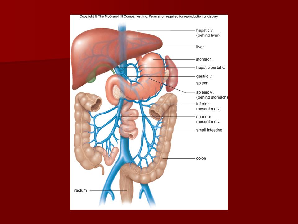
122
* hepatic portal system =
from stomach/intestines to liver

124
* cerebral arterial circle = brain

125
* fetal circulation = fetus (no circulation to lungs)
")
Similar presentations


 Location: to the left of the midline in the Thoracic Cavity –Between the lungs and above the diaphragm Function: Pump blood.>")


 Transport O 2, nutrients, hormones, cell wastes, etc…>")


>")

