Download presentation
Presentation is loading. Please wait.

2
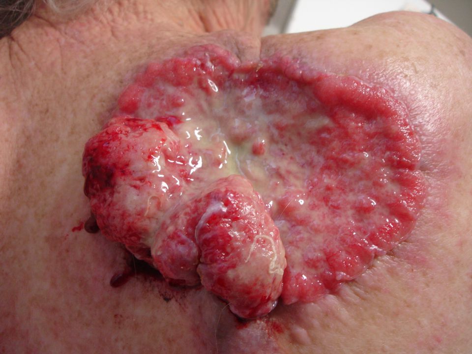
Squamous Cell Carcinoma

5
Incidence 2nd most common skin cancer
Behind BCC, accounting for 20% skin cancers Due to propensity to metastasise, makes them responsible for majority of NMSC deaths

6
Pathogenesis UV Incidence doubles with 8-10 degrees decrease in latitude Induces formation of pyrimidine dimers resulting in DNA point mutations Causes mutations in p53 tumour suppressor gene

7
Skin pigmentation Age Primary dermatoses – xeroderma pigmentosa, oculocutaneous albinism

8
Immunosuppression – due to immunosuppressive drugs, UVR, viral infection esp HPV
Reversed ratio of BCC:SCC, SCC being 3x more common in transplant patients Higher rates – cumulative risk of SCC/ BCC in heart transplant recipient is 3% at one year, 21% at 5 years, 35% at 10 years

9
Histological subtypes
Pleomorphic Adenoid/Acantholytic Simplex Small cell Verrucous Keratoacanthoma Actinic keratosis Bowenoid/Erythoplasia of Queyrat

10
Simplex Majority of SCCs
Atypical keratinocytes develop within epidermis and invade the dermis Tumour cells are enlarged, hyperchromatic, variably pleomorphic nuclei, prominent mitotic activity Keratin pearls

11
Actinic Keratosis Also SCC in situ, micro invasive SCC, as there is considerable overlap in the histology Atypical keratinocytes that have not breached the dermal barrier SCCIS is typically full thickness keratinocyte atypia

12
Rate of malignant transformation is 0.1% per lesion per year
About 16% will eventually transform Can progress to other skin cancers such as sebaceous carcinoma

13
Pleomorphic AKA spindle / sarcomatoid, RARE
Associated with previous trauma or RTX Most commonly found on face or sun exposed areas of elderly Commonly ulcerate, but may present as an exophytic mass

14
Microscopically whorls of atypical squamous cells co-mingle with collagen fibres
Pleomorphic giant/spindle cells may be present Neoplastic keratinocytes have hyperchromatic eosinophilic cytoplasm, elongated, pleomorphic and veiscular nuclei with multiple nucleoli

15
Small cell May resemble metastatic small cell neuroendocrine carcinoma or Merkel cell carcinoma Invades in cohesive nests with adjacent intense inflammatory and desmoplastic host response Stains for cytokeratin, but may stain for neuron specific enolase (NSE), a neuroendocrine marker
, a neuroendocrine marker.")
16
Verrucous Exophytic or endophytic masses growing at sites of chronic irritation Slowly locally invasive, little or no propensity to metastasise Morphologically appear well differentiated with little atypia Thickened papillae composed with well differentiated squamous cells invading into dermis

17
Verrucous 3 distinct clinicopathologic subtypes Oral
Associated with tobacco chewing, betel nut chewing, HPV, poor oral hygiene Typically wart like white/gray lesion Well differentiated Plantar Many crypt like openings Slowly enlarging, fleshy pink exophytic mass Verrucous hyper/para keratotic component, epithelial crypts with keratinaceous debris Buschke-Loewenstein Anogenital type, described by B-L in 1925 Occur most commonly in uncircumcised men under 50, associated with HPV 6 & 11 Present as caulflower like lesions most commonly on glans penis Extensive verrucous acanthosis with dermal extension, keratinocyte atypia minimal, hypergranulosis and crypt/sinus formation

18
Keratoacanthoma Period of rapid growth lasts 4-8 weeks
Potential for spontaneous involution usually within 4-6 months, sometimes with considerable scarring Clinically tend to be rapid growing smooth, firm nodule with central keratin plug

19
Histologically difficult to distinguish between benign KA and SCC KA type, so being amalgamated by histopathologists Atypical squamous proliferation with intradermal invasion Typically crateriform architecture with keratin plug and well developed collarette

20
Adenoid / Acantholytic
Form a pseudoglandular appearance Cells arranged in cords and nests with clefts produced by acantholysis of cells leaving spaces that superficially resemble glands

21
Enlarged free floating dysplastic keratinocytes found in lumina
Clinically appear as ulcer on head & neck of men in 5th to 6th decade High incidence of recurrence after radiation therapy Tend to be more locally aggressive but metastasise less

22
Bowenoid Considered to be SCC in situ
Most common site is head and neck, followed by limbs and then trunk Well demarcated, slow growing, erythematous scaly patch, usually small in size

23
Histologically shows hyperkeratosis, acanthosis, psoriasiform hyperplasia, full thickness atypia, loss of polarity reflecting cessation of maturation When neoplastic keratinocytes invade the dermis, this lesion is termed Bowenoid SCC Especially associated with HPV – HPV2 with extragenital lesions, HPV16 with genital lesions

24
Metastasis Overall risk is 2 – 6%, not 0.5%
Recurrent SCC has metastatic rate of 30%, and metastatic cases had a survival rate of 1/3 Metastases tend to be to regional lymph nodes Most mets (and local recurrences) are found within first 2 years, and 95% within first 5 years
 are found within first 2 years, and 95% within first 5 years.")
25
Risk factors for metastasis and recurrence
Recurrence rate doubled and tripled metastatic rate Size > 2cm Grade 3 & 4 vs. Grade 1 & 2 tumours Well differentiated has recurrence 7%, mod well diff 23%, poor diff 28% Tumour thickness 3 year recurrence free survival is 98% for <3.5mm, 84% for > 3.5mm (Breslow thickness) Rapid growth rate
 Rapid growth rate.")
26
Sun exposed areas tend to metastasise and recur less than mucosal SCC
Scar SCC are very aggressive Lip and ear SCC have higher metastatic rate than other head and neck sites (16 & 10%) Probably due to decreased subcutaneous fat Nose and scalp, anogenital are intermediate risk Periungal SCC has high recurrence rate but almost never metastasises Previous treatment – recurrent cancers have a metastatic rate of 25% Location – ear 45%, lip 32% metastatic rate
 Probably due to decreased subcutaneous fat. Nose and scalp, anogenital are intermediate risk. Periungal SCC has high recurrence rate but almost never metastasises. Previous treatment – recurrent cancers have a metastatic rate of 25% Location – ear 45%, lip 32% metastatic rate.")
27
Histopathology Isolated strands, infiltrative pattern, haphazard growth vs. broad pushing borders Perineural invasion (occurs in 2-14% SCC, most commonly H&N in elderly men) Has been quoted as local recurrence 47%, regional mets 35%, distant nodes 15%; so post op RTX commonly offered NO good evidence that any subtype has greater risk recurrence or metastasis
 Has been quoted as local recurrence 47%, regional mets 35%, distant nodes 15%; so post op RTX commonly offered. NO good evidence that any subtype has greater risk recurrence or metastasis.")
28
Immunosuppression Biologically more aggressive, with higher rates of lymph node metastases and deaths secondary to skin cancer

29
Tumour size Size (cm) Metastatic rate 5 yr disease free survival T1
< 2 1.4% 95-99% T2 2 – 4 9.2% 85-60% T3 > 4 > 13% 75-60% T4 Invading deep structures < 40%

30
Tumour depth and metastatic rate
< 2mm 2 – 6mm 4.5% > 6mm 15%

31
Grades Broder’s Grade Undifferentiated cells
Ratio of differentiated cells I – Well differentiated < 25% 3:1 II – Moderately well differentiated 25 – 50% 1:1 III – Poorly differentiated 50 – 75% 1:3 IV – Anaplastic or pleomorphic > 75% Nil

32
Surgical Management Tumours < 2cm diameter are successfully excised 95% of the time with a margin of 4mm, 6mm for high risk cases (Brodland & Zitelli) Tumours > 2cm diameter require margin of 10mm for equivalent local control rates Moh’s surgery
 Tumours > 2cm diameter require margin of 10mm for equivalent local control rates. Moh’s surgery.")
33
Other modalities Dessication and curettage Cryosurgery
Lesions less than 2cm diameter have cure rates of % Cryosurgery Well localised, superficial lesions on trunk or limbs 5FU & Imiquimod & Photodynamic therapy Useful for actinic keratoses

34
Radiation Therapy < 2cm tumours have a cure rate of 95%
Adjunctive RTX must be given within 8 weeks for greatest efficiency (Late) changes include : atrophy, fibrosis, hypopigmentation, telangiectasia, ulceration “As late results of RTX can be poor, it is not recommended for patients under 60 yo with uncomplicated primary SCC” May hasten natural history of KA
 changes include : atrophy, fibrosis, hypopigmentation, telangiectasia, ulceration. As late results of RTX can be poor, it is not recommended for patients under 60 yo with uncomplicated primary SCC May hasten natural history of KA.")
Similar presentations


>")


>")


