Download presentation
Presentation is loading. Please wait.

1
Initial periodontal therapy I:
Instruments and principles for instrumentation 何坤炎副教授:高雄醫學大學 口腔醫學院牙醫學系 轉 7004 , 7029

2
學習目標: 學習資源: 1. Classification of Periodontal instruments
2. General principles of instrumentation 3. Principles of scaling and root planing 學習資源: 1. Rateishak KH&EM, Wolf HE, Hassell TM: Color Atlas of Periodontology pp 2. Newman, Takai, klokkevold, and Carranza: Clinical Periodontology. 10th edition, pp 3. Egelberg J: Periodontics The scientific way, synopses of human clinical studies. 1995,pp25-71

3
Classification of Periodontal instruments:
Periodontal probe Explorer Scaling and root planing instruments Polishing instruments Surgical instruments

4
* With firm, gentle pressure to the bottom of pocket
1. Periodontal probe--- to locate, measure the depth of pockets and to determine their configuration * With firm, gentle pressure to the bottom of pocket * The shank should be aligned with long axis of tooth to be probed Marquis Michigan O WHO Newman, Takai, klokkevold, and Carranza: Clinical Periodontology. 10th edition, pp 750

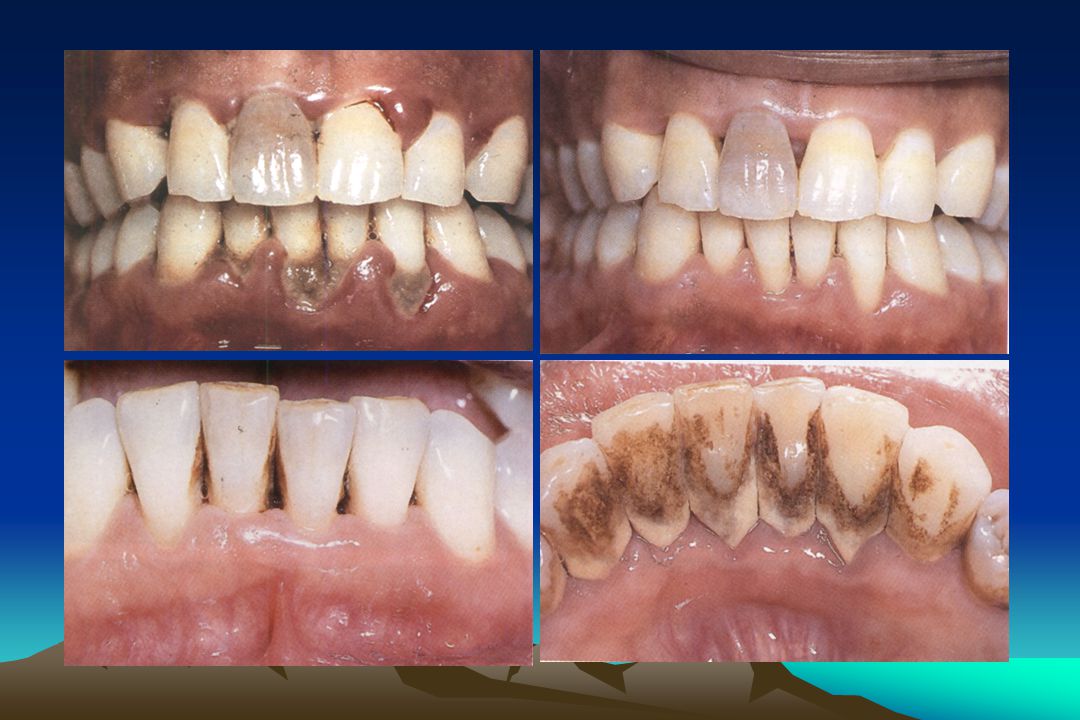
5
From KMUH

6
* Furcation area--- best be evaluated by curved, blunt, Nabers probe
Rateishak KH&EM, Wolf HE, Hassell TM: Color Atlas of Periodontology pp 60

7
* Also to check the smoothness of the root
2. Explorer--- to locate subgingival calculus deposition and caries area * Also to check the smoothness of the root surface after root planing Newman, Takai, klokkevold, and Carranza: Clinical Periodontology. 10th edition, pp 751

8
3. Scaling and root planing instruments
a) Curette b) Sickle scaler c) File scaler d) Chisel scaler e) Hoe scaler f) Ultrasonic instruments Newman, Takai, klokkevold, and Carranza: Clinical Periodontology. 10th edition, pp 751
 Curette. b) Sickle scaler. c) File scaler. d) Chisel scaler. e) Hoe scaler. f) Ultrasonic instruments. Newman, Takai, klokkevold, and Carranza: Clinical Periodontology. 10th edition, pp 751.")
9
Sickle scaler--- primary to remove the
supragingival calculus *With straight shanks for incisors and canine *With contra-angled shanks for posterior teeth From KMUH

10
* Difficult to insert the blade under the gingiva
without damaging the surrounding gingiva Small, curved sickle scaler– 204SD, and Nevi 2 posterior sickle scaler inserted several millimeter subgingivally Newman, Takai, klokkevold, and Carranza: Clinical Periodontology. 10th edition, pp 751

11
* Finer than sickle, no sharp points or corners
Curette--- used for subgingival scaling, root planing and removing soft tissue lining the pocket * Finer than sickle, no sharp points or corners can be adapted and provide better access to deep pockets with a minimal soft tissue trauma Newman, Takai, klokkevold, and Carranza: Clinical Periodontology. 10th edition, pp

12
Two basic type--- universal and Gracey curette
Universal type--- by altering and adapting finger rest, fulcrum and hand position of the operator (Barnhart curettes, Columbia curettes …) Newman, Takai, klokkevold, and Carranza: Clinical Periodontology. 10th edition, pp 755
 Newman, Takai, klokkevold, and Carranza: Clinical Periodontology. 10th edition, pp 755.")
13
Newman, Takai, klokkevold, and Carranza: Clinical Periodontology
Newman, Takai, klokkevold, and Carranza: Clinical Periodontology. 10th edition, pp

14
* Rigid Gracey has a larger, stronger and less
Gracey curettes--- rigid or finishing type of shank * Rigid Gracey has a larger, stronger and less flexible shank and blade than the standard finishing Gracey * Rigid Gracey--- to remove moderate to heavy calculus Gracey #1-2 & 3-4: anterior teeth Gracey #5-6: anteriors and premolars From KMUH

15
Extended shank curettes--- Hu-Friedy After-Five
curettes are modification of standard Gracey curettes design * The terminal shank is 3 mm longer, allowing extension into deeper periodontal pockets of 5 mm or more * Thinned blade for smoother subgingival insertion and reduced tissue extension Newman, Takai, klokkevold, and Carranza: Clinical Periodontology. 10th edition, pp 756

16
Mini-bladed curettes--- Hu-Friedy Mini-Five
curettes are modification of After Five curettes * The blade is half the length of After Five or standard Gracey curettes * Gracey Curvette Sub-0, # premolar #11-12,# posterior mesial and distal Newman, Takai, klokkevold, and Carranza: Clinical Periodontology. 10th edition, pp 756

17
* Shorter blade allows easier insertion and adaptation in deep, narrow pockets; furcations; developmental grooves; line angles; and deep, tight, facial, lingual or palatal pockets ---Gracey Curvette Newman, Takai, klokkevold, and Carranza: Clinical Periodontology. 10th edition, pp 757

18
Mini-bladed curettes should not be used
routinely 2. Large #4 handle are recommended for any mini-bladed instruments 3. Can be used to scale with toe directed either mesially or distally 4. Generally used with straight vertical stroke

19
Quétin furcation curettes, MD1,BL1---0. 9 mm; BL2,MD2—1
Quétin furcation curettes, MD1,BL mm; BL2,MD2—1.3 mm blade width 2. Diamond-coated files, no cutting edge for final finishing of root surface, used with light, even pressure against to root surface Newman, Takai, klokkevold, and Carranza: Clinical Periodontology. 10th edition, pp 759 Fig.51-37,38

20
Mini-Five curettes for anterior teeth has
proven to be more effective than conventional curette in debriding narrow root surface Singer et al. J. Clin. Periodontol 1992

21
Plastic instruments for implant
* Avoid scarring and permanent damage to implants * Plastic probes * Implacare implant instruments Newman, Takai, klokkevold, and Carranza: Clinical Periodontology. 10th edition, pp 758

22
Hoe scalers--- to remove tenacious subgingival
calculus and necrotic cementum * The blade is bent at a 99 degree angle, the cutting edge is beveled at 45 degrees * The blade is slightly bowed so that it can maintain contact at two points on a convex surface --- stabilize the instrument Newman, Takai, klokkevold, and Carranza: Clinical Periodontology. 10th edition, pp 759

23
Chisels--- the end of blade is beveled at 45
degrees to form the cutting edge * With a modified pen grasp, push stroke File --- periodontal surgery to fracture or crush large deposits Newman, Takai, klokkevold, and Carranza: Clinical Periodontology. 10th edition, pp 759

24
POWER-DRIVEN SCALERS Sonic Ultrasonic Magnetostrictive Piezoelectric
Manual tune Auto tune

25
Oscillating scaler system (振動潔牙系統)
* Sonic scaler: rotating cam generates vibration with frequencies 2000 to 6500 Hz, vibrations depending on the air pressure input, with an amplitude of up to 1000 m, plaque and calculus are removed by tapping motion (輕敲) * Ultrasonic scaling instruments Newman, Takai, klokkevold, and Carranza: Clinical Periodontology. 10th edition, pp 760
 * Ultrasonic scaling instruments. Newman, Takai, klokkevold, and Carranza: Clinical Periodontology. 10th edition, pp 760.")
26
* Frequency ranging from 20,000 to million cycles per second
Ultrasonic instruments --- for scaling, curetting, and removing stain * Frequency ranging from 20,000 to million cycles per second * The spray is directed at the end of the tip to dissipate the heat generated by the ultrasonic vibration * Apply by slight tactile force

27
* The cavitating water spray also serves to
flush calculus, plaque, and debris dislodged by the vibrating tip from the pocket

28
Ultrasonic scaling instruments
* Magnetostrictive (磁振式)--- are driven by nickel-iron alloy strips or a Ferrite Insert inserted into a hand-piece, vibration frequencies to Hz, vibration of tip is elliptical All side of tip are active and work when adapted to tooth surface. Hammering (錘敲打) or scraping motion (刮削) Newman, Takai, klokkevold, and Carranza: Clinical Periodontology. 10th edition, pp 760
--- are driven by nickel-iron alloy strips or a Ferrite Insert inserted into a hand-piece, vibration frequencies to Hz, vibration of tip is elliptical. All side of tip are active and work when adapted to tooth surface. Hammering (錘敲打) or scraping motion (刮削) Newman, Takai, klokkevold, and Carranza: Clinical Periodontology. 10th edition, pp 760.")
29
Ultrasonic scaling instruments
* Piezoelectric (壓電式)--- vibration is generated by changes in the dimension of a quartz crystal, vibration of tip is linear, or back and forth, only two sides of tip are active Tapping (輕敲) or scraping motion (刮削) Newman, Takai, klokkevold, and Carranza: Clinical Periodontology. 10th edition, pp 761
--- vibration is generated by changes in the dimension of a quartz crystal, vibration of tip is linear, or back and forth, only two sides of tip are active. Tapping (輕敲) or scraping motion (刮削) Newman, Takai, klokkevold, and Carranza: Clinical Periodontology. 10th edition, pp 761.")
30
Newman, Takai, klokkevold, and Carranza: Clinical Periodontology
Newman, Takai, klokkevold, and Carranza: Clinical Periodontology. 10th edition, pp 760

31
壓電式 VS 磁致伸縮式 壓電式 VS 磁致伸縮式洗牙機 直線型 橢圓形 壓電式 磁致伸縮式 手機內壓電陶瓷(crystal)造成電流改變
機頭體上有扁平金屬條彈簧片及手機上有線圈 量改變導致機頭尖端呈直線型運動,頻率為每秒25k-50k 電流磁化線圈,導致彈簧片伸縮及接觸產生快速振動。電能轉換機械能 機頭尖端只有二邊作用,限制了效率 機頭橢圓形360度路徑,可輕易觸及每個部位和角落 所有機器的頻率都是自動調整 機器上可選用手動或自動調整 不會產生熱,水是用來沖洗 會產生熱:水是用來冷卻及沖洗 頻率範圍從18k–42k Hz 壓電式 VS 磁致伸縮式 直線型 橢圓形

32
Safety and Efficacy of Oscillating Scalers
* Hand instruments depends on the numbers of scaling stroke and lateral force applied * Oscillating scalers depends on instrumentation time, lateral force, scaler tip angulation, and instrument power setting

33
Safety and Efficacy of Oscillating Scalers
* If scaler tip is angulated parallel to root surface and force applied do not exceed 2 N 50 m/year (critical defect depth 臨界缺損深度), 40 second instrumentation --- acceptable
, 40 second instrumentation --- acceptable.")
34
Safety and Efficacy of Oscillating Scalers
* Magnetostrictive type ---tip angulation, lateral forces have identical influence on substance removal. The critical defect depth 50 m can be maintained if tip is angulated absolutely parallel to root surface and forces used do not exceed 1 N

35
* Piezoeletric type --- mostly influenced by scaler
tip angulation. If scaler tip is angulated parallel to root surface, CDD can be maintained below 50 m even forces up 2N * Sonic scaler is comparable to the efficacy of magnetostrictive scaler at low power setting or to the efficacy of piezoelectric scaler at medium power setting

36
Newman, Takai, klokkevold, and Carranza: Clinical Periodontology
Newman, Takai, klokkevold, and Carranza: Clinical Periodontology. 10th edition, pp

37
FREQUENCY Active Tip Area Active tip area 30 K = 4.2 mm 50 K = 2.3 mm
1. affected by frequency. 2. higher frequency = smaller active tip area Active tip area 30 K = 4.2 mm 50 K = 2.3 mm

38
Stroke – ie. Power in Scaling
The tip of the insert is tracing an elliptical path or “track” 30,000 times each second. BUT – how big is the track? The “Power” setting on a scaler defines how big the track is and thus defines the “stroke” High Power Med. Power Low Power (Blue Zone) ~ in ~ in ~~.0001 “ 30,000 laps per second 30,000 laps per second 30,000 laps per second
 ~ in. ~ in. ~~ ,000 laps. per second. 30,000 laps. per second. 30,000 laps. per second.")
39
Power = The length of the stroke of the insert

40
Power Adjustment - New Insert
A new insert tip moving at moderate power creates a cone shape ---the very tip moves along this bottom of this cone and trace an ellipse whose size (stroke) is dictated by power setting
 is. dictated by power setting.")
41
* Light pressure, 15 degree to tooth surface
How to use ultrasonic scaling instruments * Position * Light pressure, 15 degree to tooth surface * Cooling system, cc/min * Not be used in pt’ with cardiac pacemaker transmissible disease

42
Ultrasonic scaling instruments * Less tissue trauma
* Useful for initial debridement * Bulky and blunt--- subgingival insertion to base of pocket is limited * Fracture calculus and remove it * Diminish tactile sensation

43
ULTRASONICS ADVANTAGES Reduced clinician fatigue
Increased access and adaptability Less tissue distension and more patient comfort Less chair time Promotes faster healing Bactericidal effect Sharpening eliminated Benefits of lavage

44
Ultrasonic debridment is not recommended in
Titanium implants Restorative materials Areas of demineralization Hypersensitive teeth

45
ULTRASONICS CONSIDERATONS Contaminated aerosol production
Less tactile sense Requires water and suction Effects of noise, vibration Handpiece sterilization

47
Ultrasonic debridement vs. hand scaling * Microbial plaque removal
To be significant more effective in * Microbial plaque removal * Class II or III furcation involve Newman, Takai, klokkevold, and Carranza: Clinical Periodontology. 10th edition, pp 761

48
Use of Modified Ultrasonic Inserts at Furcation area

49
Ultrasonics vs. Hand Instruments: Calculus Removal
Most literature shows both are equally effective Drisko (1993): modified ultrasonic inserts (slimlines®) produced smoother roots, better access to the bottom of the pocket, better calculus and plaque removal, less operator time, and less operator fatigue.
: modified ultrasonic inserts (slimlines®) produced smoother roots, better access to the bottom of the pocket, better calculus and plaque removal, less operator time, and less operator fatigue.")
50
Ultrasonic devices (with thin tip)
As effective as hand-held curette in * Maintaining clinical attachment levels * Significantly reduced time Copulos et al. JP 1993

51
* Significantly less percentage of residual
Mini-bladed curettes vs. slim ultrasonic tip * Significantly less percentage of residual deposits --- fine curettes * The potential value of small, thin blade curettes in debriding involved furcation during initial therapy Francisco et al. JP 1997

53
Sonic units do not release heat the way
ultrasonic units do, but still have water for cooling and flushing away debris

57
Dental Endoscope --- Perioscopy system
For use subgingivally in diagnosis and treatment of periodontal disease, also evaluate subgingival caries, root fracture, defect restorations, and resorption Newman, Takai, klokkevold, and Carranza: Clinical Periodontology. 10th edition, pp 762

58
Dental Endoscope --- Perioscopy system
* It consist of a 0.99 mm diameter reusable fiberoptic endoscope over which is fitted a disposable, sterile sheath. Newman, Takai, klokkevold, and Carranza: Clinical Periodontology. 10th edition, pp 762

59
Fiberoptic endoscope fit
onto periodontal probes and ultrasonic instrument The sheath delivers water irrigation that flushes the pocket while the endoscope is in use and keeps the field clear Newman, Takai, klokkevold, and Carranza: Clinical Periodontology. 10th edition, pp 762

60
Cleaning and polishing instrument
Rubber cup, brushes and dental tape --- for clean and polish tooth surface Newman, Takai, klokkevold, and Carranza: Clinical Periodontology. 10th edition, pp 763

61
Air-powder polishing --- Prophy-Jet
An air-powdered slurry of warm water and sodium bicarbonate. The slurry remove stains rapidly and efficiently by mechanical abrasion and provides warm water for rinsing and lavage Newman, Takai, klokkevold, and Carranza: Clinical Periodontology. 10th edition, pp 764

62
* Damage to gingival tissue is transient and
Study shows that tooth substance (cementum and dentin) can be lost by Prophy-Jet * Damage to gingival tissue is transient and insignificant clinically * Composite restoration can be roughened Newman, Takai, klokkevold, and Carranza: Clinical Periodontology. 10th edition, pp 764
 can be lost by Prophy-Jet. * Damage to gingival tissue is transient and. insignificant clinically. * Composite restoration can be roughened. Newman, Takai, klokkevold, and Carranza: Clinical Periodontology. 10th edition, pp 764.")
63
* Pt’ with medical history of respiratory disease
Contraindications * Pt’ with medical history of respiratory disease * Those with sodium-restricted diets * Individuals on medications affecting the electrolyte balance * Infectious diseases aerosol created

64
* Excisional and incisional instruments * Surgical curette
Surgical Instruments * Excisional and incisional instruments * Surgical curette * Periosteal elevator * Hoe, chisel, file and rongeur * Tissue and thread scissors * Hemostats and tissue forceps Newman, Takai, klokkevold, and Carranza: Clinical Periodontology. 10th edition, pp 45-98

65
a) Excisional and incisional instruments
* Surgical blade--- No. 15, 12, and 11 * Electrosurgery: Newman, Takai, klokkevold, and Carranza: Clinical Periodontology. 10th edition, pp

66
Electrosurgery (surgical diathermy)
* Using controlled frequency electrical currents million cycles/second * Three active electrodes: 1. Single-wire electrodes for incision 2. loop electrodes for planing tissue 3. Heavy, bulkier electrodes for coagulation procedure Newman, Takai, klokkevold, and Carranza: Clinical Periodontology. 10th edition, pp 45-98

67
* Deep resection close to bone, can produce gingival
Most important basic rule of electrosurgery--- always to keep the tip moving, 5-10 second for cooling * Deep resection close to bone, can produce gingival recession, bone necrosis and sequestration, loss of bone height, furcation exposure and tooth mobility * Contraindicated for patients who have poorly shielded cardiac pacemakers

68
Four types electrosurgical technique:
1. Electrosection---performs incision, excision, and tissue planing 2. Electrocoagulation--- can prevent bleeding at initial entry into tissue, but cannot stop bleeding after blood is present 3. Electrofulguration--- burning of the tissue 4. Electrodesication--- drying of the tissue

69
* Water Laser (Er-YAGG) ---Frenectomy, tooth preparation
* Lukki Laser --- detoxication, desensitization

70
* Surgical curette--- for the removal of granulation
tissue, fibrous interdental tissue and tenacious subgingival deposits * Periosteal elevator--- to reflect and remove the flap after the incision has been made for flap surgery

71
Surgical chisel, rongeur and proximal bone file
For removal of sharp bone and osteoplasty Rateishak KH&EM, Wolf HE, Hassell TM: Color Atlas of Periodontology pp 60

72
* Pocket marker, Kirkland and interproximal
Surgical instruments for gingivectomy * Pocket marker, Kirkland and interproximal gingival knife Rateishak KH&EM, Wolf HE, Hassell TM: Color Atlas of Periodontology pp 60

73
* Pocket marker, Kirkland and interproximal
Surgical instruments for gingivectomy * Pocket marker, Kirkland and interproximal gingival knife Rateishak KH&EM, Wolf HE, Hassell TM: Color Atlas of Periodontology pp

74
Gingival enlargement gingivectomy

75
Gingival enlargement gingivectomy

Similar presentations


>")
 (cont.)>")


: Access, Visibility and Isolation JANET WEBER, RDH, M.Ed.>")


>")


 何坤炎副教授:高雄醫學大學 口腔醫學院牙醫學系 07-3121101 轉 7004, 7029>")


