Download presentation
Presentation is loading. Please wait.

2
SIAscope Training Course Example lesions

3
Training lesions This course trains you in identifying SIAgraph features, as well as linking them with ELM (dermoscopy) diagnosis. Answer the questions on the different kinds of lesions in the following slides. Use the evidence in the features to make a diagnosis.

4
Feature checklist The three features which research has identified as the most useful in melanoma diagnosis are as follows: –Dermal melanin –Displaced blood with erythematous blush –Holes in collagen These features will now be defined.

5
Dermal Melanin Shown on the SIA-DM (dermal melanin) SIAgraph Areas of colour (green – blue – red) Goes from least (green) to most (red) involvement (combination of depth + concentration) A contiguous area of dermal melanin within the lesion –NB excluding hairs.
 SIAgraph Areas of colour (green – blue – red) Goes from least (green) to most (red) involvement (combination of depth + concentration) A contiguous area of dermal melanin within the lesion –NB excluding hairs.")
6
Displaced blood Shown on the SIA-B (blood) SIAgraph A contigious absence of blood within the lesion Area of white
 SIAgraph A contigious absence of blood within the lesion Area of white")
7
Erythematous blush Shown on the SIA-B (blood) SIAgraph Increased vascularity around the periphery of the lesion for at least ¾ of its circumference (when compared to surrounding skin, within confines of lesion.) Seen as darker red area within a lesion
 SIAgraph Increased vascularity around the periphery of the lesion for at least ¾ of its circumference (when compared to surrounding skin, within confines of lesion.) Seen as darker red area within a lesion")
8
Holes in Collagen Shown on SIA-C (collagen) SIAgraph A contiguous absence of collagen within the lesion but discounting hair follicles. Black “holes” in collagen

9
Example lesions The following slides contain SIAgraphs of different lesions, in addition to a few case notes. Using the definitions from the previous slide, try to decide whether or not a feature is present in a lesion.

10
Example lesions Then press one of the bottom buttons to answer the question. Click in the centre of the slide for an explanation of the feature. Turn your computer’s sound on if possible.

11
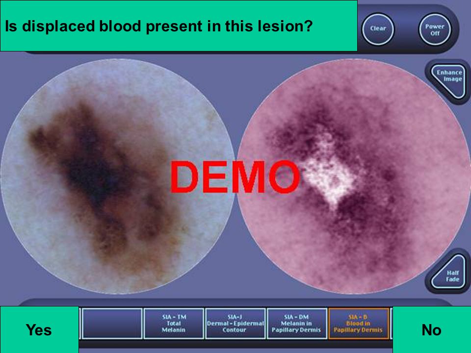
26_05_00_16_56 This lesion presented on the chest of a 53 year old male.

12
Are collagen holes present in this lesion? YesNo

13
No. There is no contiguous absence of collagen within the lesion. (Black areas)
")
14
Is displaced blood present in this lesion? YesNo

15
No. There is no contigious absence of blood within the lesion. (White areas)
")
16
Is dermal melanin present in this lesion? YesNo

17
No. There is no blue on the SIA-DM.

18
What is your diagnosis? No collagen holes No displaced blood No dermal melanin

19
Diagnosis Compound Naevus Remember, although they have melanocytes in the dermis, most compound naevi do not have dermal melanin. –Melanocytes have stopped producing melanin.

20
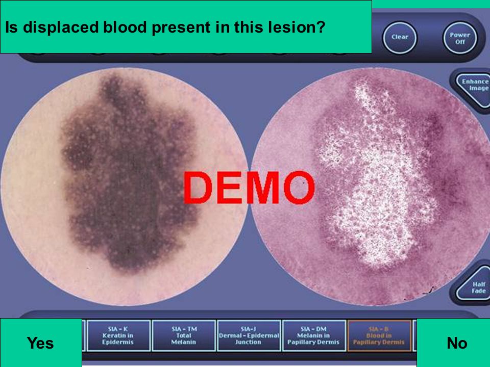
19_05_00_16_47 This lesion presented on the back of a 43 year old male.

21
Are collagen holes present in this lesion? YesNo

22
Yes. There are black areas within the body of the lesion on the SIA-C, which are not hair follicles. Using a SIAscope would allow you to zoom in on areas of doubt, to clarify whether or not the feature exists.

24
Is displaced blood present in this lesion? YesNo

25
Yes. There is a large white area in the centre of the lesion.

26
Is Erythematous blush present in this lesion? YesNo

27
Yes. There is increased vascularity around the periphery of the lesion for at least ¾ of its circumference. Compared to the surrounding skin. This is a strong example of both displaced blood and erythematous blush.

28
Is dermal melanin present in this lesion? YesNo

29
Yes. This is shown in several colours, showing that the lesion has a large dermal component.

30
What is your diagnosis? Holes in collagen Displaced blood plus erythematous blush Dermal melanin

31
Diagnosis Melanoma. Clark’s level 3+ –Holes in the collagen SIAgraph show that the melanoma has penetrated to the bottom of the papillary dermis. –The SIAscope cannot image any further than the bottom of the papillary dermis. As the reticular dermis does not scatter light sufficiently.

32
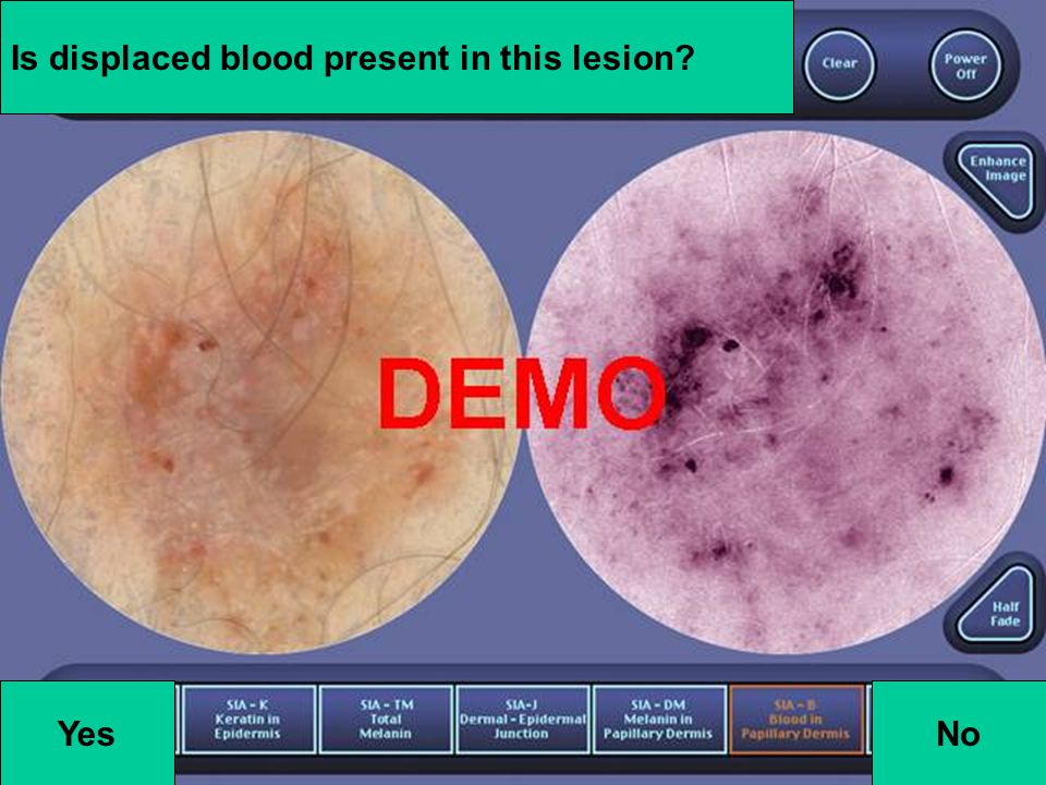
1491688_a An 18 year old male presented with this lesion on the back.

33
Is displaced blood present in this lesion? YesNo

34
No. Displaced blood is not present in this lesion. –WHY? There are no areas demonstrating an absence of blood (white).
..")
35
Are collagen holes present in this lesion? NoYes

36
No. There are no black areas within the lesion. In this case, matching fluid has not been used, and the collagen view is slightly distorted. –In difficult cases, rescanning the lesion using matching fluid may help improve the clarity of the collagen image.

37
Is dermal melanin present in this lesion? YesNo

38
No. Areas of dermal melanin are shown in blue on the SIA – DM. Areas of blue ARE visible in a rim around the top right of the SIA-DM. –However, they are not within the lesion, and are probably a result of light leakage when taking the handset. Remember to use matching fluid – –Lesions without dermal melanin can appear to have dermal melanin if scanned without fluid

39
Would you excise this lesion? No collagen holes No displaced blood No dermal melanin

40
Diagnosis Benign Dysplastic Naevus

41
728834_a This lesion presented on the forearm of an 84 year old female.

42
Are collagen holes present in this lesion? YesNo

43
No. There is no contiguous absence of collagen within the lesion.

44
Is displaced blood present in this lesion? YesNo

45
No. However, note that the lesion consists of blood, making the diagnosis of haemangioma most likely.

46
Is dermal melanin present in this lesion? YesNo

47
Yes. Dermal melanin is shown on the SIAgraph. However, this may be haemasiderin, a breakdown product of blood that mimics dermal melanin on dermal melanin SIAgraph. If dermal melanin is shown in a haemangioma, it is normally only green in colour, and not blue or red.

48
Diagnosis Haemangioma No collagen holes No displaced blood Dermal melanin indicated (Haemasiderin)
")
49
05_06_00_08_59 This lesion presented on the face of a 54 year old female.

50
Are collagen holes present in this lesion? YesNo

51
Yes. All over the lesion are large black areas – these are holes.

53
Is displaced blood present in this lesion? YesNo

54
Yes. There are large areas where there is significantly less blood than in the surrounding tissue.

55
Is erythematous blush present in this lesion? YesNo

56
Yes. There is increased vascularity around the periphery of the lesion for at least ¾ of its circumference.

57
Is dermal melanin present in this lesion? YesNo

58
Yes. Throughout the lesion. Note the blue around the edge of the lesion, caused by grease/dirt on the lens or light leakage. Note that for the majority of the lesion, a red colour is shown, implying a large amount of melanin in the dermis.

59
Would you remove this lesion? Holes in collagen Displaced blood and erythematous blush Dermal melanin

60
Diagnosis Melanoma Clark’s level 3+ Large areas of invasion with a great deal of melanin in the dermis indicate a poor prognosis in this case.

61
30_05_00_11_41 This lesion presented on the lower leg of an 89 year old male.

62
Are collagen holes present in this lesion? YesNo

63
Yes. Black areas are present, although they are not as obvious as in the last case.

65
Is displaced blood present in this lesion? YesNo

66
Yes.

67
Is erythematous blush present in this lesion? YesNo

68
Yes. The SIAscope handset is unable to image the entirety of this lesion. However, we can see increased vascularity at the periphery of the lesion for a significant proportion of the visible area.

69
Is dermal melanin present in this lesion? YesNo

70
Yes. Note that there is less red colour than in the previous lesion, implying less invasion.

71
What is your diagnosis? Holes in collagen Displaced blood plus erythematous blush Dermal melanin

72
Diagnosis Melanoma

73
06_06_00_11_50 This lesion presented on the forearm of a 55 year old male.

74
Are collagen holes present in this lesion? YesNo

75
No.

76
Is displaced blood present in this lesion? YesNo

77
Yes.

78
Is erythematous blush present in this lesion? YesNo

79
Yes

80
Is dermal melanin present in this lesion? YesNo

81
Yes. Once again, three levels are shown even though the lesion is small.

82
What is your diagnosis? No collagen holes Displaced blood plus erythematous blush Dermal melanin

83
Diagnosis Although this lesion is small, using the SIAgraphs gives us additional information to aid diagnosis. The lesion is a melanoma. Clark’s level 2 –Melanin in the dermis but no holes in the collagen.

84
670577_a This lesion presented on the abdomen of a 56 year old male.

85
Are collagen holes present in this lesion? YesNo

86
No. A difficult case though, as collagen is thinner in small areas inside the circle. These areas could be the start of holes.

87
Is displaced blood present in this lesion? YesNo

88
No. But it’s not easy to see. This is an unusual case where disagreement is possible. Using the “image enhance” button can help in cases like this.

89
Is erythematous blush present in this lesion? YesNo

90
Yes. Although it is less pronounced than in some lesions.

91
Is dermal melanin present in this lesion? YesNo

92
Yes. 3 levels.

93
What is you diagnosis? And would you remove this lesion? No collagen holes No displaced blood plus erythematous blush Dermal melanin

94
Diagnosis Melanoma –This is a case where there are no holes in the collagen or displacement/blush combination. –However, if a lesion has dermal melanin, the doctor should consider precautionary excision in any case. –Especially if the dermal melanin is deep.

95
564891_b This lesion presented on the face of a 46 year old female.

96
Are collagen holes present in this lesion? YesNo

97
No. The collagen within the lesion is very smooth.

98
Is displaced blood present in this lesion? YesNo

99
No. There is very little change in vascularity within the lesion, implying that the lesion is largely epidermal in nature.

100
Is dermal melanin present in this lesion? YesNo

101
No.

102
What is your diagnosis?

103
Diagnosis Seborrhoeic Keratosis

104
749590_a This lesion presented on the shoulder of a 13 year old female.

105
Are collagen holes present in this lesion? YesNo

106
No.

107
Is displaced blood present in this lesion? YesNo

108
No. There are areas of white in the blood SIAgraph of the lesion, but these are not contiguous. This is a very difficult example.

109
Is dermal melanin present in this lesion? YesNo

110
Yes.

111
What is your diagnosis? And would you excise this lesion? No collagen holes No displacement plus blush Dermal melanin (3 levels)
.")
112
Diagnosis Compound Naevus –Only one of the diagnostic indicators is present in this lesion. –Seeing compound naevi with such large levels of dermal involvement is quite rare. –Melanoma is rare in people 13 years of age, although the shoulder may be an area of high sun exposure.

113
749590_c This lesion presented on the shoulder of a 13 year old female.

114
Are collagen holes present in this lesion? YesNo

115
No.

116
Is displaced blood present in this lesion? YesNo

117
No. The lesion has an even increase in vascularity throughout.

118
YesNo Is dermal melanin present in this lesion?

119
No.

120
What is your diagnosis? No collagen holes No displacement plus blush No dermal melanin

121
Diagnosis Junctional Naevus This should be an easy diagnosis.

122
1002256_b This lesion presented on the forehead of a 36 year old female.

123
Are collagen holes present in this lesion? YesNo

124
No.

125
Is displaced blood present in this lesion? Yes No

126
No.

127
Is dermal melanin present in this lesion? YesNo

128
Yes. Note the areas of light leakage around the edges of this SIAgraph. This lesion was on the patient’s forehead, a difficult area to examine without light leakage occurring. Loosening the skin as much as possible around the lesion may help in cases like this.

129
What is your diagnosis? No collagen holes No displaced blood plus erythematous blush Dermal melanin

130
Diagnosis Intra-dermal Naevus Unusually, the melanocytes in the dermis are still producing melanin.

131
564891_a This lesion presented on the face of a 46 year old female.

132
Are collagen holes present in this lesion? YesNo

133
No.

134
Is displaced blood present in this lesion? YesNo

135
No. Note the hair, which shows up white. Comparison to the ELM / dermoscopy view helps to clarify this. The half fade button is useful when trying to match up hairs etc.

136
YesNo Is dermal melanin present in this lesion?

137
No.

138
Would you excise this lesion? No collagen holes No displacement plus blush No dermal melanin

139
Diagnosis Seborrhoeic Keratosis

140
16_05_00_11_29 This lesion presented on the lower leg of a 51 year old female.

141
Are collagen holes present in this lesion? YesNo

142
No. The zoom function is useful in cases like this, where some areas within the lesion seem to have approximately the same amount of collagen as the surrounding skin.

143
Is displaced blood present in this lesion? YesNo

144
Yes.

145
Is erythematous blush present in this lesion? YesNo

146
Yes.

147
Is dermal melanin present in this lesion? YesNo

148
Yes.

149
What is your diagnosis? No collagen holes Displaced blood plus erythematous blush Dermal melanin

150
Diagnosis Melanoma Small holes in the collagen make this a Clark’s 3+. However, as the holes are small it is unlikely that the melanoma has progressed beyond Clark’s level 3.

151
16_05_00_09_45 This lesion presented on the back of a 22 year old male.

152
Are collagen holes present in this lesion? YesNo

153
No.

154
Is displaced blood present in this lesion? YesNo

155
No.

156
Is dermal melanin present in this lesion? YesNo

157
Yes.

158
What is your diagnosis? No collagen holes No displaced blood plus blush Dermal melanin

159
Diagnosis Melanoma –A thin melanoma, with some dermal component. –There is a great deal of vascularity in this lesion, and a significant amount of fibrosis. –Once again, a doctor would think hard before sending home any lesion with dermal melanin.

160
A more difficult case… This case contains some more complicated histology, some of which may be confusing for doctors beginning to use the SIAscope. One example of a histological feature that may appear confusing in SIAgraphs is haemasiderin, a breakdown product of blood that mimics dermal melanin.

161
382263_a This lesion presented on the lower leg of a 63 year old male.

162
Are collagen holes present in this lesion? Yes No

163
Yes. This is an infiltrative BCC. Remember, to ensure proper identification of collagen holes, matching fluid must be used. The amount of artifact caused by each hair in this scan implies that matching fluid has not been used.

165
Is displaced blood present in this lesion? YesNo

166
No. However, note the lacunes of blood.

167
Is dermal melanin present in this lesion? YesNo

168
No. As you know, sometimes haemasiderin can be detected as dermal melanin. In a lesion of this sort, sometimes dermal melanin is detected in the areas where there are lacunes of blood.

169
Diagnosis Pigmented Basal Cell Carcinoma

170
761817_a This lesion presented on the lower leg of a 16 year old male.

171
Are collagen holes present in this lesion? Yes No

172
Yes. There are collagen holes in this lesion.

173
Is displaced blood present in this lesion? YesNo

174
Yes. Displaced blood is present in this lesion. Note the lacunes of blood, with clearly demarcated edges, in this lesion.

175
Is erythematous blush present in this lesion? YesNo

176
Yes. Increased vascularity around the periphery of the lesion for at least ¾ of its circumference (when compared to surrounding skin, within confines of lesion.)
.")
177
Is dermal melanin present in this lesion? YesNo

178
Yes. Dermal melanin is present.

179
Diagnosis Haemangioma The SIAscope helps more in some cases and less in others!

180
End of Example Lesions By now you should feel able to comment intelligently on the SIAgraphs of various clinical conditions including melanoma. Use the SIAscope (or an emulator) for more practice.
 for more practice..")
Similar presentations


![Area under curves Consider the curve y = f(x) for x [a, b] The actual area under the curve is 83.60 units 2 The approximate area is the sum of areas.](/9/2418662/big_thumb.jpg "Area under curves Consider the curve y = f(x) for x [a, b] The actual area under the curve is 83.60 units 2 The approximate area is the sum of areas.>")


