Download presentation
Presentation is loading. Please wait.

1
LECTURE 3, DISEASES OF THE JAW
Fatima Obeidat, MD

2
I.Cysts of the Jaw

3
EPITHELIAL CYSTS Epithelium-lined cysts of the mandible and maxilla are common disease. A majority of these cysts are derived from remnants of odontogenic epithelium. Clinico-radiologic and pathologic correlation is essential to reach a specific diagnosis

4
Two types: Odontogenic cysts: Arise from odontogenic epithelium and located in the jaw Non odontogenic cysts : - Arise from epithelium inclusions in soft tissues or bony portions of the region along embryonal tissue lines .

5
1.ODONTOGENIC CYSTS ii. Eruption cysts iii. Gingival cysts.
i. Dentigerous cysts. ii. Eruption cysts iii. Gingival cysts. vi. Radicular cysts v. keratocysts

6
i. The dentigerous cyst - Originates around the crown of an un-erupted tooth and is thought to be the result of a degeneration of the dental follicle (primordial tissue that makes the enamel surface of teeth

7
Clinically Affects young adults Manifests with pain ad swelling
Dysplasia and carcinomas can arise in these cysts. Surgical excision is the treatment of choice Recurrence is unusual

8
On radiographic evaluation: - Are unilocular lesions most often are associated with impacted third molar (wisdom) teeth.
 teeth.")
9
Histopathology Thin fibrous wall lined by keratinized stratified squamous epithelium. Secondary changes: Inflammation, ulceration, hyperplasia, calcifications and clusters of histiocytes

10
Dentigerous cyst

11
ii. ERUPTION CYST A subtype of dentigerous cyst
Arise above erupting primary teeth or rarely above permanent teeth Patients present with gingival swelling . - Pathologic examination shows hemorrhagic cyst wall lined by thin non-keratinizing stratified squamous epithelium

12
iii. GINGIVAL CYSTS Affect newborn and infants Are minute cystic structures - Seen in most neonates and gradually disappear within weeks.

13
iv. RADICULAR OR PERIAPICAL CYSTS
- Are the most common jaw cyst Result from inflammation Are more common in the third and fourth decades If are seen after tooth extraction are called residual cysts

14
Histopathology Are lined by stratified squamous epithelium Ulceration is common Calcification can occur along with many cholesterol clefts.

15
histopathology

16
v. KERATOCYSTS - Can occur at any age but are most frequent in persons between 10 and 40 years of age, Have a male predominance, Account for 10% of the jaw cysts.

17
Are solitary in 90% of cases
Are multiple in 10% of cases- Associated with Gorlin’s syndrome Differentiation of the odontogenic keratocysts from other odontogenic cysts is important because these are locally aggressive and have a high recurrence rate.

18
Clinically Patients present with swelling
Most common sites: Typically are located within the posterior mandible--third molar region of the mandible Recurrence rates of up to 60% are associated with inadequate resection

19
On radiographic evaluation,
- Odontogenic keratocysts are seen as well-defined unilocular or multilocular radiolucencies.

20
On histologic examination - The cyst lining consists of a thin layer of parakeratinized or orthokeratinized stratified squamous epithelium with a prominent basal cell layer and a corrugated luminal epithelial surface

21
keratocyst

22
ii.Nasopalatine cysts iii.Dermoid cysts iv.Palatal cysts
2.NONODONTOGENIC CYSTS i. Nasoalveolar cysts ii.Nasopalatine cysts iii.Dermoid cysts iv.Palatal cysts

23
Nasopalatine cyst is the most common non- odontogenic cyst
Is lined by squamous or respiratory epithelium.

24
Nasopalatine cyst
25
II.ODONTOGENIC TUMOURS
- Are tumors of the jaw which differentiate towards tooth structures. Are classified into 1.Benign 2.Borderline 3. Malignant tumors

26
1.BENIGN ODONTOGENIC TUMOURS
Squamous odontogenic tumor Adenomatoid odontogenic tumor others

27
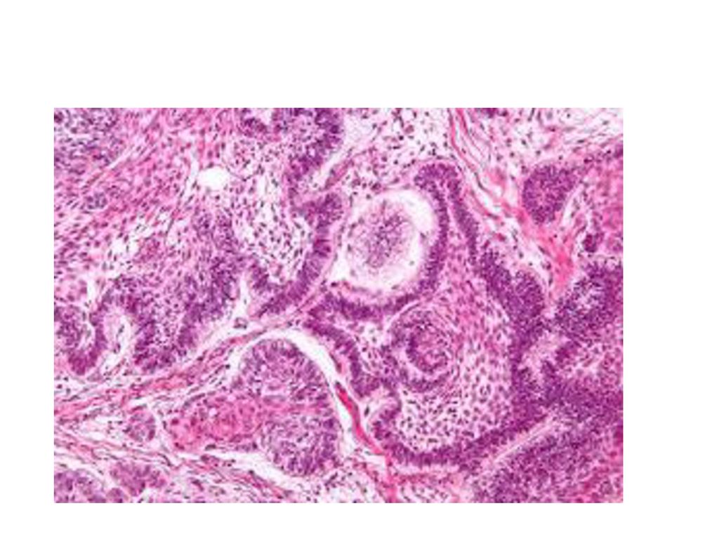
2.BORDERLINE TUMOUR AMELOBLASTOMA
Is the most common epithelial odontogenic tumor Affect people in the third to fifth decades of life 80% arise in the mandible. - Radiography : lytic expansile lesion.

28
Clinically Characterized by an abnormal growth in
the jaw, often at the site of the third molar. It may be aggressive and may spread to the nose, eye socket and skull. It is important for ameloblastoma to be diagnosed and treated early in order to stop growth of the tumors and possible progression to cancer.

29
Histopathology Several histological patterns
The most common patterns are the follicular and plexiform - .Follicular type: - Shows nests of loose network of cells resembling stellate reticulum and surrounded by an outer palisaded layer of tall columnar epithelium

31
3. MALIGNANT TUMOURS - Ameloblastic carcinoma is a tumor that has histological features similar to ameloblastoma but also shows malignant features such a as nuclear atypia and increased mitotic rate and necrosis

Similar presentations


 {AJCC} from Cummings.please see handouts as well for updated AJCC Tx Minimum requirements.>")


. arise from remnants.>")
 DR.SHAHZADI TAYYABA HASHMI>")
 a malignancy of mesenchymal cells that have the ability to produce osteoid or.>")

