Download presentation
Presentation is loading. Please wait.

1
Oral Cavity By Dr. Shawky M. Tayel Dr. Safaa Ahmed - Professor of Anatomy, Embryology & Human Genetics Anatomy Dep., Alexandria Faculty of Medicine, Anatomy Dep., Alexandria Faculty of Medicine, - Genetics Consultant, Clinical Genomic Center www.alexmedicine.com/alexgenomics.htm, www.alexmedicine.com/alexgenomics.htm,www.alexmedicine.com/alexgenomics.htm - Genetics Consultant, Suzanne Mubarak Regional Centre for Women's Health & Development www.smcalex.org, www.smcalex.org - Fellow, Medical College of Ohio, USA, - Member, American Society of Human Genetics.

2
1- Vestibule of mouth: 1- Vestibule of mouth: It is a slit like space bound externally by the lips and cheeks, internally by gums and teeth. It is a slit like space bound externally by the lips and cheeks, internally by gums and teeth.

3
2- Mouth cavity proper It is bounded by: It is bounded by: Anterior and laterally: the gums and teeth. Anterior and laterally: the gums and teeth. Posteriorly: it communicates with the pharynx through the isthmus. This isthmus is bounded by the palato-glossal arch on each side. Posteriorly: it communicates with the pharynx through the isthmus. This isthmus is bounded by the palato-glossal arch on each side. Roof: is formed by the hard and soft palate. Roof: is formed by the hard and soft palate. Floor: is formed by the anterior 2/3 of the tongue. Floor: is formed by the anterior 2/3 of the tongue.

4
Frenulum linguae= A median fold of mucosa connects the under surface of the tongue to the floor of the mouth. Frenulum linguae= A median fold of mucosa connects the under surface of the tongue to the floor of the mouth. The sublingual fold= a small ridge on each side of the frenulum. Lateral to the frenulum there are the lingual vein and lateral to it are the fimbriated folds (one on each side). The sublingual fold= a small ridge on each side of the frenulum. Lateral to the frenulum there are the lingual vein and lateral to it are the fimbriated folds (one on each side).
. The sublingual fold= a small ridge on each side of the frenulum. Lateral to the frenulum there are the lingual vein and lateral to it are the fimbriated folds (one on each side)..")
5
Muscles of the floor of the mouth Mylohyoid Mylohyoid Geniohyoid Geniohyoid Stylohyoid Stylohyoid Digastric Digastric

6
Mylohyoid muscle Origin: Origin: Mylohyoid line. Mylohyoid line. Insertion: Insertion: Hyoid bone and mylohyoid raphe. Hyoid bone and mylohyoid raphe. Nerve supply: Nerve supply: Mylohyoid nerve. Mylohyoid nerve. Action: Action: 1. Elevation of hyoid bone,Floor of mouth and tongue. 2. Depression of mandible

7
Geniohyoid muscle Origin: Inferior genial tubercle. Origin: Inferior genial tubercle. Insertion: Hyoid bone. Insertion: Hyoid bone. Nerve supply: C1 (component of hypoglossal nerve) Nerve supply: C1 (component of hypoglossal nerve) Actions: 1- Elevates hyoid bone. Actions: 1- Elevates hyoid bone. 2- Depresses the mandible. 2- Depresses the mandible.
 Nerve supply: C1 (component of hypoglossal nerve) Actions: 1- Elevates hyoid bone. Actions: 1- Elevates hyoid bone. 2- Depresses the mandible. 2- Depresses the mandible..")
8
Stylohyoid Origin: Styloid process. Origin: Styloid process. Insertion: Hyoid bone. Insertion: Hyoid bone. Nerve supply: Facial nerve. Nerve supply: Facial nerve. Action: Elevates the hyoid bone. Action: Elevates the hyoid bone.

9
Digastric muscle Origin: Anterior belly, digastric fossa of mandible. Anterior belly, digastric fossa of mandible. Posterior belly, Mastoid notch. Posterior belly, Mastoid notch.Insertion: Intermediate tendon. Nerve supply: Anterior belly, mandibular nerve. Anterior belly, mandibular nerve. Posterior belly, facial nerve. Posterior belly, facial nerve.Actions: Elevation of hyoid bone. Elevation of hyoid bone. Depression of mandible. Depression of mandible.

10
Sensory Innervation of the Mouth Roof: The greater palatine and nasopalatine nerves from the maxillary division of the trigeminal nerve Roof: The greater palatine and nasopalatine nerves from the maxillary division of the trigeminal nerve Floor: The lingual nerve (common sensation), a branch of the mandibular division of the trigeminal nerve. The taste fibers travel in the chorda tympani nerve, a branch of the facial nerve. Floor: The lingual nerve (common sensation), a branch of the mandibular division of the trigeminal nerve. The taste fibers travel in the chorda tympani nerve, a branch of the facial nerve. Cheek: The buccal nerve, a branch of the mandibular division of the trigeminal nerve (the buccinator muscle is innervated by the buccal branch of the facial nerve) Cheek: The buccal nerve, a branch of the mandibular division of the trigeminal nerve (the buccinator muscle is innervated by the buccal branch of the facial nerve)
, a branch of the mandibular division of the trigeminal nerve. The taste fibers travel in the chorda tympani nerve, a branch of the facial nerve. Cheek: The buccal nerve, a branch of the mandibular division of the trigeminal nerve (the buccinator muscle is innervated by the buccal branch of the facial nerve) Cheek: The buccal nerve, a branch of the mandibular division of the trigeminal nerve (the buccinator muscle is innervated by the buccal branch of the facial nerve).")
11
Tongue Skeletal muscles covered by mucosa. Skeletal muscles covered by mucosa. Lies in the proper mouth cavity and the oropharynx. Lies in the proper mouth cavity and the oropharynx. Functions: Taste, speech, chewing & swallowing. Functions: Taste, speech, chewing & swallowing. Surfaces: 2 = Surfaces: 2 = Dorsum: Faces the palate Dorsum: Faces the palate Divided into oral and pharyngeal parts by the V-shaped sulcus terminalis. Divided into oral and pharyngeal parts by the V-shaped sulcus terminalis. Inferior surface: lies in the oral cavity. Inferior surface: lies in the oral cavity. The tongue also has a margin, tip and root attaching it to the floor of the mouth. The tongue also has a margin, tip and root attaching it to the floor of the mouth.

12
Dorsum of the tongue The Oral part; The Oral part; = the 2/3 anterior to sulcus terminalis. = the 2/3 anterior to sulcus terminalis. Covered by papillae (which bear taste buds); Covered by papillae (which bear taste buds); - Vallate (anterior to sulcus terminalis) - Vallate (anterior to sulcus terminalis) - Fungiform (at the edges) - Fungiform (at the edges) - Filiform (scattered) - Filiform (scattered)
; Covered by papillae (which bear taste buds); - Vallate (anterior to sulcus terminalis) - Vallate (anterior to sulcus terminalis) - Fungiform (at the edges) - Fungiform (at the edges) - Filiform (scattered) - Filiform (scattered).")
13
Pharyngeal part of the dorsal surface: Pharyngeal part of the dorsal surface: = The 1/3 posterior to sulcus terminalis. = The 1/3 posterior to sulcus terminalis. Covered by aggregations of lymphoid tissue called Lingual tonsil. Covered by aggregations of lymphoid tissue called Lingual tonsil. Foramen caecum = Blind pouch at the apex of the sulcus terminalis (site of origin of the thyroid diverticulum (thyroglossal duct). Foramen caecum = Blind pouch at the apex of the sulcus terminalis (site of origin of the thyroid diverticulum (thyroglossal duct).
. Foramen caecum = Blind pouch at the apex of the sulcus terminalis (site of origin of the thyroid diverticulum (thyroglossal duct)..")
14
Inferior surface of the tongue; Inferior surface of the tongue; Covered by mucous membrane, showing: Covered by mucous membrane, showing: - Frenulum lingulae, - Frenulum lingulae, - Deep lingual vein on each side. - Deep lingual vein on each side. - Fimbriated fold (laterally). - Fimbriated fold (laterally).
. - Fimbriated fold (laterally)..")
15
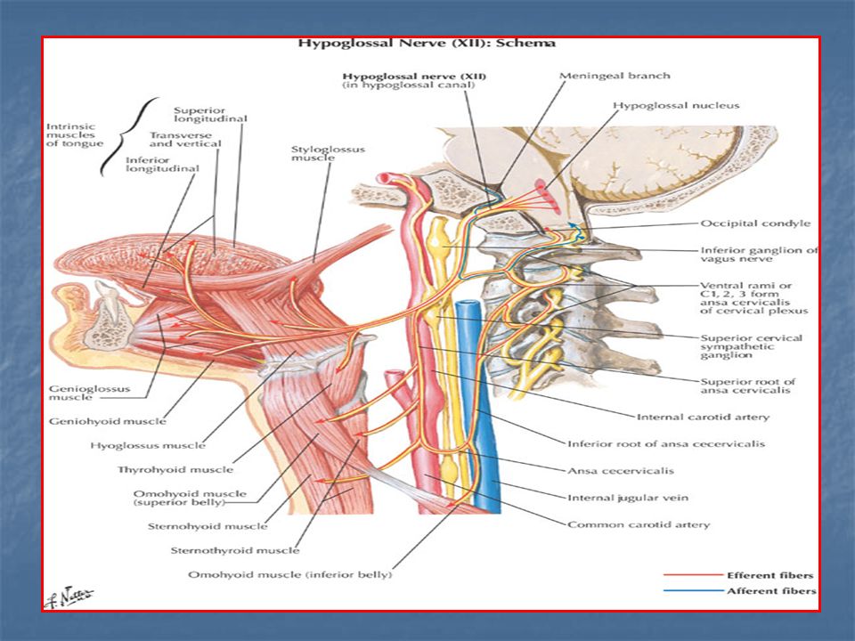
Muscles of the tongue The muscles of the tongue are divided into two types: The muscles of the tongue are divided into two types: Intrinsic Intrinsic Extrinsic Extrinsic Intrinsic: Longitudinal Longitudinal Transverse Transverse Vertical Vertical These muscles are confined to the tongue and are not attached to bone. Action: Alter the shape of the tongue

16
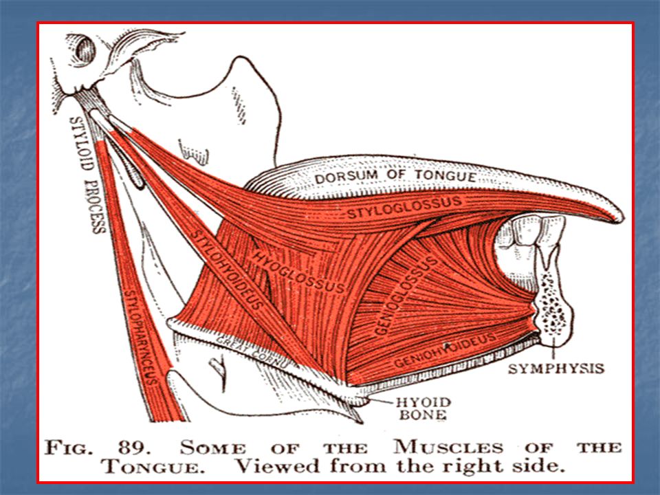
Extrinsic muscles of the tongue These muscles are attached to bones and the soft palate. They are: These muscles are attached to bones and the soft palate. They are: Genioglossus, Genioglossus, Hyoglossus, Hyoglossus, Styloglossus Styloglossus Palatoglossus. Palatoglossus.

17
Genioglossus: Origin: Sup. Genial tubercle of the mandible Insertion: Into the side of the tongue and hyoid bone Action: Depresses, protrudes, and deviates the tongue to the opposite side. N. Supply Hypoglossal nerve (XII)
.")
18
Hyoglossus: Hyoglossus: Origin: hyoid bone Origin: hyoid bone Insertion:side of tongue Insertion:side of tongue Action: depresses the tongue Action: depresses the tongue N. Supply: XII nerve N. Supply: XII nerve

19
Styloglossus: Styloglossus: Origin: styloid process behind ear Origin: styloid process behind ear Insertion: into side of the tongue Insertion: into side of the tongue Action: pulls the tongue upward and back Action: pulls the tongue upward and back N. supply: XII N. supply: XII

20
Palatoglossus: Palatoglossus: Origin: palatine aponeurosis Origin: palatine aponeurosis Insertion: Lateral surface of the back of the tongue Insertion: Lateral surface of the back of the tongue Action:pulls the tongue back to narrows the oropharyngeal ismuthus, Depresses the palate. Action:pulls the tongue back to narrows the oropharyngeal ismuthus, Depresses the palate. N. Supply: Pharyngeal plexus of vagus (X) nerve N. Supply: Pharyngeal plexus of vagus (X) nerve
 nerve N. Supply: Pharyngeal plexus of vagus (X) nerve.")
21
Movements of the Tongue Protrusion: The genioglossus muscles on both sides acting together. Protrusion: The genioglossus muscles on both sides acting together. Retraction: Styloglossus and hyoglossus muscles on both sides acting together. Retraction: Styloglossus and hyoglossus muscles on both sides acting together. Depression: Hyoglossus muscles on both sides acting together. Depression: Hyoglossus muscles on both sides acting together. Retraction and elevation of the posterior third: Styloglossus and palatoglossus muscles on both sides acting together. Retraction and elevation of the posterior third: Styloglossus and palatoglossus muscles on both sides acting together. Shape changes: Intrinsic muscles Shape changes: Intrinsic muscles

22
Nerve supply of the tongue Sensory: Sensory: Anterior 2/3: Anterior 2/3: Gerneral sensation: Lingual nerve of mandibular of trigeminal. Gerneral sensation: Lingual nerve of mandibular of trigeminal. Taste sensation: Chorda tympani (the parasympathetic part of facial (VII) nerve. Taste sensation: Chorda tympani (the parasympathetic part of facial (VII) nerve. Posterior 1/3: IX (glossopharyngeal) nerve General and taste). Posterior 1/3: IX (glossopharyngeal) nerve General and taste). Posterior end of the dorsum: Internal laryngeal n. (from X). Posterior end of the dorsum: Internal laryngeal n. (from X). Motor: XII to all muscles except palatoglossus. Motor: XII to all muscles except palatoglossus.
 nerve. Taste sensation: Chorda tympani (the parasympathetic part of facial (VII) nerve. Posterior 1/3: IX (glossopharyngeal) nerve General and taste). Posterior 1/3: IX (glossopharyngeal) nerve General and taste). Posterior end of the dorsum: Internal laryngeal n. (from X). Posterior end of the dorsum: Internal laryngeal n. (from X). Motor: XII to all muscles except palatoglossus. Motor: XII to all muscles except palatoglossus..")
25
A) Arterial supply of the tongue: Lingual artery (which gives 2 dorsalis linguae and deep lingual), Tonsillar branch of the facial artery Ascending pharyngeal artery B) Venous drainage: Lingual vein --- I J V.
 Arterial supply of the tongue: Lingual artery (which gives 2 dorsalis linguae and deep lingual), Tonsillar branch of the facial artery Ascending pharyngeal artery B) Venous drainage: Lingual vein --- I J V.")
26
Lymphatic drainage Tip: Submental lymph nodes Tip: Submental lymph nodes Sides of the anterior two thirds: Submandibular and deep cervical lymph nodes Sides of the anterior two thirds: Submandibular and deep cervical lymph nodes Posterior third: Deep cervical lymph nodes Posterior third: Deep cervical lymph nodes

27
Lymphatic drainage Tip: Submental lymph nodes Tip: Submental lymph nodes Sides of the anterior two thirds: Submandibular and deep cervical lymph nodes Sides of the anterior two thirds: Submandibular and deep cervical lymph nodes Posterior third: Deep cervical lymph nodes Posterior third: Deep cervical lymph nodes

28
The Teeth The teeth are collectively called the dentition. The teeth are collectively called the dentition. Adults normally 32 permanent teeth16 teeth in the mandible and 16 in the maxilla. Adults normally 32 permanent teeth16 teeth in the mandible and 16 in the maxilla. On each side of the midline, there are: 2 incisors, a canine, 2 premolars, and 3 molars in each jaw. On each side of the midline, there are: 2 incisors, a canine, 2 premolars, and 3 molars in each jaw. The incisors are chisel-like cutting teeth used to bite off a piece of food. The incisors are chisel-like cutting teeth used to bite off a piece of food. The canines are more pointed and act to puncture and shred it. The canines are more pointed and act to puncture and shred it. The premolars and molars have relatively broad surfaces adapted to crushing and grinding. The premolars and molars have relatively broad surfaces adapted to crushing and grinding. 20 deciduous teeth (milk teeth or baby teeth) erupt from the ages of 6 to 30 months, beginning with the incisors. 20 deciduous teeth (milk teeth or baby teeth) erupt from the ages of 6 to 30 months, beginning with the incisors. These are replaced by the 32 permanent teeth(6 and 25 years of age) These are replaced by the 32 permanent teeth(6 and 25 years of age)
 erupt from the ages of 6 to 30 months, beginning with the incisors. 20 deciduous teeth (milk teeth or baby teeth) erupt from the ages of 6 to 30 months, beginning with the incisors. These are replaced by the 32 permanent teeth(6 and 25 years of age) These are replaced by the 32 permanent teeth(6 and 25 years of age).")
29
Human Deciduous and Permanent Teeth

Similar presentations


 and posterior 1/3 (the soft palate).>")