Download presentation
Presentation is loading. Please wait.

2
Regional anatomy of thorax Boundaries Superior - jugular notch, sternoclavicular joint, superior border of clavicle, acromion, spinous processes of C7 Inferior - xiphoid process, costal arch, 12th and 11th ribs, vertebra T12 Regions Thoracic wall Thoracic cavity

3
THORACIC CAGE Thoracic cage is an osteo-cartilagenous conical cage which has a narrow inlet & a wide outlet …

4
Ant: Sternum, Costal cartilages and Ribs. Post: Thoracic vertebrae and ribs. Lat: Ribs.

5
Thoracic Inlet/SUPERIOR APERTURE Ant: Upper border of manubrium sterni. Post : sup.sur. 1 st thoracic vertebra. On each side: 1 st rib & 1 st costal cartilage. It is sloping downwards & forward.

6
OUTLET OF THORAX Def.-the broad lower end of thorax which is continuous with the abdominal cavity is called the outlet of thorax BOUNDARIES : In front-by : the xiphoid process & 7-10 th costal cartilage forming an intrasternal angle Behind - inf.surface of body of T12 On Sides - 11th & 12th ribs Inferiorly : Diaphragm

9
Thoracic vertebrae. They are 12 vertebra. From 2 to 8 they are called Typical. Character of typical thoracic vertebrae: Body: Heart shape & carries 2 demi-facet at its side. Transverse process: has a facet for rib tubercle of the same number. Spine: Long, pointed & directed downward and backward. Vertebral foramen: Small & circular.

10
Articulation between Thoracic vertebrae and the ribs

11
Atypical (Non typical ) thoracic vertebrae. 1 st, 10 th,11 th and 12 th T1: Has a complete Superior costal facet. One very small inferior demifacet. Spine nearly horizontal Has costal facet in transverse process for the tubercle of first rib. It has a small body, looks like a cervical vertebra.

13
12 pairs. Articulates with thoracic vertebrae. - Number might ↑ or ↓. Uppers ribs – oblique (max at 9 th ). Lower ribs – less oblique. Length – maximum in 7 th and decreases towards both ends.
. Lower ribs – less oblique. Length – maximum in 7 th and decreases towards both ends..")
14
Classification: True,false,typical and atypical ribs. True and false ribs. -True ribs – 1-7 as they are connected to the sternum through their respective cartilages.(vertebro sternal ribs). - False ribs – 8, 9, 10, 11, 12. Connected to next higher cartilages to reach sternum but 11, 12 are free in the anterior ends, called floating ribs. Typical ribs: 3 – 9 Atypical ribs: 1,2 & 10, 11, 12
. - False ribs – 8, 9, 10, 11, 12. Connected to next higher cartilages to reach sternum but 11, 12 are free in the anterior ends, called floating ribs. Typical ribs: 3 – 9 Atypical ribs: 1,2 & 10, 11, 12.")
15
Parts of typical rib: Anterior,posterior ends & shaft. - Anterior end : Oval, concave articulates with costal cartilage. -Posterior end: - head with 2 facets articulating with the body of thoracic vertebra. - small neck. - tubercle: junction of neck & shaft; articulates with transverse process of thoracic vertebra.

17
Shaft: 2 surfaces (outer and inner)& angle - outer surface: convex - inner surface: concave, covered by pleura; & inner costal groove above the inferior border.
& angle - outer surface: convex - inner surface: concave, covered by pleura; & inner costal groove above the inferior border.")
18
TYPICAL RIB

19
Atypical ribs ● 1st, 2nd, 10th, 11th, 12th ribs. First rib Shortest, flattest and most curved. Articulate with T1 only. 2 groove: -Anterior- subclavian vein -Posterior-lowest trunk of the brachial plexus and subclavian artery 1 tubercle (inner); attachment of scalenus anterior muscle.
; attachment of scalenus anterior muscle..")
20
Atypical ribs… Second rib -Less curved -2x long as 1 st rib. -tubercle (ext lower border) Tenth rib 1 articular facet on its head. Eleventh rib -1 articular facet on its head. - NO tubercle for articulation with the transverse process. Twelfth rib -1 articular facet on its head. -NO tubercle and subcostal groove.
 Tenth rib 1 articular facet on its head. Eleventh rib -1 articular facet on its head. - NO tubercle for articulation with the transverse process. Twelfth rib -1 articular facet on its head. -NO tubercle and subcostal groove..")
22
Forking or duplication of the ribs

23
Frontal chest radiograph shows a bone bridge joining the right anterior first and second ribs. Pseudoarticulation is also present (arrows). This finding is typical in pseudarthrosis and should not be mistaken for a fracture.
. This finding is typical in pseudarthrosis and should not be mistaken for a fracture..")
24
● Cervical ribs: Bony or fibrous bands between C7 and the 1st rib. 1-2% of subjects. 10% of these cause compressive symptoms, ranging from nerve compression or to subclavian artery post stenotic aneurysm. The remainder are asymptomatic. May be large or small, single or bilateral, and may articulate with the 1 st rib. If they consist only of a fibrous band they will not be visible on radiographs. They can be confused with hypoplastic first ribs.

25
*The 7th cervical transverse processes point downwards, 1st thoracic transverse processes are angled upwards.

26
Cervical ribs in an asymptomatic patient. Frontal chest radiograph shows bilateral cervical ribs (arrowheads); the left one fuses anteriorly to the first rib (arrow).
; the left one fuses anteriorly to the first rib (arrow)..")
27
● Lumbar ribs: transverse process (costal element of the lumbar vertebra) may fail to fuse with the vertebral body and retain asynovial joint with the neural arch. Asymptomatic.

28
COSTAL CARTILAGES ● unossified anterior ends of the ribs. ● Slope upwards to the sternum 1 st – 7 th ribs articulate with sternum (sternochondral joints) ● 8th – 10th ribs articulate with the costal cartilages of the ribs above. ● 11th and 12th costal cartilages have pointed ends and end in the muscles of the abdominal wall.
 ● 8th – 10th ribs articulate with the costal cartilages of the ribs above. ● 11th and 12th costal cartilages have pointed ends and end in the muscles of the abdominal wall..")
29
Sternum

30
Sternum is a flat bone, present in front of the thoracic cage. Parts: Manubrium, Body and Xiphoid process. Manubrium is the - upper part of the sternum. - joins with medial end of clavicle and first costal cartilage. - Jugular or interclavicular or suprasternal notch. Sternum

31
Body : - larger part of the sternum. - on each side, it receives ribs through their costal cartilages (3rd to 6th rib). Xiphoid process: - the most variable part of the sternum. - cartilaginous structure in adults. Sternum
. Xiphoid process: - the most variable part of the sternum. - cartilaginous structure in adults. Sternum.")
32
THE STERNUM ● Manubrium -opposite T3 and T4 -articulates with clavicle and with 1½ costal Cartilages ● Sternal angle - secondary cartilaginous joint,lies opposite T 4/5 disc space ● Body -opposite T 5 -T 9, made up of four stenebrae which articulate with 5 ½ costal cartilages ● Xiphoid process -remains cartilaginous into adult life.

33
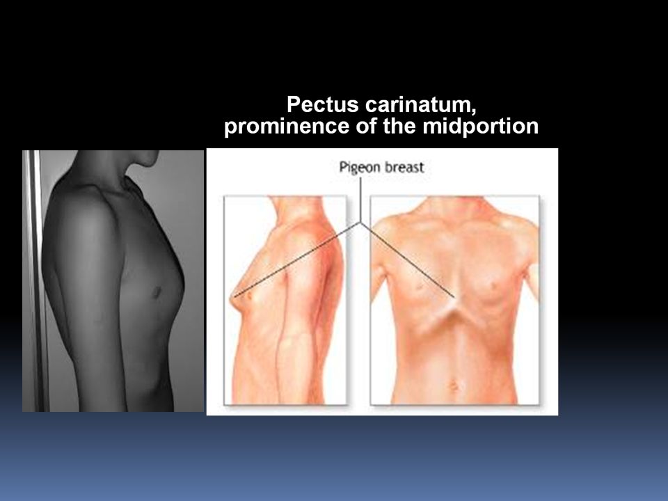
Variation in Sternal configuration: Pectus excavatum, depression of the lower end. Pectus carinatum, prominence of the midportion

Similar presentations


 are the smallest, lightest vertebrae C3-C7 are distinguished with an oval body, short spinous processes, and.>")


