Download presentation
Presentation is loading. Please wait.

1
Nose Anatomy of the Nose

2
External nose External nose It is a projecting triangular pyramid directed downwards. It has apex, root connected to the forehead and base perforated by two nostrils.

3
Bones and Cartilages of the nose Nasal bones. Maxillae. Frontal bone (nasal process). Upper lateral cartilages. Lower lateral cartilages. Septal cartilage. The muscles of the nose are a part of facial muscles and are supplied with facial nerve. The muscles of the nose are a part of facial muscles and are supplied with facial nerve.
. Upper lateral cartilages. Lower lateral cartilages. Septal cartilage. The muscles of the nose are a part of facial muscles and are supplied with facial nerve. The muscles of the nose are a part of facial muscles and are supplied with facial nerve..")
6
Nasal Cavity Nasal Cavity Nasal Vestibule Nasal Vestibule It is the entrance to the nasal cavity, lined with skin which is hair bearing. Nasal cavity proper They are two cavities separated by the nasal septum, extending from the anterior nares to the nasopharynx.The mucosa is Ciliated Columnar Epithelium with Olfactory epithelium at the roof.

7
The nasal septum Medial Wall of the nasal cavity is composed of the following: 1-Quadrilateral Cartilage ((Septal Cartilage)). 2-Perpendicular Plate of the Ethmoid bone. 3-The Vomer bone. 4-Nasal Crests of the Maxilla and the Palatine bones.

10
The Lateral Wall The Inferior Turbinate: is a separate bone attached to the maxilla. The Middle Turbinate. The Superior Turbinate. The middle and the superior turbinates are parts of the ethmoid bones.

13
Bellow the inferior turbinate is the inferior meatus which receives the nasolacrimal duct opening. The middle meatus lies bellow the middle turbinate and receives the openings of the maxillary, frontal and the anterior ethmoidal sinuses. The superior meatus receives the opening of the posterior ethmoidal cells. Above the superior turbinate is the Sphenoethmoidal Recess which receives the sphenoid sinus ostium.

15
The Roof of the nose is formed from anterior to posterior from: the nasal bones, the cribriform plate of the ethmoid bone and sphenoid bone. The olfactory cleft area is lined with olfactory epithelium (special sensory epithelium) and occupies the area of the cribriform plate, the superior turbinate and the corresponding area of the septum. The olfactory cleft area is lined with olfactory epithelium (special sensory epithelium) and occupies the area of the cribriform plate, the superior turbinate and the corresponding area of the septum. The floor is formed of the maxilla and the palatine bones.
 and occupies the area of the cribriform plate, the superior turbinate and the corresponding area of the septum. The olfactory cleft area is lined with olfactory epithelium (special sensory epithelium) and occupies the area of the cribriform plate, the superior turbinate and the corresponding area of the septum. The floor is formed of the maxilla and the palatine bones..")
18
The Blood Supply The external nose is supplied by branches of the facial, the maxillary and the ophthalmic arteries. The venous drainage is through the facial, maxillary and the ophthalmic veins, the latter drains to the cavernous sinus. The venous drainage is through the facial, maxillary and the ophthalmic veins, the latter drains to the cavernous sinus.

20
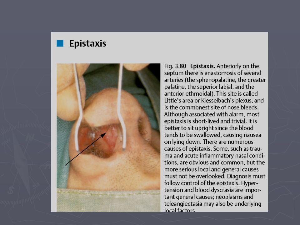
The blood supply to the nasal cavity is coming from the maxillary, facial, the anterior and the posterior ethmoidal arteries. Little's area is the anteroinferior part of the nasal septum where anastomosis of vessels called Kiesselbach's plexus is located and is the commonest site of bleeding.

23
Nerve supply The sensory innervations of the nose is supplied by the trigeminal nerve, mainly through the maxillary and the ophthalmic divisions. The olfactory area is supplied by the olfactory nerve. The nose also has sympathetic supply from the upper deep cervical ganglion. The parasympathetic supply comes from the geniculate ganglion of the facial nerve. The parasympathetic supply comes from the geniculate ganglion of the facial nerve.

27
THE PARANASAL SINUSES THE PARANASAL SINUSES They are Air Filled cavities within the bones surrounding the nose and have openings or ducts draining into the nose. They are arranged in pairs and lined with respiratory mucus membrane. They comprise the maxillary, the frontal, the ethmoid and the sphenoid sinuses.

28
The Maxillary Sinus This is the largest Para nasal sinus; it occupies the body of the Maxilla. It is also called the ANTRUM. It has a roof which is the floor of the orbit, a base or the medial wall, a floor which is the alveolar process of the maxilla and an apex. The ostium is situated high on the medial wall and it opens into the middle meatus, so the drainage is dependant on the ciliary action of the mucosa, not on gravity.

31
The Frontal Sinuses They are situated in the frontal bone and are divided into two parts by a septum. The frontonasal duct of each sinus opens into the middle meatus.

33
The Ethmoid Sinuses They are situated in between the nasal cavity medially and the orbit laterally where a very thin bone (lamina papyraceae) separates it from the orbit, superiorly the sinuses are bounded by the cranial cavity. The sinuses are divided into two groups, an anterior group which drains into the middle meatus and posterior group which drains into the superior meatus.

36
The Sphenoid Sinuses These occupy the body of the sphenoid bone and are divided by a septum into two, each sinus drains into the sphenoethmoidal recess.

38
The Physiology of the Nose The Physiology of the Nose 1-It is an airway passage which moistens and heats the inspired air due to high vascularity of the mucus membrane. 2-The mucus contains antibodies which act as a defense mechanism. 3-It filters the inspired air from foreign bodies. 4-It adds resonance to sound. 5-Olfaction, the sense of smell.

39
Symptoms and Signs of Nasal diseases Nasal block. Nasal discharge ((Rhinorrhoea)) and postnasal drip. Bleeding from the nose ((Epistaxis)) Sneezing and itching. Nasal pain, facial pain and headache. External deformity.
) and postnasal drip. Bleeding from the nose ((Epistaxis)) Sneezing and itching. Nasal pain, facial pain and headache. External deformity..")
40
Disorders of smell Anosmia. Hyposmia. Hyperosmia (increased sense of smell). Cacosmia (perception of bad smell).
..")
41
Signs like external deformity, scars, masses and other skin lesions are readily seen by simple examination. Examination of the nose is done by using Nasal Speculum and Good light.This is Called Anterior Rhinoscopy. Deviated nasal septum, abnormality of the mucosa, bleeding vessels, and character of the secretions, nasal masses and polyps.

43
Postnasal examination is done by Nasopharyngeal Mirror. This is called Posterior Rhinoscopy. ENDOSCOPIC EXAMINATION OF THE NOSE IS POSSIBLE BY USING FLEXIBLE AND RIGID ENDOSCOPES.

44
Investigations of the nose X-ray paranasal sinuses. CT scan. MRI scan. Skin prick test for allergy.

47
TRAUMA TO THE NOSE Nasal bone Fracture Caused by external force, blow and fall from height or assault. Presented with Pain, Swelling, Bruises, Epistaxis, Nasal block, External deformity or Deviation.

49
On Examination: Septum for the presence of septal haematoma, especially in Children. Septal haematoma is accumulation of blood between the mucus membrane(the muco- perichondrium) and the cartilage of the nasal septum. When present, the haematoma needs urgent drainage; otherwise septal abscess may develop which may result in cartilage necrosis.
 and the cartilage of the nasal septum. When present, the haematoma needs urgent drainage; otherwise septal abscess may develop which may result in cartilage necrosis..")
53
The correction of nasal bone fracture is needed when there is recent and apparent deformity or deviation of the external nose. This is usually done after 5 to 7 days after the subsidence of edema and good assessment of the nose is possible and before healing of the fracture which makes its reduction difficult.

54
EPISTAXIS It is defined as Bleeding from the nose. It is usually Anterior bleeding. Can be posterior or both anterior and posterior bleeding depending on the site and severity of bleeding. The commonest site of bleeding is Little's area which has high vascularity.

56
CAUSES A-Local causes: 1-Trauma like fracture nose and nose picking. 1-Trauma like fracture nose and nose picking. 2-Upper respiratory tract infections. 2-Upper respiratory tract infections. 3-Acute or Chronic rhinitis. 3-Acute or Chronic rhinitis. 4-Postoperative. 4-Postoperative. 5-Foreign bodies. 5-Foreign bodies. 6-Tumours ((benign or malignant)) of the nose and para nasal sinuses like Angiofibroma. 6-Tumours ((benign or malignant)) of the nose and para nasal sinuses like Angiofibroma.
) of the nose and para nasal sinuses like Angiofibroma. 6-Tumours ((benign or malignant)) of the nose and para nasal sinuses like Angiofibroma..")
57
B-Systemic Causes: 1- Hypertension, atherosclerosis and blood vessels abnormalities. 1- Hypertension, atherosclerosis and blood vessels abnormalities. 2- Clotting mechanism defects like hemophilia and thrombocytopenia. 2- Clotting mechanism defects like hemophilia and thrombocytopenia. 3- Anticoagulant drugs like heparin and warfarin. 3- Anticoagulant drugs like heparin and warfarin. 4-Antiplatelet drugs like aspirin. 4-Antiplatelet drugs like aspirin. 5- Hormonal Changes like in pregnancy and puberty. 5- Hormonal Changes like in pregnancy and puberty. The cause may be unknown, this is called Idiopathic

58
MANAGEMENT OF EPISTAXIS MANAGEMENT OF EPISTAXIS 1. Local treatment 1. Local treatment Mild and intermittent bleeding: pinching of the nose and application of ice on the forehead. Mild and intermittent bleeding: pinching of the nose and application of ice on the forehead. Local antibiotic cream or ointment is applied locally. Local antibiotic cream or ointment is applied locally. Cautery is done when there is obvious area of dilated vessels and this can be either chemical cautery or electrical cautery. If the bleeding is severe and not controlled with the above measures, then PACKING of the nose is needed. Packing can be either anterior OR posterior and anterior packing.

61
2. Treatment of the underlying cause when present, stop or decrease the dose of the anticoagulant drug, treat sinusitis … etc. 3. Resuscitation in case of shock because of the bleeding. I.V. fluid, blood transfusion may be needed.

62
4. Other methods to control epistaxis We may rarely need ligation of the artery to control epistaxis If facilities are available, embolization of the bleeder under radiographic control may be of great benefit.

63
Vestibulitis Inflammation of the vestibular skin. Usually secondary to conditions causing long term or chronic discharge from the nose. There is excoriation of the vestibular skin and sometimes painful fissuring and bleeding (epistaxis). There is excoriation of the vestibular skin and sometimes painful fissuring and bleeding (epistaxis). Treatment of the underlying cause and topical antibiotic cream or ointment till subsidence of the condition. Treatment of the underlying cause and topical antibiotic cream or ointment till subsidence of the condition. Another form of vestibulitis is the BOIL, which is a staphylococcal infection of hair follicles. In addition to local treatment; it may need anti-staphylococcal antibiotic like cloxacillin.
. There is excoriation of the vestibular skin and sometimes painful fissuring and bleeding (epistaxis). Treatment of the underlying cause and topical antibiotic cream or ointment till subsidence of the condition. Treatment of the underlying cause and topical antibiotic cream or ointment till subsidence of the condition. Another form of vestibulitis is the BOIL, which is a staphylococcal infection of hair follicles. In addition to local treatment; it may need anti-staphylococcal antibiotic like cloxacillin..")
67
Foreign Bodies in the Nose This is a problem of young children who tend to push objects into the nose. F.B. can be organic or non organic. Manifested by nasal block, discomfort and sometimes if the F.B. is present for long time, there is unilateral foul smelling discharge which is characteristic for F.B. Management is removal which sometimes needs general anesthetic when the F.B. is deep in the nose and difficult to remove in the uncooperative child.

69
Acute Rhino sinusitis Acute Rhino sinusitis The Common Cold or Coryza It is usually viral infection of the mucus membrane of the nose Accompanied by general inflammation of the nose and sinuses. Predisposing factors include exposure to cold, fatigue, poor nutrition, nasal obstruction and chronic nasal and sinus infections. All ages are affected with higher incidence in children. Spread of infection is by droplet, dust and eating.

70
Clinical Features Incubation period of 1 to 3 days. Sensation of discomfort in the nose and attacks of sneezing, chills and low grade pyrexia. Nasal discharge and nasal block (inflammation and swelling of the nasal mucus membrane. The discharge to start with is watery from, and then it changes to mucopurulent when secondary bacterial infection ensues. Mucosal swelling results in obstruction of sinus ostia, causing Sinusitis and associated headache.

72
Management The disease is self limiting and needs supportive measures like good nutrition and bed rest together with simple analgesics and local or systemic nasal decongestants. Antibiotics are indicated when there are complications like: acute otitis media, acute tonsillitis, acute sinusitis, and chest infection.

73
Acute Sinusitis Acute Sinusitis Acute infection and inflammation of Para nasal sinuses. It is usually caused by acute rhinitis but it can be dental in origin (spread of infection from the teeth). The commonest sinuses to be involved are the maxillary and the ethmoids, but all sinuses can be affected and this is called Pan Sinusitis. Predisposing factors include nasal block from nasal septal deviation, adenoids, polyps and allergic rhinitis.
. The commonest sinuses to be involved are the maxillary and the ethmoids, but all sinuses can be affected and this is called Pan Sinusitis. Predisposing factors include nasal block from nasal septal deviation, adenoids, polyps and allergic rhinitis..")
74
Clinical features are similar to those of acute rhinitis (nasal block, nasal discharge of mucopurulent material) but the symptoms are more severe, there may be headache and tenderness on pressure on the affected sinuses. Diagnosis is done by the clinical features and aided by radiology ((X-ray of the Para nasal sinuses)). Treatment includes rest, antibiotic and nasal decongestants.
). Treatment includes rest, antibiotic and nasal decongestants..")
78
Complications of Sinusitis 1. Orbital complications Spread of infection to the eye is usually from the ethmoid sinuses through the Lamina Papyracea which is very thin bone separating the ethmoid from the eye. It is the commonest complication which is mainly in children. If the condition is early then is treated with hospital admission, observation and antibiotics. If the situation is severe with abscess then surgery is needed.

81
2. Osteomyelitis It affects diploic bones like the frontal sinus. It is treated with antibiotics and surgery of no response. 3-Intracranial complications Meningitis, Cortical venous thrombosis, Cavernous Sinus thrombosis and Brain Abscess.

Similar presentations


 Root Ala Dorsum>")


