Download presentation
Presentation is loading. Please wait.

1
TUMORS OF URINARY TRACT

2
RENAL PARENCHYMAL NEOPLASMS

3
BENIGN RENAL TUMOURS Angiomyolipoma (Renal Hamartoma)
❏rare benign tumour ❏round, oval, expansible ❏characterized by 3 major histologic components: blood vessels, smooth muscle and fat cells ❏many asymptomatic, may spontaneously rupture, especially in pregnant females ❏found in approximately 45-80% of patients with tuberous sclerosis which is characterized by • epilepsy • mental retardation • sebaceous adenoma • hamartomas of brain and kidney ❏diagnose by CT ––> fat (negative density on CT) observed in kidneys is pathognomonic for angiomyolipoma ❏treatment: <4cm, >4cm
 observed in kidneys is pathognomonic for angiomyolipoma. ❏treatment: <4cm, >4cm.")
4
Renal Adenoma ❏commonly found incidentally at autopsy or after nephrectomy for an unrelated disease ❏10-20% of the population ❏asymptomatic ❏need tissue diagnosis to definitively differentiate from renal cell carcinoma Renal Oncocytoma ❏ occur in variant organ (adrenal, salivary gland, thyroid,…) represent about 3% of kidney tumor ❏has a characteristic findings on imagingstudies but need histopathology for diagnosis
 represent about 3% of kidney tumor ❏has a characteristic findings on imagingstudies but need histopathology for diagnosis")
5
MALIGNANT RENAL TUMORS
ADENOCARCINOMA (Renal Cell Carcinoma, Grawitz’s Tumour) ❏also known as hypernephroma ❏eighth most common malignancy (accounts for 3% of all newly diagnosed cancers) ❏85% of primary malignant tumours in kidney ❏male:female = 3:1 ❏called the “internist’s tumour” because of paraneoplastic symptomatology
 ❏also known as hypernephroma. ❏eighth most common malignancy (accounts for 3% of all newly diagnosed cancers) ❏85% of primary malignant tumours in kidney. ❏male:female = 3:1. ❏called the internist’s tumour because of paraneoplastic symptomatology.")
6
ETIOLOGY ❏ The cause is unknown.
❏ There are various theories of risk factors: • environmental and occupational factors: • smoking (results in 2x increased relative risk), cadmium exposure, employment in leather industry • familial incidence seen with von Hippel Lindau syndrome which is characterized by: • RCC (present in 2/3) • headache, ataxia, and blindness due to cystic lesions of cerebellum and retinal vessel aneurysms • aquired cystic disease
, cadmium exposure, employment in leather industry. • familial incidence seen with von Hippel Lindau syndrome which is characterized by: • RCC (present in 2/3) • headache, ataxia, and blindness due to cystic lesions. of cerebellum and retinal vessel aneurysms. • aquired cystic disease.")
7
PATHOLOGY The tumor occur in equal frequency in either kidney
originates in the cortex, grow out in the perinephric tissue originates from proximal convoluted tubule epithelial cell it is characteristically yellow to orange because of high lipid content

8
PATHOGENESIS ❏methods of spread
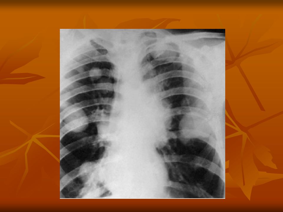
• direct • venous • lymphatic RCC is a vascular tumor, tend to spread by direct invasion Vascular invasion is through renal vein About 1\3 of patients have metastasis at presentation The most common site of distant metastasis is lung, opposite kidney, followed by liver, bone.

10
TNM Classification

11
TUMOR STAGING Stage I: tumor is confined within kidney parenchyma
StageII:tumor involve perinephric fat but is confined within Gerota’s fascia, or adrenals Stage IIIa : tumor involve the main renal vein or inferior vena cava, byeond Gerotas Stage IIIb :tumor involve regional lymph node

12
Stage IIIc : tumor involve both local vessels and regional lymph node
Stage IVa : tumor involves adjacent organs (colon,pancreas,….) Stage IVb :distant metastasis
 Stage IVb :distant metastasis.")
14
CLINICAL PICTURE It has a wide variety of presentation
❏symptoms and signs • increasingly diagnosed incidentally with U/S and CT • poor prognostic indicators include • weight loss • weakness • anemia • bone pain • classic triad (too late triad!) found in 10-15% • gross hematuria 50% • flank pain < 50% • palpable mass < 30% • 30% have metastases when first seen: dyspnea, cough, headache, bone pain • Abd pain, abd mass,
 found in 10-15% • gross hematuria 50% • flank pain < 50% • palpable mass < 30% • 30% have metastases when first seen: dyspnea, cough, headache, bone pain. • Abd pain, abd mass,")
15
Para-neoplastic syndrome
3-10% of RCC present by paraneoplastic syndrome hematopoietic disturbances: anemia, polycythemia; raised ESR: RCC is the most common cause of paraneoplastic erythrocytosis endocrinopathies: hypercalcemia 20% (production of PTH), production of other hormones including erythropoietin, renin, prolactin, gonadotropins, TSH, insulin, and cortisol. PUO hemodynamic alterations: systolic hypertension 40% (due to AV shunting, production of renin), and peripheral edema (due to caval obstruction) Non-metastatic hepatic dysfunction: “Staufer’s syndrome”: abnormal liver function tests, decreased WBC count, fever, areas of hepatic necrosis; reversible following removal of primary tumour
, production of other hormones including erythropoietin, renin, prolactin, gonadotropins, TSH, insulin, and cortisol. PUO. hemodynamic alterations: systolic hypertension 40% (due to AV shunting, production of renin), and peripheral edema (due to caval obstruction) Non-metastatic hepatic dysfunction: Staufer’s syndrome : abnormal liver function tests, decreased WBC count, fever, areas of hepatic necrosis; reversible following removal of primary tumour.")
16
Laboratory FINDINGS • routine labs for paraneoplastic syndromes
• CBC anemia (30%) • High ESR • urinalysis (60-75% have hematuria)
 • High ESR. • urinalysis (60-75% have hematuria)")
17
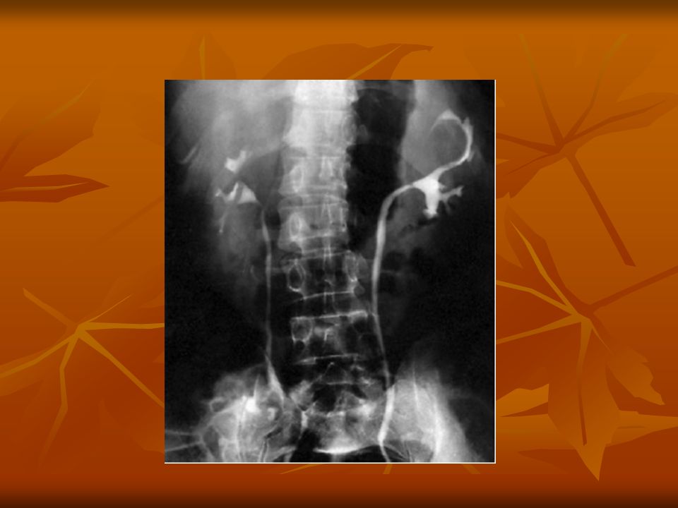
Imaging U\S: Doppler IVP 75% accurate
CT scan: it is the leader for diagnosis and staging and detect distant metastasis MRI Renal angiography: no longer routinely done Fine needle aspiration indicated in : metastatic disease, planned for nonsurgical management establishing diagnosis in patients who are not surgical candidate

21
Differential diagnosis
Carcinoma of renal pelvis Renal lymphoma Adrenal cancer Benign renal tumor Renal cysts Renal abscess

22
TREATMENT LOCALISED DISEASE • surgical (mainstay):
Stage (I, II , IIIa )→ • surgical (mainstay): • partial Nx: • small, polar, or bilateral tumors • radical nephrectomy: • en bloc removal of kidney, tumour, ipsilateral adrenal gland and intact Gerota’s capsule and periaortic lymphadenectomy
→ • surgical (mainstay): • partial Nx: • small, polar, or bilateral tumors. • radical nephrectomy: • en bloc removal of kidney, tumour, ipsilateral adrenal gland and intact Gerota’s capsule and periaortic lymphadenectomy.")
24
Immunotherapy→15% response rate
DISSEMINATED DISEASE 30% of RCC are metastatic Surgical: the role of radical nephrectomy is limited. It is a palliative therapy • surgical removal of solitary metastasis may be considered Immunotherapy→15% response rate cytokine interleukin-2 α- INF Tyrosin kinase inhibitors---monoclonal Abs Radiotherapy (RCC is a radioresistant): • radiation for palliation • for painful bony lesions Chemotherapy (is also chemoresistant )
: • radiation for palliation. • for painful bony lesions. Chemotherapy (is also chemoresistant )")
25
Prognosis • stage at diagnosis is the single most important predictor of survival: • 5 year survival of T1 is % • 5 year survival of T2-T3 is approximately 60% • 5 year survival of patients presenting with metastasis is 0-20%

26
CARCINOMA OF THE RENAL PELVIS AND URETER
❏incidence • rare, accounts for 4% of all urothelial cancers • frequently multifocal • papillary transitional cell cancer 85% • male:female = 3:1 ❏relative incidence • bladder:pelvis:ureter = 100:10:1 ❏predisposing factors • chemical exposure (industrial dyes and solvents) • smoking • analgesic abuse (acetaminophen, aspirin, and phenacetin) • Balkan nephropathy Age group > 65 years Patients with single upper tract carcinoma are at risk of developing bladder carcinoma (30-50%) and contralateral upper tract (2-4%).
 • smoking. • analgesic abuse (acetaminophen, aspirin, and phenacetin) • Balkan nephropathy. Age group > 65 years. Patients with single upper tract carcinoma are at risk of developing bladder carcinoma (30-50%) and contralateral upper tract (2-4%).")
28
• gross painless hematuria (70-90% of patients)
❏symptoms and signs • gross painless hematuria (70-90% of patients) • microscopic hematuria found incidentally • flank pain 50% • tenderness over kidney • flank mass caused by either tumour or associated hydronephrosis (10-20% of patients) • irritative symptoms • weight loss
 • microscopic hematuria found incidentally. • flank pain 50% • tenderness over kidney. • flank mass caused by either tumour or associated hydronephrosis (10-20% of patients) • irritative symptoms. • weight loss.")
29
LABORATORY FINDINGS Hematuria is identified in majority of cases
Anemia Elevated liver function Urine cytology

30
IMAGING • diagnosis is made by noting a radiolucent filling defect on IVP • differential diagnosis of filling defect • transitional cell carcinoma (differentiate via cytology and CT scan) • uric acid stone (differentiate via cytology and CT scan) • blood clot • pyelitis cystica • papillary necrosis • fungus ball • gas bubble from gas producing organisms RETROGRADE PYELOGRAPHY is more accurate CT scan identify soft tissue abnormality of renal pelvis URETEROPYELOSCOPY allow direct visualization of upper urinary tract and tissue sampling
 • uric acid stone (differentiate via cytology and CT scan) • blood clot. • pyelitis cystica. • papillary necrosis. • fungus ball. • gas bubble from gas producing organisms. RETROGRADE PYELOGRAPHY is more accurate. CT scan identify soft tissue abnormality of renal pelvis. URETEROPYELOSCOPY allow direct visualization of upper urinary tract and tissue sampling.")
34
TREATMENT Based on: grade, stage, position and multiplicity The standard therapy is radical ureteronephrectomy with cuff of bladder

36
Tumor of distal ureter :
distal ureterectomy and ureter reimplantation

37
BLADDER CARCINOMA ❏incidence • male:female = 3:1
• usually > 55 years • average age is 65 years • may be characterized by frequent recurrences • common in whites than in blacks • is the second most common cancer of genitourinary tract • 85% are localized, 15% have distant sites

38
❏classification • transitional cell carcinoma (TCC) 92% • squamous cell carcinoma (SCC) 7% • adenocarcinoma 1% • others < 1%
 92% • squamous cell carcinoma (SCC) 7% • adenocarcinoma 1% • others < 1%")
39
PATHOGENESIS AND ETIOLOGY
Cigarette smoking account for 50 % of men and 30% of women, the causative agent are to be alpha and beta naphthylamine which are secreted in urine of smokers Occupational exposure to certain chemicals as •naphthylamines, benzidine, tryptophan metabolites (rubber ,petroleum, printing industries) Cyclophosphamide phenacetin metabolites Schistosoma hematobium (associated with SCC) Artificial sweeteners Calculi and infection: chronic irritation (cystitis) Genetic predisposition
 Cyclophosphamide. phenacetin metabolites. Schistosoma hematobium (associated with SCC) Artificial sweeteners. Calculi and infection: chronic irritation (cystitis) Genetic predisposition.")
40
STAGING

41
HYSTOPATHOLOGY PAPILOMA: is uncommon Transitional cell carcinoma:
Represent about <2 % of all transiotional cell Tumor , has a very good prognosis Transitional cell carcinoma: Accounts for 90% of all bladder cancer Appears as papillary exophytic lesion May be sessile or ulcerated

42
❏stages of transitional cell carcinoma at diagnosis
• superficial papillary (75%) • 15% of these will progress to invasive TCC • the majority of these patients will have recurrence • invasive (25%) • 85% have no prior history of superficial TCC • 50% have occult metastases at diagnosis • carcinoma in situ • may progress to invasive TCC
 • 15% of these will progress to invasive TCC. • the majority of these patients will have recurrence. • invasive (25%) • 85% have no prior history of superficial TCC. • 50% have occult metastases at diagnosis. • carcinoma in situ. • may progress to invasive TCC.")
43
1: ADENOCARCINOMA : NONTRANSITIONAL CELL CARCIMOMA
Accounts for <2% of bladder cancer Mucous secreting tumor Arise along the floor of bladder Muscle invasion is usually present 5 years survival <40%

44
2 SQUAMOUS CELL CARCINOMA
Accounts for 5-10% of bladder tumor Often associated with H\O bilharzial infection, vesical calculi , chronic catheterisation In Egypt represent about 60% of bladder cancer

45
3 UNDIFFERENTIATED CARCINOMA:
Is rare , represent < 2% of bladder carcinoma 4 MIXED CARCINOMA: Constitute 4-6% of all bladder carcinoma Composed of transitional , squamous, or undifferentiated carcinoma

46
Clinical picture A :SYMPTOMS:
• hematuria is the presenting symptom in 85-90% May be gross or microscopic Intermittent rather than constant • pain 50% • clot retention 17% • no symptoms 20% • occult hematuria • irritative urinary symptoms - consider carcinoma in situ • symptoms of advanced disease

47
B: SIGNS: The majority of patients have no pertinent physical signs. patients with advanced disease may have a palpable mass. Hepatomegaly and supraclavicular lymph node indicate advanced disease

48
LABORATORY FINDINGS UA: the most common is hematuria
Azotemia in case of ureteral occlusion Anemia may be a presenting symptom due to chronic blood loss and replacement of bone marrow by metastatic cells. Urine cytology .

49
IMAGING: Used To ultrasound
Evaluate the upper urinary tract Assess the depth of muscle infiltration Presence of regional or distant metastasis ultrasound IVU: the most common imaging test for evaluation of hematuria CT scan Cystourethroscopy with bladder washings (gold standard) new advances with specific bladder tumour markers (NMP-22, BTA, Immunocyt, FDP) for invasive disease CT, chest x-ray, liver tests
 new advances with specific bladder tumour markers. (NMP-22, BTA, Immunocyt, FDP) for invasive disease. CT, chest x-ray, liver tests.")
53
TREATMENT TUR or laser vaporization : Partial cystectomy
For patients with single low grade, noninvasive tumor Partial cystectomy For solitary infiltrating tumor (T1-T3) in bladder dome, cancer of bladder diverticula Radical cystectomy with urinary diversion and/or irradiation In locally advanced disease (T2a, T2b, T3) Irradiation +/– systemic chemotherapy Metastatic disease (T4a, T4b, N+, M+)
 in bladder dome, cancer of bladder diverticula. Radical cystectomy with urinary diversion and/or irradiation. In locally advanced disease (T2a, T2b, T3) Irradiation +/– systemic chemotherapy. Metastatic disease (T4a, T4b, N+, M+)")
56
Thank You The End

Similar presentations