Download presentation
Presentation is loading. Please wait.

1
Tissue processing & sectioning

2
Biopsy: examination of tissue taken from living body (gross µscopical examination). Autopsy: examination of dead body Disease: any abnormality in the structure or function of an organ or tissue.

3
Types of biopsy 1)Incisional biopsy: a portion of tissue from a large lesion is taken-only diagnostic 2) Excisional biopsy: the entire lesion is removed with a margin of adjacent normal tissue-diagnostic & therapeutic. 3) Punch biopsy: by biopsy forceps in the uterus,cervix, oral cavity, esophagus.
 Punch biopsy: by biopsy forceps in the uterus,cervix, oral cavity, esophagus..")
4
4) Core needle biopsy: by wide bore needle used percutaneously for sampling of internal organs. 5) Curettage biopsy : for diagnosis of uterine diseases.
 Curettage biopsy : for diagnosis of uterine diseases..")
5
Multiple excised masses ( excisinal biopsy)
")
6
Large bulky well defined mass removed totally ( excisinal biopsy)
")
7
Segment of colon removed with central mass ( excisinal biopsy)
")
8
Kidney : (excisinal biopsy)
")
9
Uterus with enlarge ovaries ( excisinal biopsy)
")
10
Handling of biopsy: Once a biopsy is taken, it should be put in plastic or metal container with adequate amount of fixative ( 10% formalin) which causes rapid denaturation of cellular proteins & prevents autolysis. It should be sent to the lab with a request form including patient’ s name, age, sex, short clinical notes, type of biopsy, name of tissue submitted & findings of operation.

11
General principles for gross examination: 1)Proper identification & orientation of the specimen. 2) Place the specimen on a cutting board & record all the following data Type of specimen Dimension ( in centimeters) Weight Shape Consistency Surgical margins whether included or not involved by the tumor.
 Place the specimen on a cutting board & record all the following data Type of specimen Dimension ( in centimeters) Weight Shape Consistency Surgical margins whether included or not involved by the tumor..")
12
Histopathological techniques Deals with tissue deals with preparation of tissue for histopathological examination, the aim of these technique is to preserve microscopic anatomy of tissues & to cut tissue in very thin sections( 4-5 microns) this is achieved by passing tissue in a series of process
 this is achieved by passing tissue in a series of process")
13
Tissue processing can be done manually or mechanically & includes the following processes 1)Fixation 2)Dehydration 3)Cleaning 4)Embedding 5)Cutting 6)Staining
Fixation 2)Dehydration 3)Cleaning 4)Embedding 5)Cutting 6)Staining")
16
Tissue processor used for biopsy processing through passing the biopsy into multiple steps

17
Fixation Most fixatives act by denaturating or precipitating cellular proteins which form meshwork that hold other structures & prevent autolysis. The most widely used fixative is 10% formalin.

18
Dehydration Is removal of water molecules from tissues and is achieved by graded alcohol. Cleaning Alcohol replace water in the tissues, removal of alcohol from tissues is by Xylene which creates empty tissue spaces to be infiltrated by wax.

19
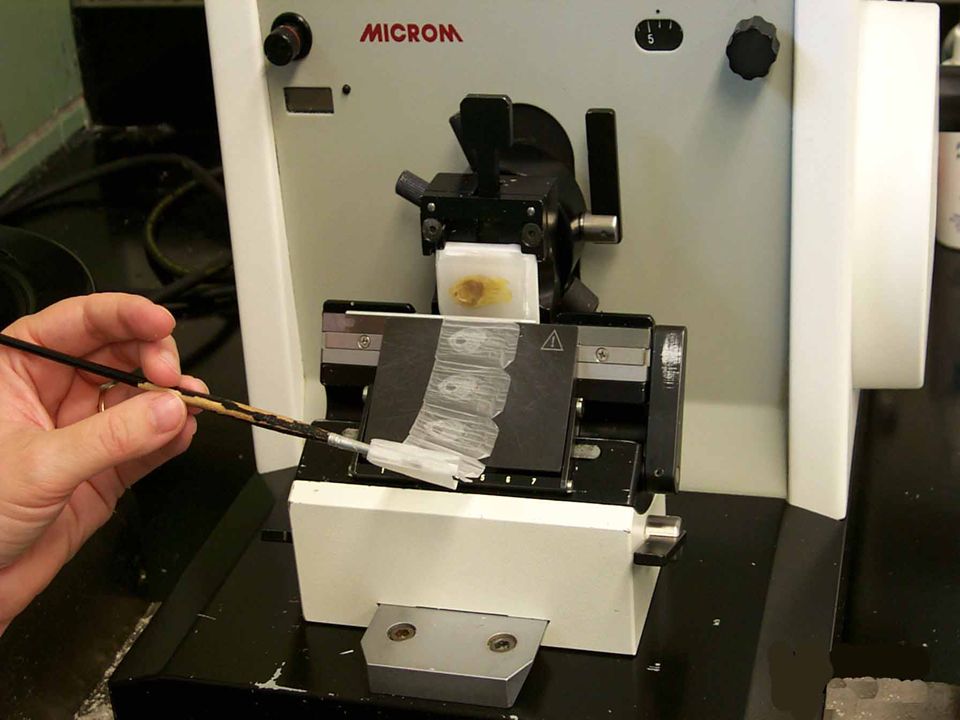
Embedding with wax Paraffin wax is used for embedding of tissue which form tissue blocks after cooling the it can be trimmed into thin sections (4- 5microns) using the microtom the sections are placed on glass slides and become ready for staining
 using the microtom the sections are placed on glass slides and become ready for staining")
21
Embedding with wax

24
Tissue blocks are trimmed into thin sections (4-5microns) using the microtom
 using the microtom")
26
sections are placed in water bath for opening of folded tissue

27
S ections are placed on glass slides and become ready for staining

28
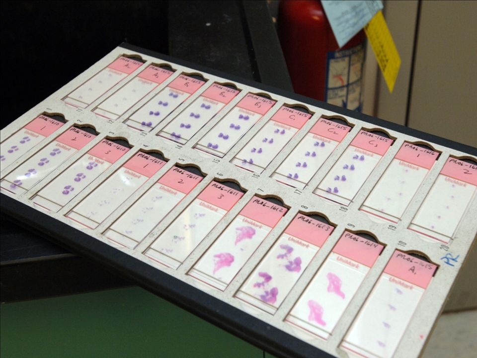
Hematoxylin & eosin is the most widely used stain in histopathology Nuclei appear dark blue Collage & cytoplasm appear pink Keratin appears pink to red

35
Special stains 1)PAS ( periodic acid schiff) stain glycogen & mucin 2)Congo-red for amyloid 3)Sudan-black for fat 4)Giamsa stain for helicobacter pylori
PAS ( periodic acid schiff) stain glycogen & mucin 2)Congo-red for amyloid 3)Sudan-black for fat 4)Giamsa stain for helicobacter pylori")
36
H-pylori stained dark blue with giamsa stain

37
Amyloid deposits stain orange-red with Congo Red stain

38
Frozen section In this technique the tissue is frozen rapidly (using cryostat) to -20ºC ten sections are cut and stained (without passing in the steps of tissue processing) so that tissue can be examined microscopically within 5- 10 minutes of removal from the body
 to -20ºC ten sections are cut and stained (without passing in the steps of tissue processing) so that tissue can be examined microscopically within minutes of removal from the body")
39
It allows rapid diagnosis of the nature of the lesion whether benign or malignant to decide the next step in surgery. All laboratory staff should be informed and all preparations should be completed before arrival of tissue

40
Cytology Is the study of cell ( normal or diseased altered cell) obtained from various sites of the body. It allows rapid diagnosis often within minutes. Cells examined by this process are collected by one of the following methods: 1) Exfoliated cell 2) Cells removed by brushing or scraping 3)Removal of cells from deep tissue ( by aspiration)
 Exfoliated cell 2) Cells removed by brushing or scraping 3)Removal of cells from deep tissue ( by aspiration).")
41
Exfoliative cytology Spontaneous shedding of cells derived from lining of cavities where they can be removed by non invasive methods e.g. vaginal smear, sputum examination, voided urine, body fluids, nipple discharge

42
Abrasive cytology Cells specimens are obtained from superficial scraping of the lesion e.g. cervical scraping (pap smear) buccal mucosal smear skin scraping of various lesions
 buccal mucosal smear skin scraping of various lesions.")
43
Fine needle aspiration cytology Cells are aspirated from deep non surfaces organs or masses e.g. Beast mass Thyroid nodules Palpable lymph nodes Internal organs like liner and kidney

44
Technique of cytology 1)using a needle and syringe to obtain cells from a mass. exfoliatied material is sprayed on a slide directly The needle is gently introduced through the skin into the mass ( while the mass is fixed in between fingers and thumb) and is moved in different directions while applying negative pressure onto the syringe with continuous suction of material through out the process, then the needle should be withdrawn gently from the mass
 and is moved in different directions while applying negative pressure onto the syringe with continuous suction of material through out the process, then the needle should be withdrawn gently from the mass.")
45
2) Smearing collected material on a glass slide 3) Immediate immersion of the slide in a fixative ( 95% ethanol) to avoid dryness of material 4) Applying a stain 5) Examine under the microscope
 Smearing collected material on a glass slide 3) Immediate immersion of the slide in a fixative ( 95% ethanol) to avoid dryness of material 4) Applying a stain 5) Examine under the microscope")
47
Palpable lymph node in the neck can be examined using fine needle aspiration cytology

48
A spiration of material from cervical mass while applying negative pressure

49
Spraying aspirated material on a glass slide

50
Immediate fixation of material in ethanol

51
Staining of slides

52
Cytology is the study of cell

54
Indications of cytopathology 1)Diagnosis of malignancy 2)Diagnosis of precancerous changes e.g. cervical atypia. 3)Detection of inflammation and pathogenic agents e.g. fungal or parasitic infection of vagina 4)Study of hormonal patterns and gonadal function e.g. examination of squamous cells in vaginal smear which are under influence of onerian hormones to assess overian function in infertility.
Detection of inflammation and pathogenic agents e.g. fungal or parasitic infection of vagina 4)Study of hormonal patterns and gonadal function e.g. examination of squamous cells in vaginal smear which are under influence of onerian hormones to assess overian function in infertility..")
55
5) Identification of sex chromosome in newborn with ambiguous genitalia, buccal smear is used as a source of cells.
 Identification of sex chromosome in newborn with ambiguous genitalia, buccal smear is used as a source of cells.")
56
Limitations of cytology 1)The nature of lesion is not so obvious as in a histological section. 2) Difficulty in identification of the exact site of lesion e.g. malignant squamous cells shaded in sputum may arise from buccal mucosa, pharynx, larynx and bronchi. 3) The size of lesion cannot be approximated by cytology.
 Difficulty in identification of the exact site of lesion e.g. malignant squamous cells shaded in sputum may arise from buccal mucosa, pharynx, larynx and bronchi. 3) The size of lesion cannot be approximated by cytology..")
Similar presentations


 B.J.THANENTRHIRAN(MBBS)>")