Download presentation
Presentation is loading. Please wait.

1
APPROACH TO PATIENT WITH SPLENOMEGALY
Thamer A. AL-TRAIKI

2
Normal Spleen The median splenic weight in adults is about 150 grams. It is not usually palpable, but may be felt in children, adolescents, and some adults, especially those of asthenic build. Patients with chronic obstructive pulmonary disease and low diaphragms commonly have palpable spleens The spleen is considered to be normal in size if its length is <13 cm or its thickness is < or = 5 cm on ultrasound examination [J Gen Intern Med 1993; 8:69.]

3
In one study, 3% of college freshmen had palpable spleens; an additional study showed that 5% of hospitalized patients with normal spleens based on scan results were thought to have palpable spleens by their physicians. Palpable spleens are Not always abnormal.

4
Enlarged Spleen The spleen must be enlarged about 3 times to be clinically palpable. The enlarged spleen may be minimally ,moderate , or massively enlarged.

5
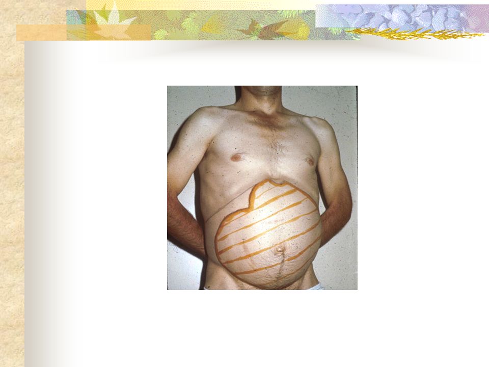
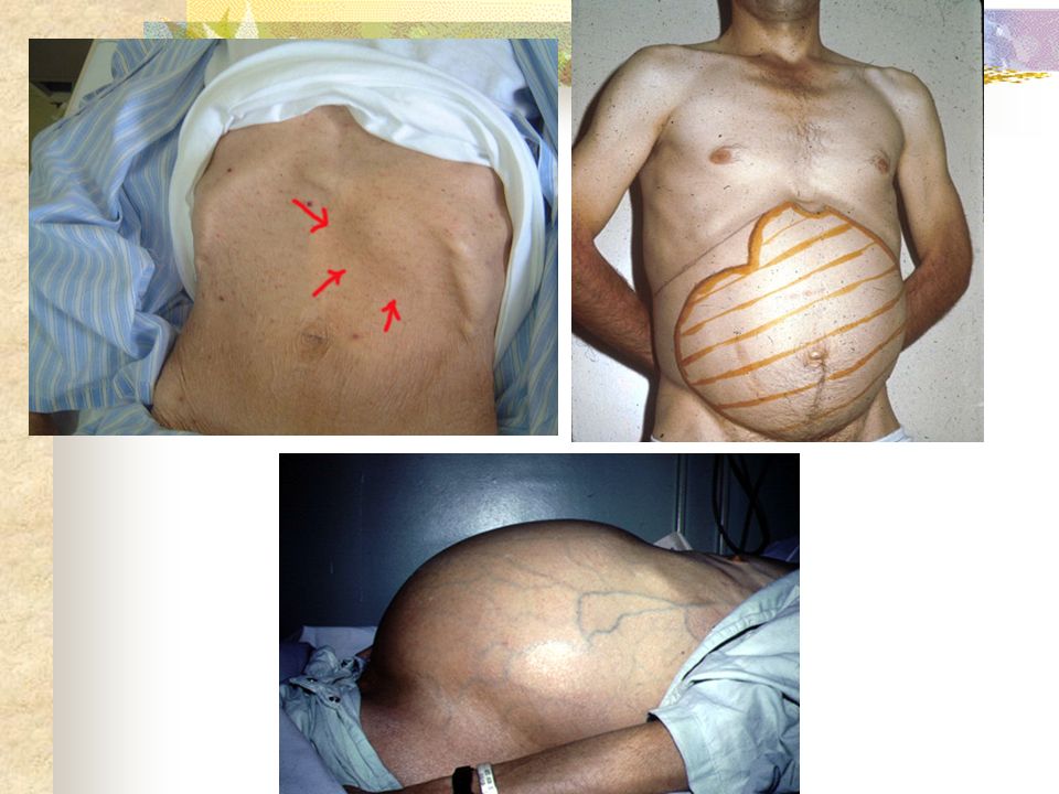
Massively Enlarged Spleen :
A spleen which is only minimally enlarged will be quite movable with respiration, and may be palpable only at the end of inspiration. Using a light touch, with the skin depressed under the left costal margin, a minimally enlarged spleen can be felt as a rounded edge with the consistency of normal liver, which slips under the examiner's fingers at the end of inspiration and back on expiration. Massively Enlarged Spleen : A spleen enlarged such that its lower pole is within the pelvis, or which has crossed the midline into the right lower or right upper abdominal quadrants. 2006 UpToDate

7
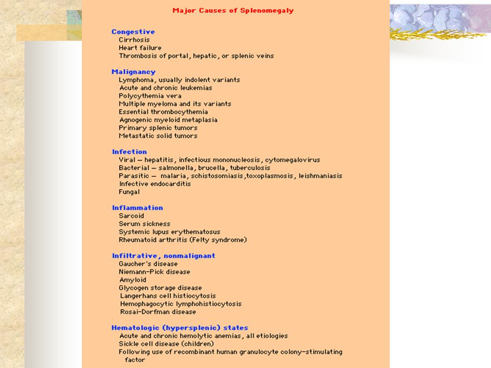
Causes of splenomegaly
1- Reticular endothelial hyperplasia 2- Gush of blood 3- Infiltration

9
Symptoms symptoms of an enlarged spleen may include one or all of the following: Pain, a sense of fullness, or discomfort in the left upper quadrant Pain referred to the left shoulder Early satiety, due to encroachment on the adjacent stomach Acute pleuritic-like pain and tenderness in the left upper quadrant in the presence of fever suggests the presence of perisplenitis or splenic abscess, most likely due to infection originating elsewhere in the body (eg, sepsis, bacterial endocarditis). The abscess may be accompanied by infarction due to septic emboli
. The abscess may be accompanied by infarction due to septic emboli.")
10
Is this mass spleen or not ?
1- we can’t get above it. 2- anteriomedial notch 3- moves inferiomedial with inspiration 4- dull to percussion 5- not ballottable unless gross ascitis

12
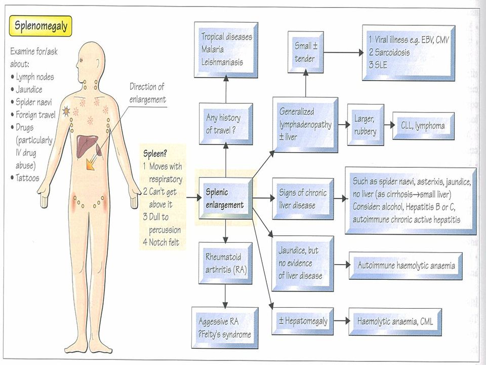
Approach to patient with splenomegaly
Hx Ex Ix Rx

13
Associated symptoms Febrile illness (infectious) headache, dry cough, constipation , rash ,then diarrhea night sweat , malaise ,cough , wt loss travel hx

14
Associated symptoms Weight loss, constitutional symptoms (neoplastic) Pallor, dyspnea, bruising, and/or petechiae (hemolytic) History of liver disease (congestive) Pancreatitis
 Pancreatitis.")
15
ON EXAMINATION Signs of cirrhosis (eg, jaundice, telangiectasias, gynecomastia, caput medusa, ascites) Temperature Heart murmur (endocarditis, congestive failure) Jaundice Scleral icterus ( cirrhosis Petechiae (any cause of thrombocytopenia)
 Jaundice. Scleral icterus ( cirrhosis. Petechiae (any cause of thrombocytopenia)")
16
ON EXAMINATION Associated hepatomegaly Associated lymphadenopathy Size of spleen

17
Massively Enlarged Spleen
Chronic myelogenous leukemia Myelofibrosis, idiopathic or post-polycythemic Gaucher disease Lymphoma, usually indolent, including hairy cell leukemia Kala-azar (visceral leishmaniasis) Hyperreactive malarial splenomegaly syndrome Thalassemia major AIDS with Mycobacterium avium complex
 Hyperreactive malarial splenomegaly syndrome. Thalassemia major. AIDS with Mycobacterium avium complex.")
18
Investigations

19
CBC and peripheral smear
Hb decrease in anemia while increase in polycythemia WBC increases in infection , abscesses & leukemia ESR increases in infection & malignancy

20
CBC and peripheral smear
Neutropenia, anemia, and/or thrombocytopenia may be present, as these formed elements can be trapped in an enlarged spleen, giving the nonspecific picture termed "hypersplenism." The term hypersplenism describes some of the sequelae often observed with splenomegaly. Criteria for a diagnosis of hypersplenism include the following: Anemia, leukopenia, thrombocytopenia, or combinations thereof, plus cellular bone marrow, splenomegaly, and improvement after splenectomy

21
Thrombocytopenia: Approximately 30% of the total platelet mass exists as an exchangeable pool in the spleen. Increased splenic platelet pooling is the primary cause of the thrombocytopenia of hypersplenism. In patients with hypersplenism, as much as 90% of the total platelet mass can be found in the spleen. In hypersplenism, the platelet count is usually 50, ,000/mL. Anemia: The etiology of the anemia observed in splenomegaly is the result of sequestration and hemodilution. Leukopenia: Increased destruction or sequestration of leukocytes causes the leukopenia observed in splenomegaly.

22
On occasion, invading organisms may be seen on the peripheral smear, either free in the plasma as in overwhelming sepsis, or within neutrophils or monocytes (bacteria, ehrlichiae) or red blood cells as occurs with malaria
 or red blood cells as occurs with malaria")
23
In systemic lupus erythematosus, circulating LE cells can occasionally be seen, while patients with neutropenia and rheumatoid arthritis can have circulating large granular lymphocytes

24
The presence of increased numbers of abnormal cells in the peripheral blood suggests the presence of a hematologic malignancy.

25
LFTs U&E Blood Culture Stool Culture Rheumatoid Factor Paul-Bunnell test Urine Culture

26
Ultrasound: This is a noninvasive, highly sensitive, and specific technique for the evaluation of spleen size.also may identify the cause eg cyst CT scan CT scan is the study of choice for identification of inflammatory changes. CT scan is sensitive for detecting mass lesions, infarcts, and cysts

27
Splenectomy or splenic aspirate/biopsy
In a series of 122 "diagnostic" splenectomies performed for unexplained splenomegaly, splenic mass lesion, or to accurately classify a lymphoproliferative disorder detected but not further characterizable on bone marrow or peripheral blood examination, the most common pathologic diagnoses were : Lymphoma/leukemia — 57 percent Metastatic carcinoma/sarcoma — 11 percent Cyst/pseudocyst — 9 percent Benign/malignant vascular neoplasm — 7 percent The spleen as a diagnostic specimen. Cancer 2001; 91:2001.

29
Thank You

30
History: The most common history is mild abdominal pain that is vague in nature. Increased abdominal girth is less common. Early satiety from gastric displacement occurs with massive splenomegaly. Associated symptoms or signs may include the following: Febrile illness (infectious) Pallor, dyspnea, bruising, and/or petechiae (hemolytic process) History of liver disease (congestive) Weight loss, constitutional symptoms (neoplastic) Pancreatitis (splenic vein thrombosis) Alcoholism, hepatitis (cirrhosis)
 Pallor, dyspnea, bruising, and/or petechiae (hemolytic process) History of liver disease (congestive) Weight loss, constitutional symptoms (neoplastic) Pancreatitis (splenic vein thrombosis) Alcoholism, hepatitis (cirrhosis)")
31
The presence or absence of symptoms due to an enlarged spleen depends on many factors, such as the acuteness and nature of the underlying illness, as well as the size of the spleen. Thus, a minimally enlarged spleen secondary to an acute viral infection may be quite tender, while a markedly enlarged spleen in one of the chronic myeloproliferative disorders (eg, polycythemia vera, agnogenic myeloid metaplasia) may be totally asymptomatic unless there is an episode of splenic infarction.
 may be totally asymptomatic unless there is an episode of splenic infarction.")
32
The ability to palpate an enlarged spleen depends upon several variables, including:
1-The size of spleen Minimally enlarged spleens may not be felt. In one study, all spleens with an estimated weight (from scanning studies) exceeding 300 grams were palpable, with the average estimated weight of a palpable spleen being 285 grams[ Ann Clin Res 1974; 6 Suppl 15:1 ]. However some spleens weighing as much as 900 grams are not palpable [ Am J Med 1972; 52:362.]
 exceeding 300 grams were palpable, with the average estimated weight of a palpable spleen being 285 grams[ Ann Clin Res 1974; 6 Suppl 15:1 ]. However some spleens weighing as much as 900 grams are not palpable [ Am J Med 1972; 52:362.]")
33
2- The body habitus of the patient
2- The body habitus of the patient. The spleen is easier to feel in thin individuals and in those who do not have an increased anterior-posterior thoracic diameter. 3- The skill of the examiner coupled with the ability of the patient to cooperate during the examination.

Similar presentations


1 CHILDHOOD LEUKAEMIA. TA OGUNLESI (FWACP)2 LEUKAEMIA Heterogenous group of malignant disorders Characterised by uncontrolled clonal.>")


 presents to the Emergency Room with a 2 day history of weakness.>")


 Myelodysplastic / myeloproliferative diseases (MDS/MPD) >")
:>")
: Principal Modality (2): PET/CT CT Faculty Reviewer:>")


