Download presentation
Presentation is loading. Please wait.

1
DEVELOPMENT OF PITUITARY, THYROID ,PARATHYROID AND SUPRARENAL GLANDS
Dr Rania Gabr

2
Objectives:- Development of Pituitary gland.
Anomalies of Pituitary gland. Development and anomalies of Thyroid gland. Development and anomalies of Parathyroid glands. Development and anomalies of suprarenal glands.

3
Development of Pituitary gland
The hypophysis, or pituitary gland, develops from two completely different parts: an ectodermal oral upgrowth of the stomodeum immediately in front of the buccopharyngeal membrane, known as Rathke’s pouch . (Hypophysial diverticulum) (b) a downward extension of the diencephalon, the Infundibulum. (Neurohypophysial diverticulum)
 (b) a downward extension of the diencephalon, the. Infundibulum. (Neurohypophysial diverticulum)")
4
Rathke’s pouch appears as an evagination of the oral cavity and subsequently grows dorsally towards the infundibulum. By the end of the second month it loses its connection with the oral cavity and is then in close contact with the infundibulum.

5
During further development, cells in the anterior wall of Rathke’s pouch increase rapidly in number and form the anterior lobe of the hypophysis, or adenohypophysis . The posterior wall of Rathke’s pouch develops into the pars intermedia, which in humans seems to have little significance. The infundibulum gives rise to the stalk and the pars nervosa, or posterior lobe of the hypophysis (neurohypophysis).
.")
6
A small extension of the anterior lobe lobe, the pars tuberalis, grows along the stalk of the infundibulum and eventually surrounds it.

7
Parts of adenohypophsis are: pars distalis, pars intermedius, pars tuberalis
Parts of neurohypophysis are: Pars nervosa, infundibulum (median eminence and stem)
")
8
Hypophyseal Anomalies:-
Pharyngeal hypophysis: Occasionally a small portion of Rathke’s pouch persists in the roof of the pharynx. Craniopharyngiomas: Arise from remnants of Rathke’s pouch. They may form within the sella turcica or along the stalk of the pituitary but usually lie above the sella. They may cause hydrocephalus and pituitary dysfunction (e.g., diabetes insipidus, growth failure).
.")
9
Development of Thyroid glands
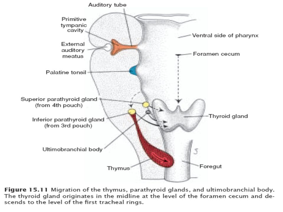
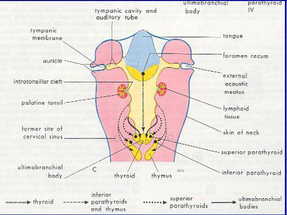
It develops from the floor of the primordial pharynx as an endodermal thickening Thyroid diverticulum develops as an outpouching of the thickening Thyroglossal duct develops as the pouching increases in size

11
The developing thyroid descends in front of the hyoid bone and larynx
Rapid proliferation of the duct obliterate the lumen By 7th week, thyroid then divides into right and left lobes connected by the isthmus Thyroglossal duct degenerates except for a small pit at the beginning called foramen cecum

13
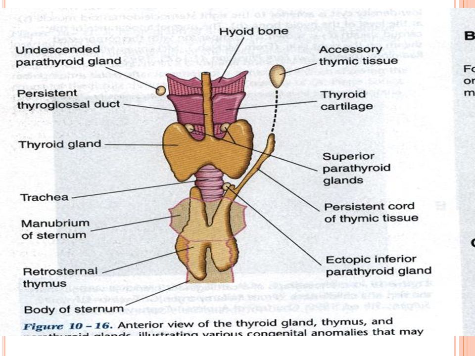
Anomalies of thyroid gland
A thyroglossal cyst. A thyroglossal fistula. Aberrant thyroid tissue Thyroglossal duct

14
Thyroglossal fistula Aberrant thyroid tissue if that tissue doesn't migrate properly, the thyroid gland is said to be ectopic. Most common is the Lingual thyroid

16
Development of Parathyroid gland
Superior parathyroid gland develops from the 4th pharyngeal pouch Inferior parathyroid gland develops from the 3rd pharyngeal pouch

17
Endodermal in origin Two pairs Inferior from 3rd pharyngeal pouch-develop with THYMUS, lose their connection with pharynx and migrate into neck. Later separate from thymus and come to lie on the dorsal surface of thyroid Superior parathyroid develops from 4th pharyngeal pouch during 7th wk

18
Anomalies of parathyroid and thymus
Ectopic Thymic and Parathyroid Tissue: Since glandular tissue derived from the pouches undergoes migration, it is not unusual for accessory glands or remnants of tissue to persist along the pathway. This is true particularly for thymic tissue, which may remain in the neck, and for the parathyroid glands. The inferior parathyroids are more variable in position than the superior ones and are sometimes found at the bifurcation of the common carotid artery.

19
Development of Suprarenal gland
The suprarenal gland develops from two components: A mesodermal portion, which forms the cortex An ectodermal portion, which forms the medulla. During the fifth week of development, mesothelial cells between the root of the mesentery and the developing gonad begin to proliferate and penetrate the-underlying mesenchyme .

20
Here they differentiate into large acidophilic organs the fetal cortex, or primitive cortex of the suprarenal gland.

21
Shortly afterward, a second wave of cells from the mesothelium penetrates the mesenchyme and surrounds the original acidophilic cell mass. These cells, smaller than those of the first wave, later form the definitive cortex of the gland.

22
While the fetal cortex is being formed, cells originating in the sympathetic system (neural crest cells) invade its medial aspect, where they are arranged in cords and clusters. These cells give rise to the medulla of the suprarenal gland. After birth the fetal cortex regresses rapidly except for its outermost layer, which differentiates into the cortical zones. The adult structure of the cortex is not achieved until puberty.

23
A–C. Transverse sections through successively older embryos showing formation of the neural groove, neural tube, and neural crest. Cells of the neural crest, migrate from the edges of the neural folds and develop into spinal and cranial sensory Ganglia. A portion of the sympathetic neuroblasts migrates toward the proliferating mesothelium to form the medulla of the suprarenal gland.

24
Abnormalities of Suprarenal gland development:-
1-Ectopic suprarenal gland. 2-Accessory Medullary Tissue. 3-Acessory Cortical Tissue. 4-Agenesis or Hypoplasia. 5-Congenital Adrenal Hyperplasia.

25
Congenital Adrenal Hyperplasia

26
Thank you 26

Similar presentations


 Glands Anatomy & Embryology>")


