Download presentation
Presentation is loading. Please wait.

1
PowerPoint Lecture Outlines to accompany
PowerPoint Lecture Outlines to accompany Hole’s Human Anatomy and Physiology Tenth Edition Shier w Butler w Lewis Chapter 3 Copyright © The McGraw-Hill Companies, Inc. Permission required for reproduction or display. 3-1

2
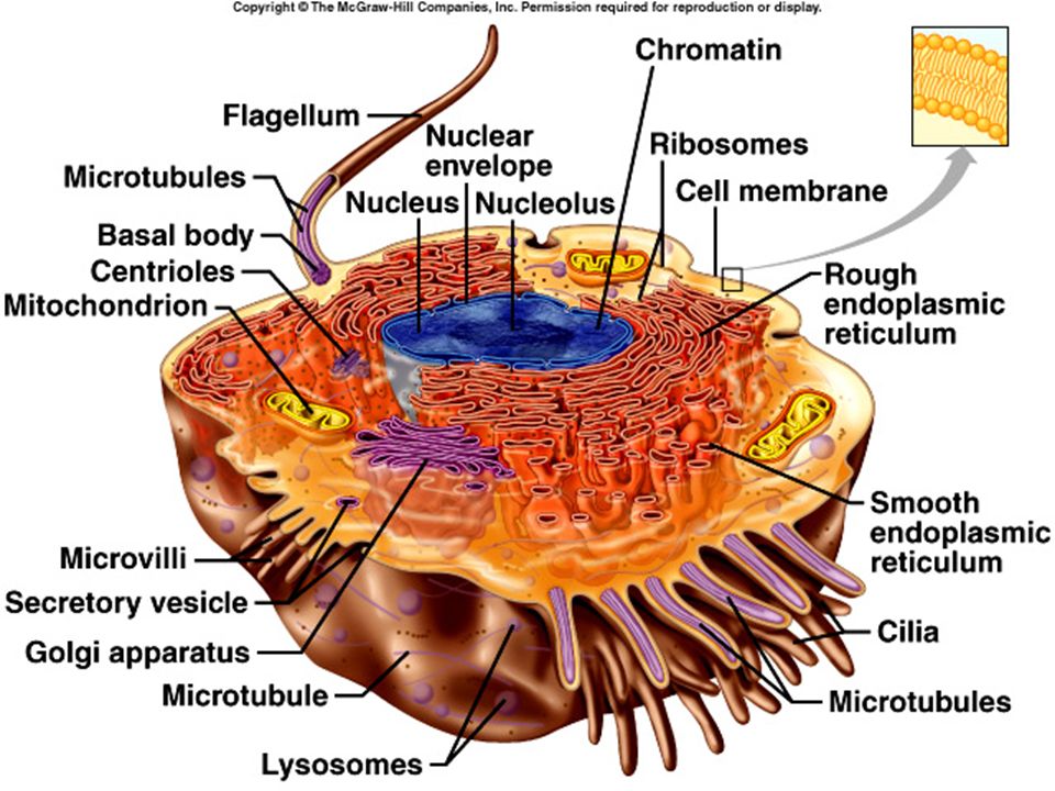
A Composite Cell Is a hypothetical cell
A Composite Cell Is a hypothetical cell Cells are differentiated (suited for their particular purpose) Examples Neurons Muscle cells Sperm Egg 3-3
 Examples. Neurons. Muscle cells. Sperm. Egg")
4
Cells….. 3 Fundamental pieces needed in a cell Plasma membrane:
phospholipid bilayer Proteins Cholesterol Cytoplasm Everything within the cell membrane except for Nucleus Contains hereditary information

5
Cell Membrane outer limit of cell
outer limit of cell controls what moves in and out of cell selectively permeable phospholipid bilayer water-soluble “heads” form surfaces water-insoluble “tails” form interior permeable to lipid-soluble substances cholesterol stabilizes the membrane proteins receptors: receive information pores, channels, carriers: allow materials to pass through enzymes: speed up chemical reactions CAMS: cell adhesion molecules self-markers: ID the cell as part of the organism 3-4

7
Cell Membrane 3-5

8
The Fluid mosaic model The components of the cell membrane are all moving randomly, switching positions

9
Intercellular Junctions
Intercellular Junctions Tight junctions close space between cells located among cells that form linings Desmosomes form “spot welds” between cells located among outer skin cells Gap junctions tubular channels between cells located in cardiac muscle cells 3-6

10
Cell Adhesion Molecules
Cell Adhesion Molecules guide cells on the move selectin – allows white blood cells to “anchor” integrin – guides white blood cells through capillary walls important for growth of embryonic tissue important for growth of nerve cells 3-7

11
Movements Into and Out of the Cell
Passive (Physical) Processes require no cellular energy simple diffusion facilitated diffusion osmosis filtration Active (Physiological) Processes require cellular energy active transport endocytosis exocytosis transcytosis 3-14
 Processes. require no cellular energy. simple diffusion. facilitated diffusion. osmosis. filtration. Active (Physiological) Processes. require cellular energy. active transport. endocytosis. exocytosis. transcytosis")
12
Simple Diffusion movement of substances from regions of higher concentration to regions of lower concentration oxygen, carbon dioxide and lipid-soluble substances 3-15

13
Facilitated Diffusion
diffusion across a membrane with the help of a channel or carrier molecule glucose 3-16

14
Cells and Solutions Cells have many materials dissolved in their cytoplasm, as does the extracellular environment around the cell. Water may move in or out of the cell in response to the concentration of these materials, called solutes. Nature works to dilute solutions by moving water where there is a high concentration of solute and low water in comparison to an adjacent area.

15
Solutions Solvent: does the dissolving
usually water in living things Solute: gets dissolved in the solvent can be just about anything, but common ones include Salts Ions sugars

16
Tonicity Hypertonic: when the solute concentration is higher than the surrounding environment Hypotonic: when the solute concentration is lower than the surrounding environment Isotonic: when the solute concentration is the same as the surrounding environment

17
Osmosis movement of water through a selectively permeable membrane from regions of higher concentration to regions of lower concentration water moves toward a higher concentration of solutes 3-17

18
Cells and Solutes A cell is placed in a hypertonic solution; cell shrinks A cell is placed in a hypotonic solution; cell swells

19
Osmosis & Pressure Osmotic Pressure – ability of osmosis to generate enough pressure to move a volume of water Osmotic pressure increases as the concentration of nonpermeable solutes increases hypertonic – higher osmotic pressure hypotonic – lower osmotic pressure isotonic – same osmotic pressure 3-18

20
Tonicity and RBCs hypertonic – higher osmotic pressure: cells shrink
isotonic – same osmotic pressure: cells remain the same size hypotonic – lower osmotic pressure: cells swell Animations: link

21
Filtration smaller molecules are forced through porous membranes
hydrostatic pressure important in the body Ex: molecules leaving blood capillaries 3-19

22
Active Transport carrier molecules transport substances across a membrane from regions of lower concentration to regions of higher concentration sugars, amino acids, sodium ions, potassium ions, etc. 3-20

23
Endocytosis cell engulfs a substance by forming a vesicle around the substance three types pinocytosis – substance is mostly water phagocytosis – substance is a solid receptor-mediated endocytosis – requires the substance to bind to a membrane-bound receptor 3-21

24
Endocytosis 3-22

25
Exocytosis reverse of endocytosis
substances in a vesicle fuse with cell membrane contents released outside the cell Ex: release of neurotransmitters from nerve cells 3-23

26
Transcytosis endocytosis followed by exocytosis
transports a substance rapidly through a cell Ex: HIV crossing a cell layer 3-24

27
Questions you must answer
Questions you must answer. You may use the transport mechanisms animations on the Bio II page to do so. When asked to compare and contrast, things you may want to think about include: Think in terms of types of materials involved (size, polarity, etc), energy (ATP) use, and how things get in/ out of the cell (protein channels and the types of channels vs. using the membrane itself). Answer these questions in the appropriate place in your notes packet.
, energy (ATP) use, and how things get in/ out of the cell (protein channels and the types of channels vs. using the membrane itself). Answer these questions in the appropriate place in your notes packet.")
28
PowerPoint Lecture Outlines to accompany
PowerPoint Lecture Outlines to accompany Hole’s Human Anatomy and Physiology Tenth Edition Shier w Butler w Lewis Chapter 3: Part II Organelles Copyright © The McGraw-Hill Companies, Inc. Permission required for reproduction or display. 3-1

29
Endoplasmic Reticulum
Endoplasmic Reticulum Endoplasmic Reticulum connected, membrane-bound sacs, canals, and vesicles Connected to nucleus Communicate with the cell membrane transport system 3-8

30
(ER) rough ER studded with ribosomes protein and lipid synthesis smooth ER lipid synthesis break down of drugs Break down of lipids Cell with many ER would be those responsible for making proteins, as well as those that are used to break down drugs Products of ER used to make new cell membrane pieces 3-8

31
Ribosomes free floating or connected to ER (rough ER)
Ribosomes free floating or connected to ER (rough ER) site of protein synthesis (link AAs to form proteins) Made from proteins and RNA (rRNA) 3-8
 site of protein synthesis (link AAs to form proteins) Made from proteins and RNA (rRNA) 3-8.")
32
Golgi Apparatus/ Bodies
Golgi Apparatus/ Bodies group of flattened, membranous sacs (cisternae) Connect to the ER (which coming out of the nucleus) packages and modifies proteins (adds carbohydrates, lipids etc as needed) 3-9
 Connect to the ER (which coming out of the nucleus) packages and modifies proteins (adds carbohydrates, lipids etc as needed) 3-9.")
33
Golgi Pieces bud off and become vesicles that can release contents to the cell (vesicle trafficking) or outside of the cell (secretory vesicles that break out) Common in white blood cells and liver cells, or those that make protein hormones (like insulin) 3-9
 or outside of the cell (secretory vesicles that break out) Common in white blood cells and liver cells, or those that make protein hormones (like insulin) 3-9.")
34
Golgi Pieces bud off and become vesicles that can release contents to the cell (vesicle trafficking) or outside of the cell (secretory vesicles that break out) Common in white blood cells and liver cells, or those that make protein hormones (like insulin) 3-9
 or outside of the cell (secretory vesicles that break out) Common in white blood cells and liver cells, or those that make protein hormones (like insulin) 3-9.")
36
Mitochondria membranous sacs with inner partitions
Mitochondria membranous sacs with inner partitions generate energy by making ATP from sugars and oxygen (from the foods we eat as well as the air we breathe) “Powerhouse” of the cell 3-9
 Powerhouse of the cell")
37
Mitochondria double membrane bound structure
Mitochondria double membrane bound structure Inner membrane folds are the cristae, which have enzymes on them and control the chemical reactions that release energy (ATP) The stuff in between the fold is the matrix 3-9
 The stuff in between the fold is the matrix")
38
Mitochondria Have own DNA
Mitochondria Have own DNA Get mitochondrial DNA from mom only (mitochondrial Eve- tracing ancestors) Possible once a primitive cell Resemble bacteria ? Bacteria taken into cells? Typical cell has 1700 mitochondria; more in cells that need a lot of energy (like muscles) 3-9
 Possible once a primitive cell. Resemble bacteria. Bacteria taken into cells Typical cell has 1700 mitochondria; more in cells that need a lot of energy (like muscles) 3-9.")
39
Lysosomes Lysosomes enzyme-containing sack Sack of membrane
digest worn out cell parts or unwanted substances by using enzymes (40 types, at least) Called the “suicide sack” because breaking it open would digest cell contents 3-10
 Called the suicide sack because breaking it open would digest cell contents")
40
Peroxisomes Peroxisomes sacks of phospholipid membrane
break down organic molecules (like drug, alcohol, or food molecules) by using enzymes Peroxidases- make hydrogen peroxide (H2O2) when breaking down molecules H2O2 is destructive to cells so must be removed Catalase- breaks down hydrogen peroxide 40 other types Abundant in liver 3-10
 by using enzymes. Peroxidases- make hydrogen peroxide (H2O2) when breaking down molecules. H2O2 is destructive to cells so must be removed. Catalase- breaks down hydrogen peroxide. 40 other types. Abundant in liver")
41
Centrosomes Centrosome two rod-like centrioles (made from proteins)
used to produce cilia and flagella distributes chromosomes during cell division (mitosis) in animal cells 3-10
 in animal cells")
42
Cilia Cilia short hair-like projections
propel substances on cell surface In respiratory tract (Smoker’s cough) Lining of fallopian tubes to propel egg In bacteria, they move the cell around 3-11
 Lining of fallopian tubes to propel egg. In bacteria, they move the cell around")
43
Flagellum Flagellum long tail-like projection Made from microtubules
provides motility to sperm Animation link 1 Animation link 2 3-11

44
Vesicles Vesicles membranous sacs (phospholipid membrane) formed from
Pinched in pieces of cell membrane Pieces of Golgi or ER that have broken off store substances (can be just about anything- water, nutrients, enzymes, other proteins or substances that the cell has made) 3-12
")
45
Microfilaments and Microtubules
Microfilaments (smaller) and microtubules (larger) thin rods and tubules of protein support cytoplasm Give shape, structure allows for movement of organelles (pull organelles around- like “ Mr. Gadget arms” Important during mitosis/ meiosis animation 3-12
 and microtubules (larger) thin rods and tubules of protein. support cytoplasm. Give shape, structure. allows for movement of organelles (pull organelles around- like Mr. Gadget arms Important during mitosis/ meiosis. animation")
46
Inclusions Inclusions Chemicals in the cytoplasm Stored nutrients
Glycogen, lipids Temporary- will go in or out over time Ex: melanin in skin- a tan goes away over time 3-12

48
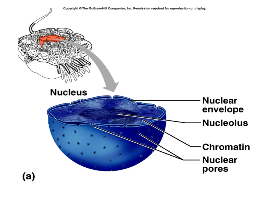
Cell Nucleus control center of cell; involved with directing the cell when to make certain materials (usually proteins) Surrounded by a nuclear envelope porous double membrane (phospholipid with over 1000 types of proteins) separates nucleoplasm from cytoplasm Allows mRNA out to make proteins (translation) Controls entry into and exit out of the nucleus 3-13
 separates nucleoplasm from cytoplasm. Allows mRNA out to make proteins (translation) Controls entry into and exit out of the nucleus")
49
Cell Nucleus Nucleoplasm nucleolus (little nucleus)
dense collection of RNA and proteins At least one per nucleus site of ribosome production Larger if protein making cells Nucleoplasm The goo that is in the nucleus and nucleolus 3-13

50
Cell Nucleus fibers of DNA and proteins (histones)
chromatin (colored substance) fibers of DNA and proteins (histones) Look like “beads on a string” stores information for synthesis of proteins 3-13
 fibers of DNA and proteins (histones) Look like beads on a string stores information for synthesis of proteins")
51
First animation From the nucleus to the golgi bodies Overall cell review of organelles

52
Links to animations, ect
Diffusion links: Osmosis Links: Interactive cellular transport: Interactive tutorial/ Quizzes

54
Cell links… Cell structure:comparisons between cell types Cell structure and function (right click on the here to start, and select play)
")
55
Just some good animation links
Similar presentations