Download presentation
Presentation is loading. Please wait.

1
RADIOLOGY ANATOMY OF THE PITUITARY GLAND
Dr. Rasheed Al Jurayyan

2
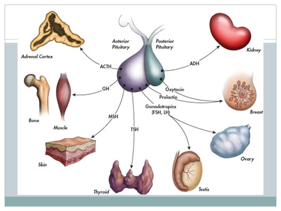
NORMAL PITUITARY GLAND
The gland is composed of two parts: Anterior lobe (adeno hypophysis) Posterior lobe (neuro hypophysis) Normal size: Weight: 0.5g Height: 4-16 mm Anterior posterior: 5-16 mm
 Posterior lobe (neuro hypophysis) Normal size: Weight: 0.5g. Height: 4-16 mm. Anterior posterior: 5-16 mm.")
3
INDICATIONS FOR IMAGING THE PITUITARY GLAND
Hormonal dysfunction Cushing syndrome Growth abnormalities e.g. Growth hormone deficiency, acromegaly Visual abnormalities headache

5
What is best modality to image the pituitary gland ?
X ray CT scan MRI US Nuclear medicine

6
What is best modality to image the pituitary gland ?
X ray CT scan MRI US Nuclear medicine

7
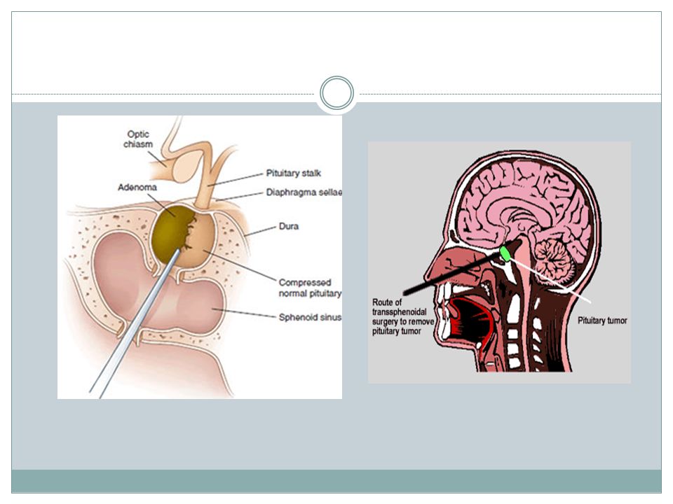
CT scan MRI

8
CT scan MRI

9
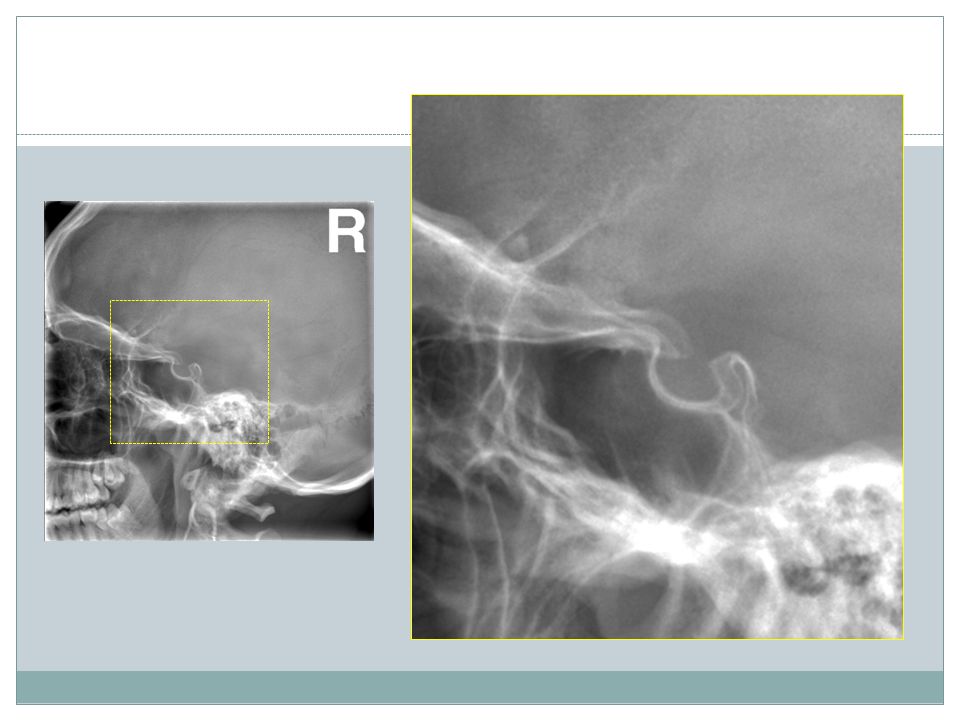
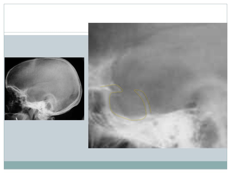
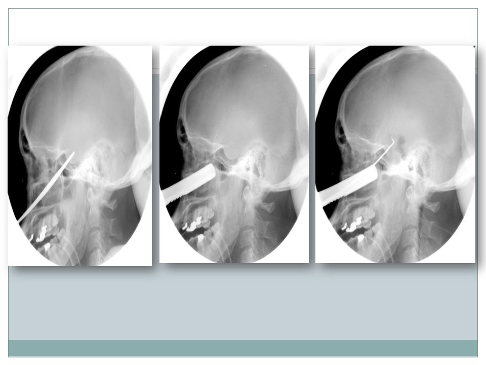
X RAY

11
1 2 3 4 5 6
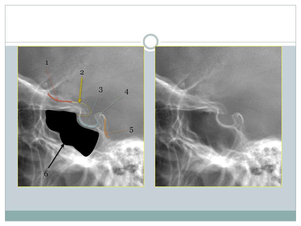
12
2- Anterior clinoid process 3-Floor of sella turcia (Pituitary fossa)
1 2 1-Optic sulcus 2- Anterior clinoid process 3-Floor of sella turcia (Pituitary fossa) 4- Posterior clinoid process 5- Dorsum sella 6- Sphenoid sinus 3 4 5 6
 4- Posterior clinoid process. 5- Dorsum sella. 6- Sphenoid sinus")
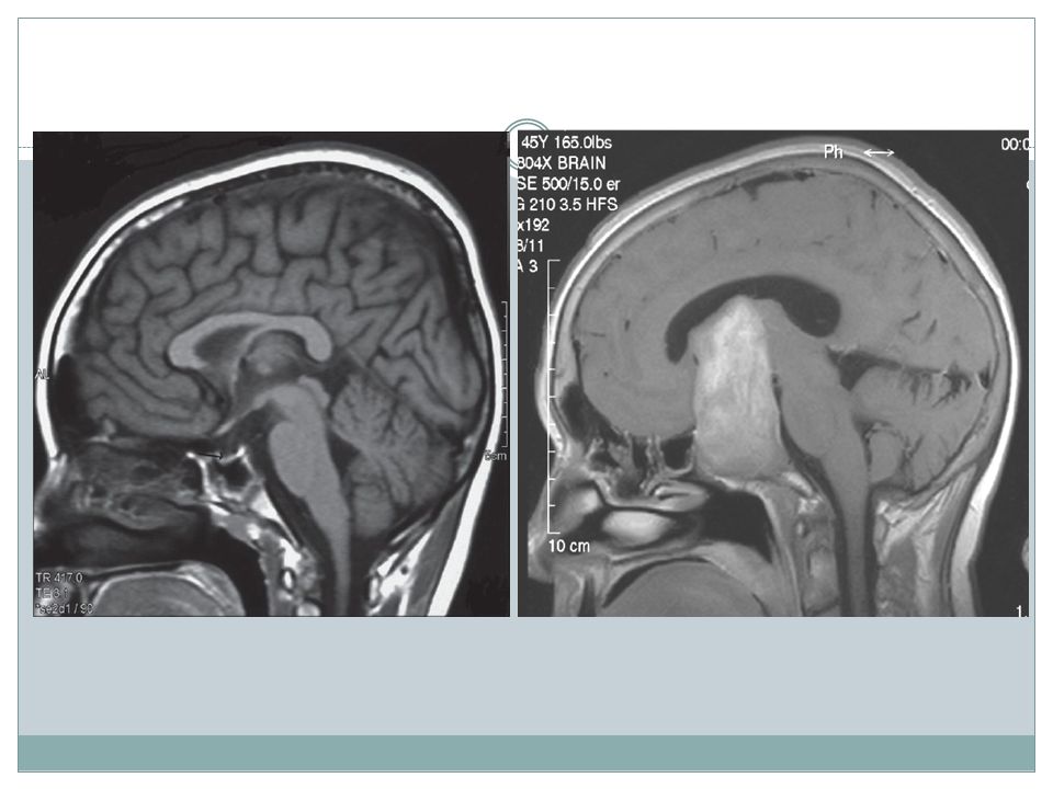
14
MRI

15
4 3 5 2 6 1
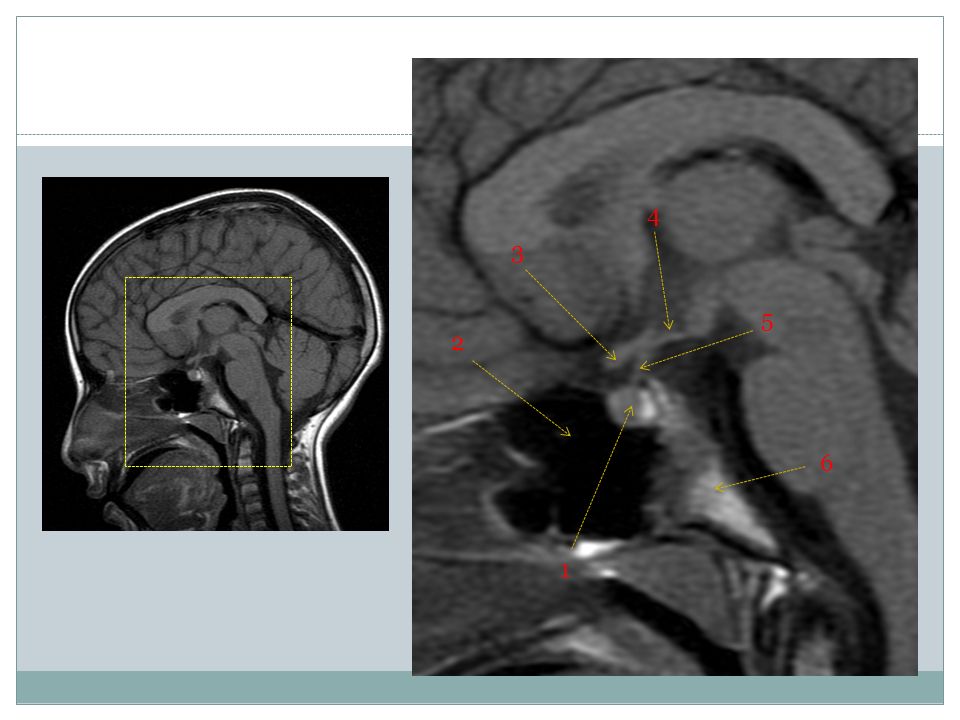
16
1- pituitary gland 2- sphenoid sinus 3- optic chiasm 4- hypothalamus
5- pituitary stalk 6- claivus 5 2 6 1

17
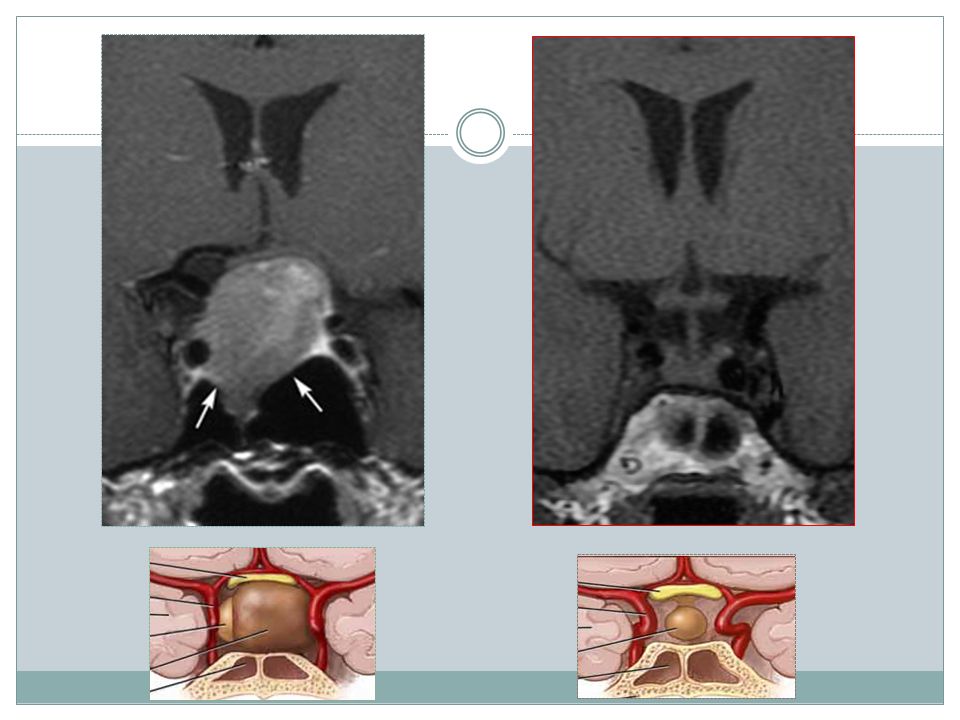
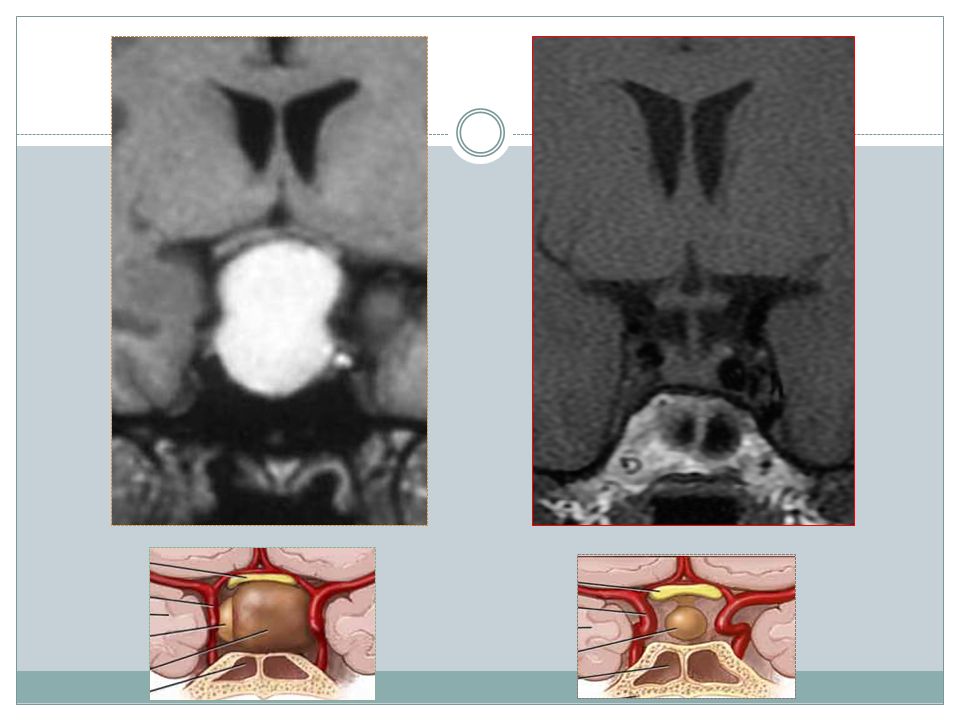
NORMAL PITUITARY ADENOMA

19
2 3 1 4 5 6
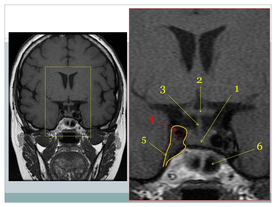
20
2 3 1 4 5 6
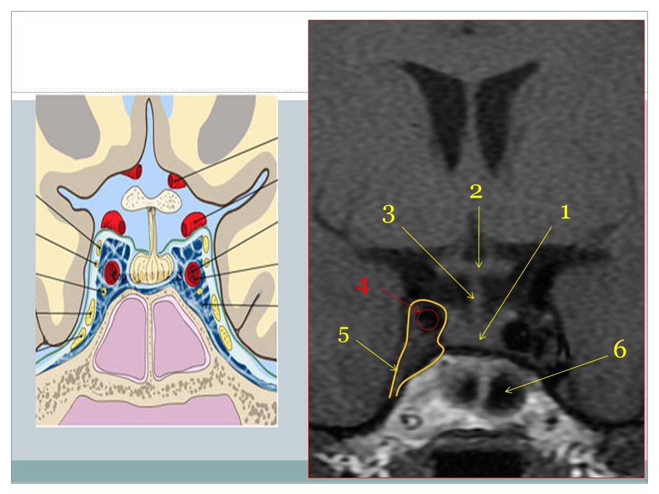
21
Optic chiasm Pituitary stalk Pituitary gland Carotid artery Cavernous sinus Sphenoid sinus

26
THE END

Similar presentations


 from pituitary disturbances? 1)Soldier # 1 2)Soldier # 2 3)Soldier # 3 4)Soldiers.>")


