Download presentation
Presentation is loading. Please wait.

1
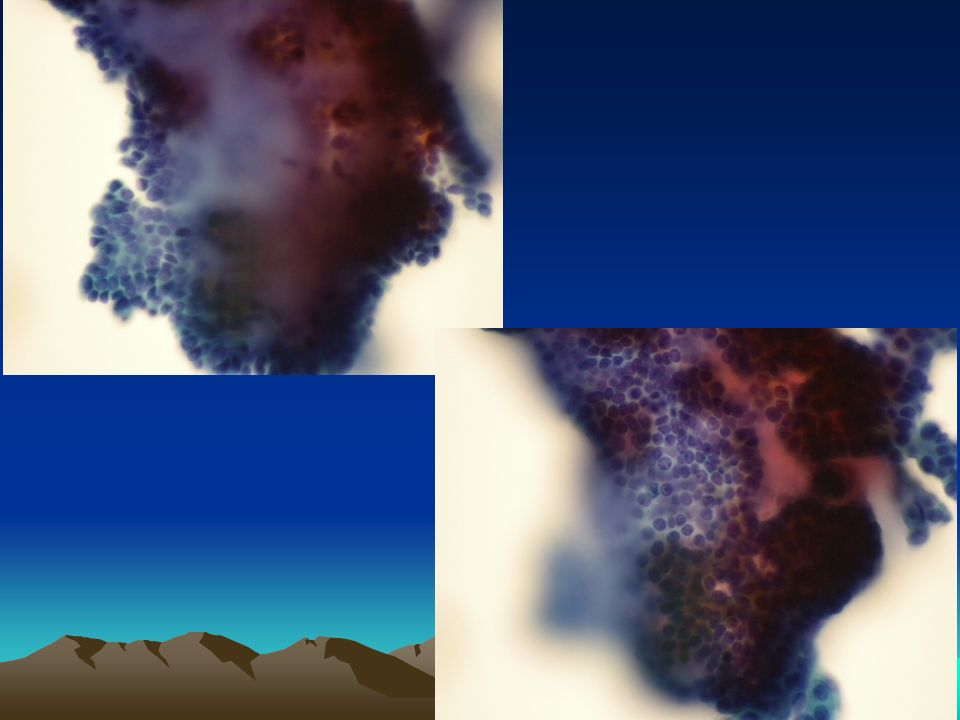
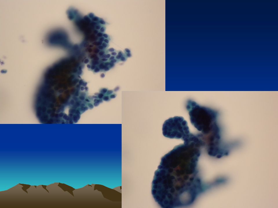
每月一例 2015 February 40 y/o, female Breast FNA Liquid-Based Preparation (SurePath) Figure 1-6
 Figure 1-6")
4
Papillary 3-D Clean background No atypia (NOTE: not apparent myoepithelial cells)
")
8
Cytomorphology Papillary, 3-D, clean background, no atypia, (NOTE: not apparent myoepithelial cells) Cytology: Papillary lesion, favor papilloma Histology: intraductal papilloma LBC: smaller area, better preservation, but too thick and less background material
 Cytology: Papillary lesion, favor papilloma Histology: intraductal papilloma LBC: smaller area, better preservation, but too thick and less background material")
9
Cytomorphology Consistent with a papillary neoplasm Typically highly cellular and contain large clusters or aggregates of ductal cells that exhibit complex folding and often a papillary configuration supported by a fibrovascular core Sheets of apocrine cells and a proteinaceous background with histiocytes and siderophages may also be present An intervening myoepithelial layer may or may not be present in these lesions

10
FNA of breast papillary lesions Papillary lesions of the breast are a heterogeneous group of breast lesions that are difficult to diagnose as benign or malignant Interpretation is challenging because of the wide morphologic spectrum encountered in the benign, atypical and malignant subtypes, which cannot typically be determined on cytology alone Papillary lesions of the breast include: Papilloma, Papillomatosis, Atypical Papilloma, Carcinoma arising in a papilloma and Intraductal papillary carcinoma (with or without invasion)
")
11
LBC in nonGYN samples More than 2 decades, though the traditional methods still widely used FDA-approved methods –ThinPrep (TP) –SurePath (SP) –Others: LiquiPrep, Turbitec, PapSpin, TACAS, Siriraj- LBC, KNA-Citoliq (Digene), GluCyte,…
 –SurePath (SP) –Others: LiquiPrep, Turbitec, PapSpin, TACAS, Siriraj- LBC, KNA-Citoliq (Digene), GluCyte,…")
12
LBC in nonGYN samples Cytological features –generally superior to conventional preparation with regard to clearer background, reduced obscuring artifacts and extracellular elements, monolayer cell preparation, and the cells being limited to smaller areas with excellent cellular preservation –Remains a diagnostic challenge because of a somewhat altered morphology and the presence of artifacts resulting from the chemical influences of the fixation medium and the physical forces of the processing techniques (familiarity with these appearances is essential for pathologists to avoid misinterpretation)
")
13
LBC in nonGYN samples Cytological changes include –Architectural changes such as smaller cell clusters and sheets and breakage of papillae –Altered cell distribution with more dyscohesion and slightly more three-dimensional clusters –Changes in cellular morphology with attenuated chromatin details, more prominent nucleoli and smaller cell size –Intranuclear inclusions difficult to visualize –Background matrix often altered in both quantity and quality

14
LBC in nonGYN samples Cytological changes include –Extracellular particles, small mononuclear cells, red blood cells, and myoepithelial cells markedly decreased in number –Lymphocytes tending to aggregate

Similar presentations