Download presentation
Presentation is loading. Please wait.

1
Exome Sequencing as Molecular Diagnostic Tool of Mendelian Diseases
BIOS 6660 Hung-Chun (James) Yu Shaikh Lab, Department of Pediatrics, University of Colorado Denver Genetic Laboratories, Children’s Hospital of Colorado 11/17/2015
 Yu. Shaikh Lab, Department of Pediatrics, University of Colorado. Denver Genetic Laboratories, Children’s Hospital of Colorado. 11/17/2015.")
2
Human Genetic Diseases
Mutation Penetrance Mutation Frequency Kaiser J. Science (2012) 338:
 338:")
3
Human Genetic Diseases
Complex Disorder Polygenic, many genes. Low penetrance/effect size. Multifactorial, environmental, dietary. Examples: heart diseases, diabetes, obesity, autism, etc. Mendelian Disorder Monogenic (mostly). Full or high penetrance/effect size. Examples: sickle cell anemia (HBB) and cystic fibrosis (CFTR).
. Full or high penetrance/effect size. Examples: sickle cell anemia (HBB) and cystic fibrosis (CFTR).")
4
Complex Diseases Multiple causes, and polygenic.
Multiple genetics factors with low penetrance individually. Coronary artery disease Coriell Institute for Medical Research.

5
Mendelian Diseases Veltman J.A. et al. Nat. Rev. Genet. (2012) 13:
 13:")
6
Mendelian Diseases Dominant Inheritance
U.S. National Library of Medicine.

7
Mendelian Diseases Recessive Inheritance
U.S. National Library of Medicine.

8
Exome Sequencing Focusing on exons or coding regions of genes Exons
Bamshad, MJ., et al. Nat. Rev. Genet. (2011) 12: Exons Complementary Baits
 12: Exons. Complementary. Baits.")
9
Exome Sequencing 3,000,000,000bp (3Gb) human genome
~45% repetitive sequence ~25% genic region ~2% exonic, coding region 20,000 – 30,000 human genes 3,000 – 5,000 disease genes ~4,000 human genetic diseases (OMIM) 114 medically actionable (treatable) genes Michael O. Dorschner., et al. Am J Hum Genet : 631–640.
 114 medically actionable (treatable) genes. Michael O. Dorschner., et al. Am J Hum Genet : 631–640.")
10
Exome Sequencing ~40Mb (coding) or 60Mb (coding + UTRs) Gene
Read Coverage Individual Reads

11
Mendelian Diseases Identified by Exome Sequencing
Timeline Gilissen C. et al., Genome Biol. (2011) 12:228.
 12:228.")
12
Mendelian Diseases Identified by Exome Sequencing
Kym M. Boycott et al. Nature Reviews Genetics (2013) 14:681–691
 14:681–691.")
13
Types of Variation What kind of variation/mutation can be detected by Exome Sequencing? SNV (single nucleotide variation) Small InDel, (insertion/deletion <25bp) Large InDel, CNV (copy number variation) Possible, but not reliable. Aneuploidy (loss/gain of entire chromosome) Possible. Translocation Difficult and not reliable. Complex rearrangement Very difficult.
 Large InDel, CNV (copy number variation) Possible, but not reliable. Aneuploidy (loss/gain of entire chromosome) Possible. Translocation. Difficult and not reliable. Complex rearrangement. Very difficult.")
14
Exome Variants SNV (single nucleotide variation)
Synonymous: (1) Silent. Nonsynonymous: (1) Missense. (2) Nonsense. (3) Stop-loss. (4) Start-gain. (5) Start-loss. (6) Splice-site.
 Silent. Nonsynonymous: (1) Missense. (2) Nonsense. (3) Stop-loss. (4) Start-gain. (5) Start-loss. (6) Splice-site.")
15
Exome Variants Small InDel (insertion/deletion <25bp) Frameshift
In-frame NHGRI Digital Media Database (DMD),
,")
16
Variant and Population Frequency
Novel/Private variant Never been reported before. Rare variant Minor allele freq. (MAF) < 1%. Databases dbSNP (NCBI): 1000 Genomes: ESP (NHLBI): ExAC:
 < 1%. Databases. dbSNP (NCBI): Genomes: ESP (NHLBI): ExAC:")
17
Exome Variants How to analyze enormous amount of variants in any given exome? Gilissen C. et al. Eur. J. Hum. Genet. (2012) 20: Private/Novel ~ Rare, MAF<1% ~ ,000 Protein altering ~4, ,000 Coding/splice-site ~10, ,000 All ~20, ,000

18
Exome Analysis Strategies
Gilissen C. et al., Eur. J. Hum. Genet. (2012) 20: Male Female Affected Heterozygous carrier Sex-linked heterozygous carrier Mating Consanguineous mating Sequenced individual
 20: Male. Female. Affected. Heterozygous. carrier. Sex-linked. heterozygous. carrier. Mating. Consanguineous. mating. Sequenced individual.")
19
Trio-based Exome sequencing
Family trio Both unaffected parents and an affected patient. Why using trio? Every inheritance model can be tested Economical, efficient, single case required. Access to samples.

20
Trio-based Exome sequencing
Autosomal dominant de novo Autosomal recessive Homozygous * * * * * Autosomal recessive Compound heterozygous X-linked recessive Hemizygous in male Gene Male Female Affected Heterozygous carrier Sex-linked heterozygous * * * * X Y X X * * X Y

21
Trio-based Exome sequencing
Candidate Genes/Variants Rare (~500-2,000) or novel (~ ) protein altering variants Plus, variants that fit inheritance model Rare Variant Novel Variant Dominant de novo 0 ~ 2 Recessive Compound Heterozygous 0 ~ 20 0 ~ 3 Homozygous X-linked 0 ~ 10 0 ~ 5
 or novel (~ ) protein altering variants. Plus, variants that fit inheritance model. Rare Variant. Novel Variant. Dominant. de novo. 0 ~ 2. Recessive. Compound Heterozygous. 0 ~ ~ 3. Homozygous. X-linked. 0 ~ ~ 5.")
22
Case 1 Clinical information
Case 1 was the result of a non-consanguineous union and he presented to care at four months of age with a seizure disorder, hypotonia and developmental delay. The patient underwent a left parietal craniotomy and partial resection of the frontal cortex without complete resolution of the seizure disorder. Initial laboratory studies included an elevated homocysteine and methylmalonic acid and a normal vitamin B12 level. Complementation analysis of the patient’s cell line placed the patient into the cblC class. Severe developmental delay, infantile spasms, gyral cortical malformation, microcephaly, chorea, undescended testes, megacolon. Sequencing and deletion/duplication analysis (microarray) the MMACHC gene was negative in both skin fibroblasts and peripheral blood.
 the MMACHC gene was negative in both skin fibroblasts and peripheral blood.")
23
Case 1

24
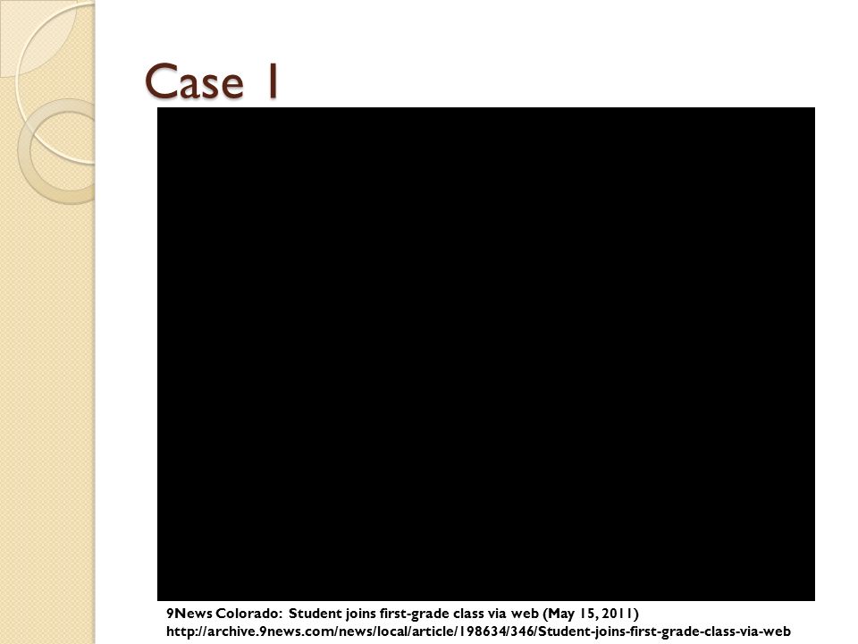
Case 1 9News Colorado: Student joins first-grade class via web (May 15, 2011)
25
Case 1 Monster Max Patient's older sister as a summer student in Shaikh Lab

26
Case 2 Clinical information
The patient was a 7-month-old boy when first evaluated. He was diagnosed with BPES by a pediatric ophthalmologist. In addition to blepharophimosis, ptosis, and epicanthus inversus normally associated with BPES, he had cryptorchidism, right hydrocele, wide-spaced nipples, and slight 2–3 syndactyly of toes. Clinical testing demonstrated a normal karyotype (46,XY), and normal FISH studies for 22q11.2 deletion, Cri-du-Chat (5p deletion) syndrome. Thyroid function was normal. Further, normal 7-dehydrocholesterol level was used to rule out Smith–Lemli–Opitz syndrome. Sanger sequencing and highresolution CNV analysis with Affymetrix SNP 500K arrays did not identify a FOXL2 mutation.
, and normal FISH studies for 22q11.2 deletion, Cri-du-Chat (5p deletion) syndrome. Thyroid function was normal. Further, normal 7-dehydrocholesterol level was used to rule out Smith–Lemli–Opitz syndrome. Sanger sequencing and highresolution CNV analysis with Affymetrix SNP 500K arrays did not identify a FOXL2 mutation.")
27
Case 2 A-D: 2-month old. note blepharophimosis, ptosis, epicanthus inversus (A), posteriorly angulated ears with thickened superior helix and prominent antihelix (B), and slight 2–3 syndactyly of toes in addition to overlapping toes (C, D) E-F: 3.5-year old. Following oculoplastic surgery to correct ptosis; note right-sided preauricular ear pit (F, indicated by arrow). G-I: 12-year old. Note the recurrence of ptosis (L>R), arched eyebrows, abnormal ears, thin upper lip vermilion, small pointed chin, downsloping shoulders, and wide- spaced and low-set nipples.
, posteriorly angulated ears with thickened superior helix and prominent antihelix (B), and slight 2–3 syndactyly of toes in addition to overlapping toes (C, D) E-F: 3.5-year old. Following oculoplastic surgery to correct ptosis; note right-sided preauricular ear pit (F, indicated by arrow). G-I: 12-year old. Note the recurrence of ptosis (L>R), arched eyebrows, abnormal ears, thin upper lip vermilion, small pointed chin, downsloping shoulders, and wide- spaced and low-set nipples.")
28
Case 3 Clinical information
The proband is a nine year old girl who presented with microcephaly, unilateral retinal coloboma, bilateral optic nerve hypoplasia, nystagmus, seizures, gastroesophageal reflux, and developmental delay including not yet saying specific words (at 29 months old). On exam, she has microcephaly with a normal height, a down-turned upper lip, and fingertip pads. A karyotype and CGH analysis have been normal. Kabuki (KMT2D and KDM6A) and Angelman (UBE3A and MECP2) syndromes were suspected in this patient.
. On exam, she has microcephaly with a normal height, a down-turned upper lip, and fingertip pads. A karyotype and CGH analysis have been normal. Kabuki (KMT2D and KDM6A) and Angelman (UBE3A and MECP2) syndromes were suspected in this patient.")
29
Case 3

30
Exome NGS Workflow Genomic DNA Exome capture Library construction
Sequencing QC Sequence read processing Mapping SAM BAM Variant calling Annotation (General) Annotation (In-house) Filtering Inheritance test, candidate genes, ect. Exome Sequencing Mapping and variant detection Variant prioritization Exome Enrichment Illumina Sequencer Galaxy/FASTX Toolkit Galaxy/BWA Galaxy/Samtool Galaxy
 Annotation. (In-house) Filtering. Inheritance test, candidate genes, ect. Exome Sequencing. Mapping and variant detection. Variant prioritization. Exome Enrichment. Illumina Sequencer. Galaxy/FASTX Toolkit. Galaxy/BWA. Galaxy/Samtool. Galaxy.")
31
Exome analysis Workflow (this class)
FASTQ sequence 2x90bp (paired-end) Variant determination BCF Filter based on Phred score, mapping quality, read depth, etc. Mapping to genome SAM Conversion Conversion VCF BAM QC: Filter duplicates, artifacts, and unpaired or unmapped reads, 100 genes “Mini” Exome ? BWA (Burrows-Wheeler Aligner) SAMtools
 Variant determination. BCF. Filter based on Phred score, mapping quality, read depth, etc. Mapping to genome. SAM. Conversion. Conversion. VCF. BAM. QC: Filter duplicates, artifacts, and unpaired or unmapped reads, 100 genes. Mini Exome. BWA. (Burrows-Wheeler Aligner) SAMtools.")
32
Data for Case Study 3 trios VCF files “Mini” Exome
A total of 3 families/cases. Each family/case includes both unaffected parents and an affected patient. VCF files Generated from 2x90bp paired-end Exome sequence reads, and at ~50X coverage Reads mapped to human GRCh37/hg19 and then familial variants calls made in VCF format “Mini” Exome 100 genes with/without known disease association. Validated causative genes and randomly selected disease genes or non-disease genes.

33
VCF Format Variant Call Format FILTER, INFO, FORMAT # Header line
## Meta-information lines FILTER, INFO, FORMAT # Header line

34
VCF Format FORMAT GT: Genoetype. 0/0: Homozygous normal
0/1: Heterozygous variant 1/1: Homozygous variant PL: the Phred-scaled genotype likelihoods (>0). 0/ / /1 , ,178 GQ : Genotype quality (1-99)
. 0/0 0/1 1/ ,0 ,178. GQ : Genotype quality (1-99)")
35
Annotation Tools Annotate variants with useful information
Mutation effect Population frequency Clinical association Genomic sequence and protein domain Pathogenicity prediction Gene expression, protein interaction. …..and many many more. SeattleSeq: VEP (Variant effect Predictor): ANNOVAR:
: ANNOVAR:")
36
Question ?

Similar presentations


