Download presentation
Presentation is loading. Please wait.

1
Surgical Wound Care

2
“Wound” Refers to any injury to the body’s tissues involving a break in the skin. Promoting wound healing is the nursing focus during the postsurgical recovery phase Patient-related factors influence wound healing: age, nutritional status, physical condition, pre-existing health status, and medication habits

3
Wound Classification Vitally important to understand the causative factors of a wound to determine the proper treatment plan In a planned surgery, a cut (incision) is made by a sharp instrument creating an opening into an organ or space in the body; or A stab wound (puncture) for a drainage system
 is made by a sharp instrument creating an opening into an organ or space in the body; or. A stab wound (puncture) for a drainage system.")
4
Surgical Wound Selection of the site for the surgical wound is based on Tissue/organ involved Nature of injury/disease process Process of inflammation/infection Strength of the site If a drainage system is required, the position of the drain may also influence the placement of the incision. Could be surgeon specific

5
Incision A=Right Upper Paramedian B=Left Lower Paramedian C=Right Subcostal D=Right Midline E=Pfannestiel

6
Wound Healing Healing process begins immediately after an injury and sometimes continues for a year or longer Follows 4 phases: Hemostasis Termination of bleeding as soon as the injury occurs Platelets adhere to the walls of the injured vessel formation of clot fibrin in the clot begins to hold the wound together

7
Wound Healing Inflammatory Phase
Increase in blood elements and water flow out of the blood vessel into the vascular space Causes cardinal signs and symptoms of inflammation: -erythema -heat -edema -pain -tissue dysfunction

8
Wound Healing 2. Inflammatory Phase cont.
Leukocytes engulf bacteria, fungi, viruses, and toxic proteins Cells in the injured tissue migrate, divide, and form new cells Blood clots dissolve Wound fills

9
Wound Healing 3. Reconstruction Phase Collagen formation occurs
(glue-like protein substance) -adds tensile strength to the wound/ tissue. Irregular, raised, purplish, immature scare Wound dehiscence risk Angiogenesis -formation of new vasculature
 -adds tensile strength to the. wound/ tissue. Irregular, raised, purplish, immature scare. Wound dehiscence risk. Angiogenesis. -formation of new vasculature.")
10
Wound Healing Remodeling Phase (maturation) -Collagen deposition
-peaks by the third week -Remodeling -can last for years after the initial injury -collagen is degraded and deposited in an equilibrium-producing fashion -no change in the amount of collagen present in the wound.

11
Process of Wound Healing
Wounds close by: primary intention, secondary intention, or tertiary intention Primary Intention Wound is made surgically with little tissue loss. Skin edges are close together. Minimal scarring results.

12
Process of Wound Healing
Secondary Intention When a wound must granulate during healing Occurs when skin edges are not close together or when pus has formed Surgeon may treat with a drainage system or by packing the wound. This gives decomposed necrotized tissue an escape. Cavity begins to fill with granulation tissue. Scarring is greater in a larger wound.

13
Process of Wound Healing
Tertiary Intention = delayed primary intention Contaminated wound is left open -sutured closed after the infection is controlled Also occurs when a primary wound becomes infected -opened -allowed to granulate -sutured

14
Keloids -Abnormal scar that grows beyond the boundary of
the original site of a skin injury – overgrowth of collagenous scar tissue -Some ethnic groups are at more risk -highly pigmented ethnic groups other than Caucasians. Parts of the body affected -upper arm, -upper back/sternum. -earlobes/back of the neck Other causes Infection at a wound site, repeated trauma to the same area, skin tension or a foreign body in a wound can also be factors.

15
Factors Affecting Wound Healing
Nutritional Needs -provide small frequent feedings -total parenteral nutrition -nasogastric feedings Hydration Offer hourly; encourage 2000 to 2400 ml in 24 hours. Intracellular Fluid accounts for 2/3 of the fluid in the body.

16
Factors Affecting Wound Healing
Blood Supply Poor circulation Age Lower metabolic state in the elderly Specialized tissue Muscle and Nerve tissue do not regenerate easily Infection Interferes with the matrix formations Rest Periods of sleep aid in healing

17
Surgical Wound A disruption of the skin integrity
Surgeon’s goal -enter the cavity involved -repair the injured/diseased area -minimize trauma as quickly as possible. Wound A disruption of the skin integrity Tissue has been disrupted so severely that it cannot heal naturally without complications or disfigurement held in approximation until the healing process provides the skin with sufficient strength to withstand stress without mechanical support

18
Wound Closure Wound closure material and techniques of using them:
Prime factors in the restoration and tensile strength of the healed tissue. -staples -sutures -clips -skin closure strips -topical adhesions

19
Sutures, (FON, pg. 330, Figure 13-4)
Sutures. A, Interrupted, or separate. B, Continuous. C, Blanket. D, Retention.

20
Staples (FON, pg. 330,Figure 13-5)
")
21
Steri-Strips (FON, pg. 330 Figure 13-6)
Butterfly Closures

22
Wound Closures Transparent Dressings Self-adhesive transparent film
-synthetic permeable membrane (breathe-able) -temporary secondary skin. Advantages contains exudates/minimize wound contamination barrier to external fluids and bacteria yet still allows the wound to breathe moist environment that speeds epithelial cell growth visualization of the wound
 -temporary secondary skin. Advantages. contains exudates/minimize wound contamination. barrier to external fluids and bacteria yet still allows the wound to breathe. moist environment that speeds epithelial cell growth. visualization of the wound.")
23
Transparent Dressing

24
Liquid Bandage Transparent Dressings

25
Care of the Incision Surgical wounds -generally heal well and quickly
-dressing may be removed within 24 – 72 hours -allow air circulation -trend – to leave sutured, clean wounds not dressed after surgery or use loose dressing Incision Coverings Gauze Permits air to reach the wound Semi-occlusive Permits oxygen but not air impurities to pass Occlusive Permits neither air nor oxygen to pass

26
Care of the Incision Securing a dressing:
Tape Ties Bandages Cloth binders Choice of anchoring depends on: Wound size Location Presence of drainage Patient’s level of activity

27
Care of the Incision Removing Dressings Sterile technique
Avoid accidental removal/displacement of underlying drains. Analgesic may need to be given at least 30 minutes before the dressing change Sterile technique Gown, mask, and protective goggles -if soiling or splashing of wound exudate is expected.

28
Dry Sterile Dressings (DSD)
For managing wounds with little exudate or drainage -keep wound dry to prevent excoriation. protects from injury, prevents introduction of bacteria, reduces discomfort, and speeds healing For Abrasions/non-draining postoperative incisions

29
Changing a sterile dry dressing.
Remove old dressing and use gloves to contain old dressing and drainage. Wash hands before and after removing dressing After washing your hand, put on new gloves. Wash wound with SNS, working from incision outwards. Use a new 2X2 each time you return to center. Apply appropriate dressing. You could consider Montgomery Straps if you are changing dressing frequently. Changing a sterile dry dressing.

30
Montgomery Straps

31
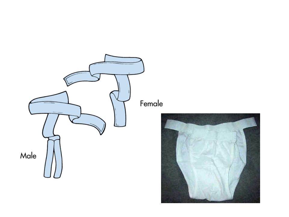
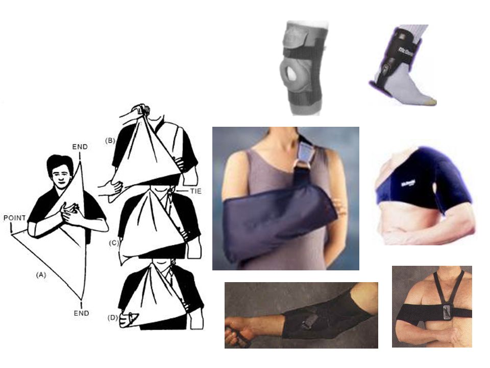
Bandages and Binders Bandage Binders
-strip or roll of cloth/other material -wrapped around a part of the body in a variety of ways -multiple purposes. -rolls of various widths/materials -gauze, elasticized knit, elastic webbing, flannel, and muslin. Binders -large pieces of material to fit a specific body part -abdominal binder or a breast binder.

32
Bandages and Binders Before a bandage or binder is applied
Inspect the skin for abrasions, edema, discoloration, or exposed wound edges. Cover exposed wounds or open abrasions with sterile dressings. Assess the condition of underlying dressings and change them if soiled. Assess the skin and underlying body parts and parts that will be distal to the bandage for signs of circulatory impairment.

33
Bandages and Binders Correctly applied bandages and binders do not cause injury to underlying and nearby body parts or create discomfort for the patient.

34
Velcro Binder

35
Cloth Binders (Do they exist?)
")
38
Care of the Incision Wet-to-Dry Dressing
Primary purpose-mechanically debride a wound. Moistened contact layer of the dressing collects exudate and wound debris. As the dressing dries, it adheres to the wound and debrides it when the dressing is removed. Normal saline and lactated Ringer’s solution, acetic acid, sodium hypochlorite solution, povidone-iodine, and antibiotic solutions.

39
Wound Irrigation Irrigations Wound cleansing and irrigation
Sterile or clean technique. Introduced directly into the wound Syringe, syringe and catheter, shower, or whirlpool Position the patient on his or her side to encourage the flow of the irrigant away from the wound Removes debris from a wound surface, Decreases bacterial counts Loosens and removes eschar.

40
Wound Irrigation Solutions used for irrigations include warm water, saline, or mild detergents. Principles of Basic Wound Irrigation Cleanse in a direction from the least contaminated area to the most contaminated area. When irrigating, all of the solution flows from the least contaminated area to the most contaminated area.

41
Complications of Wound Healing
Impaired wound healing requires accurate observation and ongoing interventions. Wound bleeding potentially indicates: -slipped suture -dislodged clot -coagulation problem -trauma to blood vessels or tissue. If internal hemorrhage occurs, the dressing may be dry while the abdominal cavity collects blood.

42
Complications of Wound Healing
Wound Infection Results when the wound becomes contaminated. “infected” when it contains purulent (pus) drainage. elevated WBC count. Purulent drainage -odor -brown, yellow, or green -depending on the pathogen.
 drainage. elevated WBC count. Purulent drainage. -odor. -brown, yellow, or green. -depending on the pathogen.")
43
Cardinal Signs of Infection and Inflammation
Erythema Edema Heat Pain Purulent drainage Loss of function

44
Complications of Wound Healing
Dehiscence Wound layers separate. Patient may say that something has given way. Result after periods of sneezing, coughing, or vomiting. Preceded by serosanguineous drainage. Patient should remain in bed and receive nothing by mouth, be told not to cough, and be reassured. The nurse should place a warm, moist sterile dressing over the area until the physician evaluates the site

45
Complications of Wound Healing
Evisceration Abdominal organs protrude through an opened incision. Patient is to remain in bed, and the wound abdominal contents should be covered with warm, sterile saline dressings. The surgeon is notified immediately. This is a medial emergency, and the wound requires surgical repair.

46
Staple and Suture Removal
Physician’s written order The time of removal -based on the stage of healing and extent of surgery. 7 to 10 days after surgery, or sooner if healing is adequate. Leaving in a suture too long -removal more difficult and increases the risk of infection. One at a time -removal of every other suture or staple and replaced with a Steri-Strip as the first phase, with the remainder removed in the second phase.

47
Staple and Suture Removal
Sutures Sutures are threads of wire or other materials Sutures are placed within tissue layers in deep wounds and superficially Deeper sutures are usually made of absorbable material Types include interrupted or separate sutures, continuous sutures, blanket sutures, and retention sutures covered with rubber tubing for strength.

48
Removing sutures

49
Staple and Suture Removal
Staples Staples are made of stainless steel wire Abdominal incisions and orthopedic surgery Removal of staples requires a sterile staple extractor

50
Removing staples.

51
Exudate/Drainage Drainage Exudate
Removal of fluids from a body cavity, wound, or other source of discharge through one or more method Exudate Fluid, cells, or other substances that have slowly exuded from cells or blood vessels through small pores or breaks in the cell membrane Exudate/drainage from organs has its own particular color. (Bile from the liver and gallbladder is green-brown.)
")
52
Exudate/Drainage Serous = clear, watery fluid that has been separated from it’s solid elements E.g exudate from a blister Sanguineous = fluid that contains blood Serosanguineous = thin, red fluid composed of both serum and blood.

53
Exudate and Drainage The type and amount
-depends on the tissue and organs involved. More than 300 ml in the first 24 hours is abnormal. When patients first ambulate, a slight increase may occur. Assess Color, amount, consistency, and odor -contained either in a drainage system or on a dressing.

54
Drainage Systems Usually used in procedures in which organs were removed or repaired. A mechanism is needed to assist gravity in removing exudates from the cavity. To facilitate drainage, an incision or a “stab” wound is made close to the incision and drains exudate away from the incision. Requires close monitoring Note the color, consistency, and amount of drainage. Note patency of tube; it should not be kinked or occluded. If blood clots or exudate have slowed drainage, record and report.

55
Drainage Systems Closed Drainage Open Drainage Suction Drainage
tubing and other apparatus attached to the body to remove fluid in airtight circuit that prevents environmental contaminants from entering the wound or cavity Open Drainage Drainage that passes through an open-ended tube into a receptacle or out onto the dressing Suction Drainage Use of a pump or other mechanical device to help extract a fluid

56
Drains

57
Jackson-Pratt

58
Hemovac

59
Care of the Patient with a T-Tube Drainage System
-surgical removal of the gallbladder -bile duct is often inflamed and edematous. -drainage tube -maintain a free flow of bile.

60
Drainage Systems T-Tube drainage system
Used after gallbladder removal when the bile duct is inflamed and edematous Goal: to maintain a free flow of bile Long end of the T-tube exits through the abdominal incision or a separate surgical wound. Fluid drains via gravity into a closed drainage system. Collection bag is emptied and measured every shift.

61
T-tube.

62
Wound Vacuum-Assisted Closure
Quicker healing times Convenient to client Discharge to home earlier Cost effective .

63
Wound V.A.C. Assists in wound closure by applying localized negative pressure to draw the edges of a wound together Accelerates wound healing by promoting the formation of granulation tissue, collagen, fibroblasts, and inflammatory cells Use of negative pressure removes fluid from the area surrounding the wound reducing local or peripheral edema and improving circulation

64
Wound VAC System ( FDA approval, 3/24/1995)
")
65
Wound V.A.C. Treat both acute and chronic wounds
Assess pt. comfort level Educate re: purpose of this device Expected outcome: Preventing infection Promoting healing Control of pain Patient and family education

66
Home Care Considerations
Is there an able and willing caregiver? Home care nurses Wound healing assessment and teaching Some insurance won’t pay for the nurse to do wound care, sometimes negotiable Medicare will usually pay if no able and willing caregiver; pt. physically unable to do wound care; or high complexity of wound care and dressing change and need for close monitoring Try to discharge patient early in the morning if going to homecare. The nurse must assess and teach. A difficult task if the client is exhausted from being in hospital all day.

Similar presentations


 Closed wound: Skin is intact (not opened) include crushing injury and contusions. Wounds A) Skin involvement: 1) Open wound: when the whole thickness.>")


