Download presentation
Presentation is loading. Please wait.

1
Chapter 18 The Cell-Division Cycle Essential Cell Biology FOURTH EDITION Copyright © Garland Science 2014 Alberts Bray Hopkin Johnson Lewis Raff Roberts Walter

2
Cycle of Cell Reproduction Fig. 18-1

3
Reproduction Rates Vary early Drosophila embryo nuclei (not cells yet) 6 minutes!
 6 minutes!")
4
Phases of Cell Cycle Fig. 18-2

5
There are quality control checkpoints along the way. Fig. 18-3

6
Cyclin/Cdk Complexes Regulate Progression Fig. 18-4

7
Cyclin Abundance Regulates Cdk Activity Cdks present at all times; cyclin levels “cycle”. Fig. 18-8

9
How We Know: Cyclin/M-Cdk was first discovered in fertilized frog and clam eggs.

10
Cytoplasm from M phase fertilized egg induces mitosis in G2-arrested oocyte Fig. 18-7 Cyclin/M-Cdk is the active agent. (called Maturation Promoting Factor (MPF) arrested in G2
 arrested in G2.")
11
Progesterone induces Cyclin/M-Cdk to promote maturation of oocytes during meiosis (Maturation Promoting Factor) Cyclin/M-Cdk
 Cyclin/M-Cdk")
12
Cyclin/Cdk Complexes Drive Progression of All Cell Cycle Stages We’ll just refer to them as S-Cyclin and M-Cyclin.

13
Cyclin Abundance Determines Cdk Activity Cyclin abundance regulated by transcription and by proteolysis. Fig. 18-8

14
Cyclin proteolysis triggered by ubiquitylation by APC allows exit from M phase Fig. 18-9

15
M-Cdk activity also regulated by phosphorylation Fig. 18-10 phosphorylation controls entry into M phase P activating phosphate and kinase (CAK)
.")
16
Positive Feedback Loop from M-Cdk on Cdc25 Makes M phase Entry Signal More Robust Fig. 18-17 Wee1 kinase phosphorylation by M-Cdk activates Cdc25

17
S-Cdk regulated by inhibitory proteins Inhibitory proteins control entry into S phase Fig. 18-11 S-

18
p27 inactive cyclin-Cdk complex UBIQUITYLATION OF p27 BY SCF p27 proteolysis triggered by ubiquitylation by SCF allows entry into S phase Fig. 18-9 modified DESTRUCTION OF p27 IN PROTEASOME

19
Replication complete? DNA damage? Chromosome attachment to spindle? Environmental cues? There are quality control checkpoints along the way. Fig. 18-12

20
Environmental Cues Can Induce S Phase Entry Through Rb Inactivation Cyclins, etc. Fig. 18-14

21
Internal Signals Can Induce Temporary Delay in S Phase Entry through p53 Activation Fig. 18-15 Checkpoint kinases (Chk) functions like p27
 functions like p27.")
22
Fig. 18-10 P activating phosphate and kinase (CAK) If DNA damage is detected during G2 phase, Cdc25 inactivation prevents entry into M phase.
 If DNA damage is detected during G2 phase, Cdc25 inactivation prevents entry into M phase..")
23
Replication Proteins Are Targets of Cyclin/S-Cdk Fig. 18-16 Cdc6 and ORC can only form pre-RC when de-phosphorylated ORC, Cdc6, and MCM phosphorylated, inactivating ORC & Cdc6 but activating MCM ensures pre-RC assembly once and only once/ cell cycle -P MCM

24
Cyclin/Cdk Complexes Regulate Progression Fig. 18-4 What regulates Cdk Activity? Cyclin activates (its level cycles) Cdk inhibitors (p27 & p21) Cdk phosphorylation -activating kinase: CAK -inhibiting kinase: Wee1 -activating phosphatse: Cdc25
 Cdk inhibitors (p27 & p21) Cdk phosphorylation -activating kinase: CAK -inhibiting kinase: Wee1 -activating phosphatse: Cdc25.")
25
Fig. 18-10 M-Cdk phosphorylation controls entry into M phase P activating phosphate and kinase (CAK) How will mutations in these phosphorylation sites affect the cell cycle?
 How will mutations in these phosphorylation sites affect the cell cycle .")
26
POLAR CHARGED POLAR UNCHARGED Ser, Thr, or Tyr mutated to Ala: can no longer be phosphorylated Ser, Thr, or Tyr mutated to Asp or Glu: mimics phosphorylation Effects of Mutating Phosphorylation Sites phosphorylation

27
Fig. 18-10 M-Cdk phosphorylation controls entry into M phase P activating phosphate and kinase (CAK) mutating inhibitory PO 4 site to Glu mutating inhibitory PO 4 site to Ala mutating activating PO 4 site to Glu mutating activating PO 4 site to Ala constitutive entry into M phase unable to enter M phase constitutive entry into M phase Mutation phenotype Dom/Rec? Dom Rec Dom
 mutating inhibitory PO 4 site to Glu mutating inhibitory PO 4 site to Ala mutating activating PO 4 site to Glu mutating activating PO 4 site to Ala constitutive entry into M phase unable to enter M phase constitutive entry into M phase Mutation phenotype Dom/Rec. Dom Rec Dom.")
28
Mitosis: The Most Visibly Dramatic Part of Cell Cycle prophase metaphase anaphase telophase

29
Panel 18.1 chromosomes condense mitotic spindle forms kinetochore forms nuclear envelope disperses chromosomes aligned chromosomes separate chromosomes decondense nuclear envelope reforms DNA replication

30
Chromosome Proteins Are Targets of Cyclin/M-Cdk in Prophase Fig. 18-18 -Cohesin and Condensin are structurally related ring- forming proteins. -Cohesin holds sister chromatids together during metaphase. -Condensin condenses chromosomes into visible bodies. target of Cyclin/M-Cdk phosphorylation

31
Mitosis requires assembly of two transient cytoskeletal structures Fig. 18-19 mitosis cytokinesis M phase

32
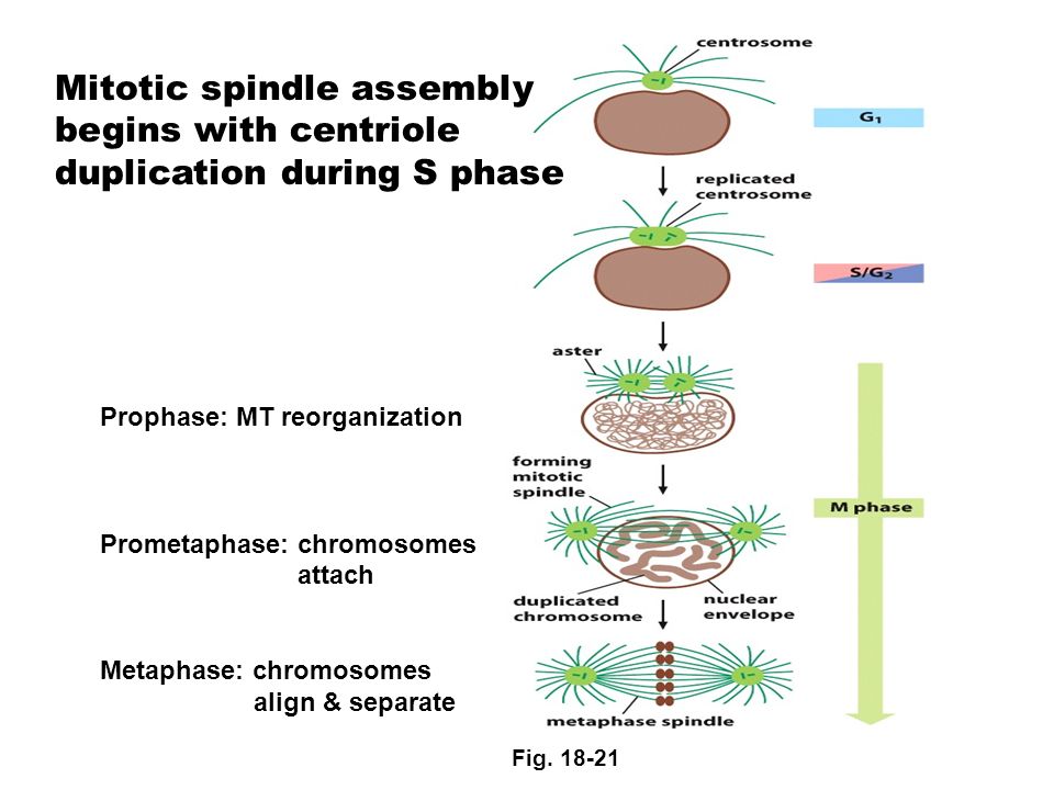
Prophase: MT reorganization Prometaphase: chromosomes attach Metaphase: chromosomes align & separate Mitotic spindle assembly begins with centriole duplication during S phase Fig. 18-21
33
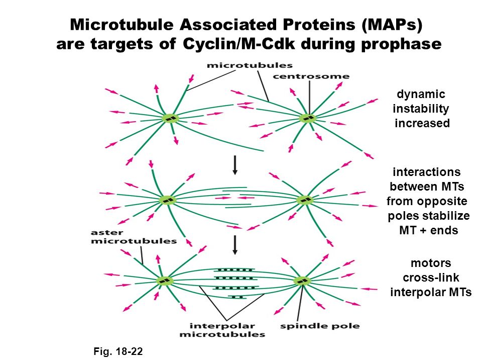
Microtubule Associated Proteins (MAPs) are targets of Cyclin/M-Cdk during prophase dynamic instability increased interactions between MTs from opposite poles stabilize MT + ends motors cross-link interpolar MTs Fig. 18-22
34
Sister chromatids bind kinetochore MTs from opposite poles during prometaphase, allowing separation during anaphase. kinetochore protein Fig. 18-24

35
Sister chromatids bind kinetochore MTs from opposite poles and separate during anaphase. Fig. 18-27 metaphase anaphase

36
Degradation of Securin and cleavage of Cohesin allow separation. Fig. 18-28

37
kinesin motors dynein motors kinesin: dynein: MT disassembly from + ends Two processes at play during sister chromatid segregation Fig. 18-29 pushes poles apart pulls poles apart

38
Kinetochore proteins bind microtubule sides, instead of ends, allowing assembly/disassembly at + end Alberts MBOC

39
Nuclear Envelope Assembly/Reassembly During Mitosis Fig. 18-30 targets of Cyclin/M-Cdk phosphatase

40
Nuclear division is followed by cytokinesis. Fig. 18-32

41
Cells lose adhesion to their substratum and change shape during cytokinesis. Fig. 18-33

42
Programmed cell death (apoptosis) is another mechanism for controlling cell numbers. apoptotic cells Fig. 18-35 Apoptosis needed for digit formation during embryonic development.

43
Apoptosis needed for tail loss during frog metamorphosis tadpole adult frog Fig. 18-36

44
Apoptosis also needed for neuron pruning during neural development Fig. 18-41

45
Necrosis Apoptosis Apoptosis in Animal Fig. 18-37 Necrosis is messy; apoptosis is neat.

46
Caspase enzymes mediate apoptosis. Fig. 18-38 cleave target proteins to kill cell allowing nuclease access to DNA

47
External signaling molecules can induce apoptosis in a developmental program. Fig. 18-40 eliminates self-recognizing T cells from immune system

48
Intrinsic signals can also induce apoptosis in response to DNA damage. Balance of pro-apoptotic and anti-apoptotic Bcl2 proteins determines outcome Fig. 18-39 pro-apoptotic DNA damage activates pro-apoptotic Bcl2 proteins

49
Survival factors increase expression of anti-apoptotic Bcl2 proteins Fig. 18-42

50
Growth Factors Control Cell Size (Growth) by Regulating Protein Synthesis & Degradation Fig. 18-43 Insulin is growth factor (Lab 4B) Cells remain in G 1 or G 0
 Cells remain in G 1 or G 0.")
51
Extracellular signal proteins can control cell growth, division and/or survival. Muscle cells: control of both cell division and growth Neurons: primarily growth Liver cells: primarily division

52
Chapter 19 Sexual Reproduction and the Power of Genetics Essential Cell Biology FOURTH EDITION Copyright © Garland Science 2014 Alberts Bray Hopkin Johnson Lewis Raff Roberts Walter

53
Fig. 19-4 Most multicellular organisms reproduce sexually. fertilization of egg by sperm

54
After fertilization, the diploid zygote then undergoes rounds of mitosis to generate a new multicellular adult. Fig. 19-5

55
Meiosis differs from mitosis in also having a reductive division. Fig. 19-6 reductive division non-reductive division non-reductive division

56
Physical Basis of reductive division: separation of chromosome homologues Fig. 19-7

57
Physical Basis of reductive division: separation of chromosome homologues Fig. 19-8

58
Fig. 19-9 Paired chromosome homologues after duplication (one from each parent) Synaptonemal Complex holds homologues together Cohesin holds sister chromatids together
 Synaptonemal Complex holds homologues together Cohesin holds sister chromatids together.")
Similar presentations


