Download presentation
Presentation is loading. Please wait.

1
Chromosomes, Mitosis, Meiosis
Cell cycle Chromosomes Cell Division (Mitosis, Meiosis) Csaba Bödör, Chapter 9, pages:
 Csaba Bödör, Chapter 9, pages:")
2
Introduction: DNA, chromatin, chromosomes
Prokaryotes: circular DNA (no histones) (Fig: 9-4, book) Eukaryotes: linear DNA + histones = chromatin Chromosome is the condensed form of chromatin Chromosomes are visible during cell division („transport” form of the genetic material) Chromosomes are not visible in non-dividing cells (but present !!!!, in extended form: chromatin) Role in chromosome packaging histon proteins and scaffolding proteins (help maintain chromosome structure)
 (Fig: 9-4, book) Eukaryotes: linear DNA + histones = chromatin. Chromosome is the condensed form of chromatin. Chromosomes are visible during cell division („transport form of the genetic material) Chromosomes are not visible in non-dividing cells (but present !!!!, in extended form: chromatin) Role in chromosome packaging histon proteins and scaffolding proteins (help maintain chromosome structure)")
3
Nucleosomes: 8 histones + DNA

5
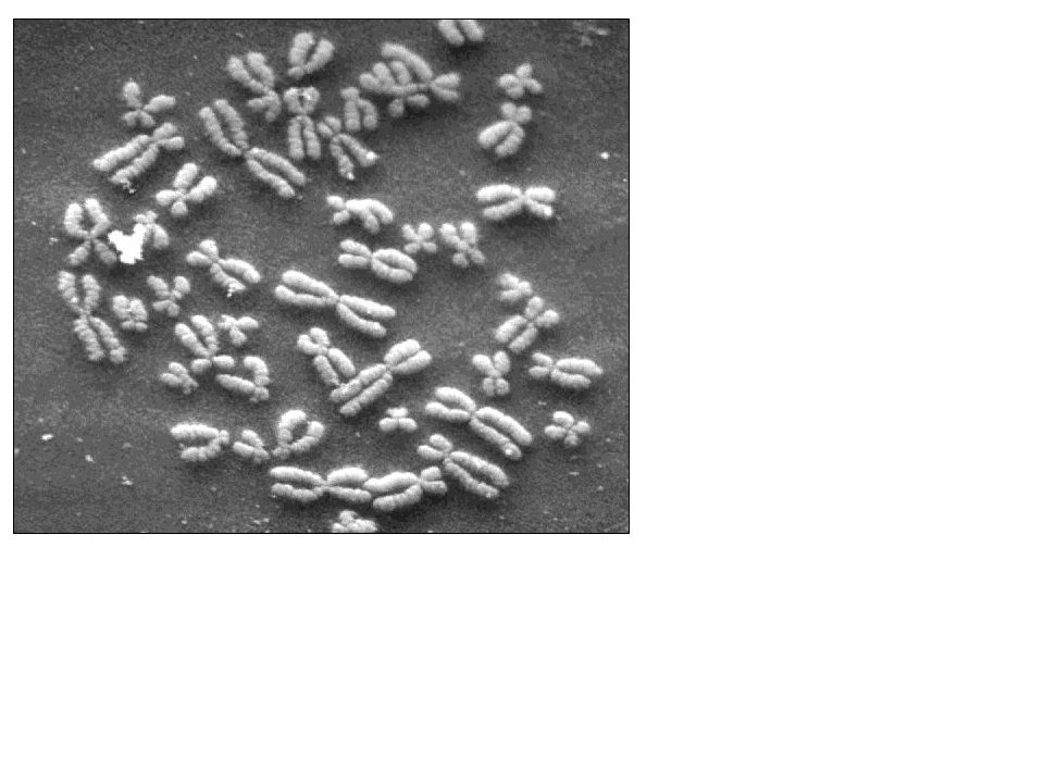
Chromosomes Number of chromosomes is different among species
In humans 46 chromosomes (23 paternal, 23 maternal) This is the human KARYOTYPE (chromosomal constitution) Structure of a duplicated chromosome (after replication) 2 sister chromatids ! with the same genetic material ! associated at the regions of centromeres ! kinetochores are attached to each centromere (they are binding sites for microtubules) before replication only one chromatid !!!!
 This is the human KARYOTYPE (chromosomal constitution) Structure of a duplicated chromosome (after replication) 2 sister chromatids ! with the same genetic material ! associated at the regions of centromeres ! kinetochores are attached to each centromere (they are binding sites for microtubules) before replication only one chromatid !!!!")
6
The cell cycle from beginning of one division to beginning of next division interphase and M phase (division) most of the cell’s life is spent in interphase (no division) IN INTERPHASE: G1 phase (first gap phase) normal metabolism, growth S phase (synthesis) DNA replication G2 phase (second gap phase) preparation for division IN M PHASE: Mitosis nuclear division (two nuclei produced) Cytokinesis division of the cell cytoplasm (two daughter cells are produced)
 IN INTERPHASE: G1 phase (first gap phase) normal metabolism, growth. S phase (synthesis) DNA replication. G2 phase (second gap phase) preparation for division. IN M PHASE: Mitosis nuclear division (two nuclei produced) Cytokinesis division of the cell cytoplasm (two daughter cells are produced)")
7
Regulation of the cell cycle
Is it possible to leave the cycle ? Yes, G0 phase: resting Is it controlled ? Yes, very strictly 3 important checkpoints checkpoint 1 First one in the G1 phase Is the DNA intact (OK) ? If not, first to correct it ! G0 phase Second checkpoint in G2 phase Is all DNA replicated ? Third checkpoint in M phase Are all chromosomes attached to the spindle ? (Molecules called cyclins and cyclin dependent kinases are important regulators of the cell cycle) checkpoint 3 checkpoint 2
 If not, first to correct it ! G0 phase. Second checkpoint in G2 phase Is all DNA replicated Third checkpoint in M phase Are all chromosomes attached to the spindle (Molecules called cyclins and cyclin dependent kinases are important regulators of the cell cycle) checkpoint 3. checkpoint 2.")
8
Interphase: S phase In synthesis (S) phase DNA replication takes place
Chromosomes are not visible in interphase (present in form of extended chromatin) centrioles are also duplicated !! centrioles (9 x 3) form the centrosome, and they are the MTOCs (microtubule organizing centers)
 centrioles are also duplicated !! centrioles (9 x 3) form the centrosome, and they are the MTOCs (microtubule organizing centers)")
9
Mitosis: Introduction
is the division of body cells, 2 genetically identical daughter cells are produced (with same number of chromosomes !!!) 46 in humans: 46 46 before replication chromosomes composed of one chromatid !!!! after replication chromosomes composed of two chromatids !!!! 1 chromatid = 1 DNA molecule G1 S G2 M Number of chromosomes 46 46 46 46 Number of chromatids 46 92 92 92 Number of DNA molecules 46 92 92 92 Number of centromeres 46 46 46 46
 46. in humans: before replication chromosomes composed of one chromatid !!!! after replication chromosomes composed of two chromatids !!!! 1 chromatid = 1 DNA molecule. G1. S. G2. M. Number of chromosomes Number of chromatids Number of DNA molecules Number of centromeres")
10
Mitosis: Prophase prophase is the first stage
chromosome condensation starts (packaging), condensin, ATP is needed chromosomes become visible (in microscope) nucleolus and nuclear envelope disappear centrioles move to the poles of the cell mitotic spindle formed between centrioles Microtubules (spindle fibers) kinetochores begin attaching to microtubules Kinetochores (proteins)
, condensin, ATP is needed. chromosomes become visible (in microscope) nucleolus and nuclear envelope disappear. centrioles move to the poles of the cell. mitotic spindle formed between centrioles. Microtubules (spindle fibers) kinetochores begin attaching to microtubules. Kinetochores (proteins)")
11
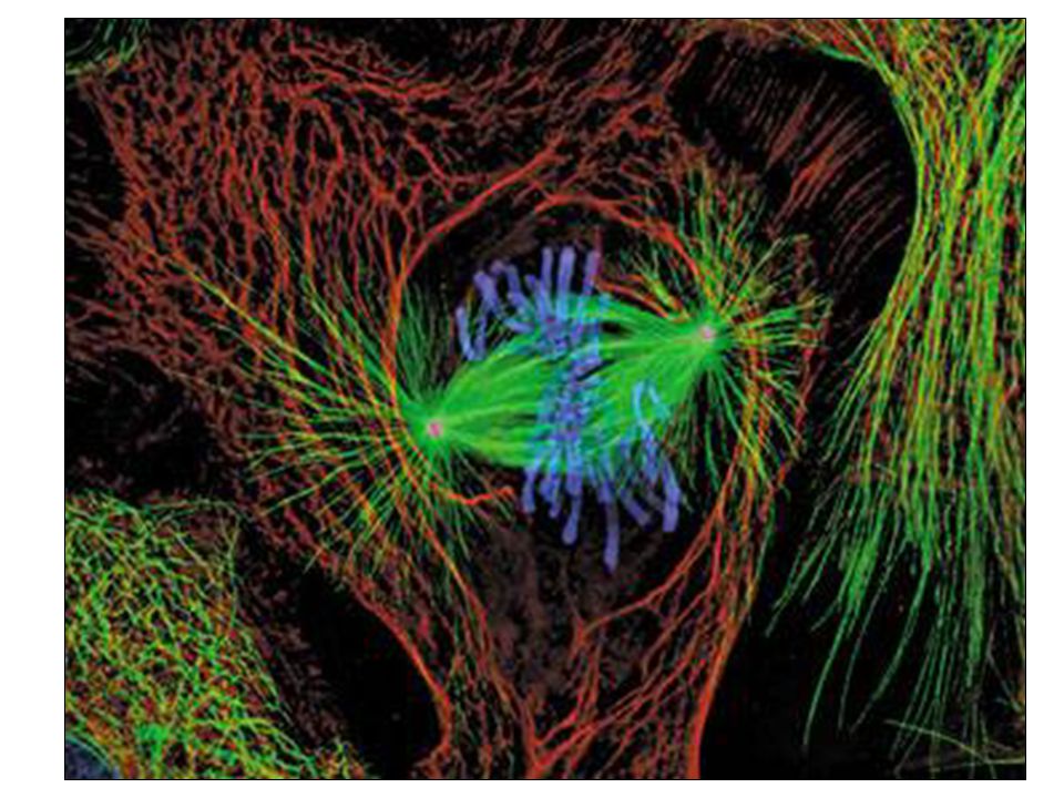
Mitosis: Metaphase metaphase is the second stage
chromosomes line up along the cell’s midplane chromosomes attached to the spindle fibers This is the best stage for chromosome analysis, most visible, organized, microtubules don’t touch the centrioles, they end in the pericentriolar material Microtubules (spindle fibers) Centrioles Kinetochores (proteins) Pericentriolar material Details on the next slide
 Centrioles. Kinetochores (proteins) Pericentriolar material. Details on the next slide.")
12
The mitotic spindle Polar microtubules Centrioles Asters
Kinetochores (proteins) Pericentriolar material Kinetochore microtubules midplane
 Pericentriolar material. Kinetochore microtubules. midplane.")
13
Mitosis: Metaphase chromosomes lined up along the cell’s midplane (or metaphase plate) centrioles: 9x3 structure of microtubules pericentriolar material: protein fibers pericentriolar material + centrioles form the centrosome mitotic spindle: centrosomes and further microtubules asters (astral microtubules): positioning of the cell pole kinetochore microtubules (or spindle fibers): separation of chromatids polar microtubules: push the poles apart Kinetochores (proteins) Kinetochore microtubules Centrioles Pericentriolar material Polar microtubules Asters midplane
: positioning of the cell pole. kinetochore microtubules (or spindle fibers): separation of chromatids. polar microtubules: push the poles apart. Kinetochores (proteins) Kinetochore microtubules. Centrioles. Pericentriolar material. Polar microtubules. Asters. midplane.")
14
Mitosis: Anaphase anaphase is the third stage
sister chromatids are separated at centromeres chromosomes move toward the opposite poles they use spindle fibers as tracks polar microtubules push the poles apart anaphase ends when chromosomes have arrived to the poles

16
Mitosis: Telophase telophase is the fourth stage
chromosomes arrived to the poles two new nuclei are formed new nuclear envelopes, nucleoli are reformed chromosomes start to decondense (uncoil) the spindle disappears CYTOKINESIS usually starts during the telophase and produces 2 daughter cells this is the actual division of the cytoplasm two genetically identical daughter cells organelles are distributed more or less equally between the two daughter cells (and mitochondria ?) Cleavage furrow
 the spindle disappears. CYTOKINESIS usually starts during the telophase and produces 2 daughter cells this is the actual division of the cytoplasm two genetically identical daughter cells organelles are distributed more or less equally between the two daughter cells (and mitochondria ) Cleavage furrow.")
19
Sexual and asexual reproduction
There are so called somatic (body) cells in our organism, they contain chromosomes (23 pairs) They are diploid (2n) They contain 2 sets of chromosomes (1 set = 23 chromosomes (in humans)) (maternal and paternal) There are some cells in our reproductive organs, called gametes, they contain 23 chromosomes They are haploid (n) They contain 1 set of chromosomes (either maternal and or paternal) Gametes (cells needed for sexual reproduction) are sperms and egg cells „n” refers to haploid chromosome number, n=23 in humans gametes fuse to form a zygote >>> This is the basis of sexual reproduction In asexual reproduction >>> there are no gametes, basis is mitosis Somatic cells are generated by mitosis (2n>2n) Gametes are generated by meiosis (2n>n)
 cells in our organism, they contain 46 chromosomes (23 pairs) They are diploid (2n) They contain 2 sets of chromosomes (1 set = 23 chromosomes (in humans)) (maternal and paternal) There are some cells in our reproductive organs, called gametes, they contain 23 chromosomes. They are haploid (n) They contain 1 set of chromosomes (either maternal and or paternal) Gametes (cells needed for sexual reproduction) are sperms and egg cells. „n refers to haploid chromosome number, n=23 in humans. gametes fuse to form a zygote >>> This is the basis of sexual reproduction. In asexual reproduction >>> there are no gametes, basis is mitosis. Somatic cells are generated by mitosis (2n>2n) Gametes are generated by meiosis (2n>n)")
20
Homologous pairs Each chromosome has its pair !!
Maternal chromosomes Paternal chromosomes Each chromosome has its pair !! There are 23 homologous pairs (similar size, shape and genetic material)
")
21
Meiosis During meiosis, gametes (haploid cells) are produced Meiosis reduces the chromosome number by half („make smaller”) A single diploid cell (2n) produces four haploid cells (n) 23 23 23 46 23 23 23 During meiosis two cell divisions takes place, but the DNA is duplicated only once, that is why is the chromosome number reduced after meiosis Phases of meiosis: Meiosis I. (prophase I, metaphase I, anaphase I, telophase I.) Meiosis II. (prophase II, metaphase II, anaphase II, telophase II.) (between meiosis I and meiosis II, there is a short phase called interkinesis (similar to the interphase), but there in no DNA duplication !!!!!!!
 produces four haploid cells (n) During meiosis two cell divisions takes place, but the DNA is duplicated only once, that is why is the chromosome number reduced after meiosis. Phases of meiosis: Meiosis I. (prophase I, metaphase I, anaphase I, telophase I.) Meiosis II. (prophase II, metaphase II, anaphase II, telophase II.) (between meiosis I and meiosis II, there is a short phase called interkinesis (similar to the interphase), but there in no DNA duplication !!!!!!!")
22
Meiosis I: Prophase I Meiosis I (or first meiotic division)
Prophase I is a very specific phase of the whole meiotic division >>>> homologous chromosomes are physically attached (synapsis) The process is called synapsis, the structure is the synaptonemal complex Sister chromatids Sister chromatids The place where the crossing over occurs is called chiasma (ta) In prophase I of meiosis there is an exchange of genetic material between homologous chromosomes, so new combinations of parental genes are created !!!! This is the process of genetic recombination Homologous pair (tetrade, bivalent)
 The process is called synapsis, the structure is the synaptonemal complex. Sister chromatids. Sister chromatids. The place where the crossing over occurs is called chiasma (ta) In prophase I of meiosis there is an exchange of genetic material between homologous chromosomes, so new combinations of parental genes are created !!!! This is the process of genetic recombination. Homologous pair (tetrade, bivalent)")
23
Meiosis I: Metaphase I The homologous pairs are aligned along the midplane In mitosis there is no connection between the members of the homologous pairs

24
Meiosis I: Anaphase I The homologous pairs are separated (not sister chromatids !!!!!) and are distributed to the two poles In mitosis the sister chromatids were separated In TELOPHASE I two haploid cells with duplicated chromosomes are produced

25
Meiosis II: The same phases like in meiosis I (P, M, A, T)
In meiosis II the sister chromatids are separated (like in mitosis) Diploid cell (2n), duplicated chromosomes DNA content: 4C (after replication) Meiosis I. Haploid cells (n), duplicated chromosomes DNA content: 2C Meiosis II. Haploid cells (n), chromosomes with one chromatid DNA content: C From 1 diploid cell, 4 genetically different haploid daughter cells are produced !!
 Diploid cell (2n), duplicated chromosomes DNA content: 4C (after replication) Meiosis I. Haploid cells (n), duplicated chromosomes DNA content: 2C. Meiosis II. Haploid cells (n), chromosomes with one chromatid DNA content: C. From 1 diploid cell, 4 genetically different haploid daughter cells are produced !!")
26
MEIOSIS versus MITOSIS
2n 4C 2n 4C Meiosis I. 2n 2C n 2C Mitosis Meiosis II. n C

27
MEIOSIS versus MITOSIS
Two genetically identical daughter cells, diploid (2n) one division, one replication > chromosome number the same chromosomes arranged in a line along the midplane of the cell no crossing over in prophase sister chromatids separate during anaphase division of somatic (body) cells MEIOSIS 4 genetically different daughter cells, haploid (n) 2 consecutive divisions, only 1 replication > chromosome number reduced homologous pairs attached to each other crossing over occurs in prophase I. members of the homologous pairs separate in anaphase I during meiosis, gametes are created sister chromatids separate in anaphase II.
 one division, one replication > chromosome number the same. chromosomes arranged in a line along the midplane of the cell. no crossing over in prophase. sister chromatids separate during anaphase. division of somatic (body) cells. MEIOSIS. 4 genetically different daughter cells, haploid (n) 2 consecutive divisions, only 1 replication > chromosome number reduced. homologous pairs attached to each other. crossing over occurs in prophase I. members of the homologous pairs separate in anaphase I. during meiosis, gametes are created. sister chromatids separate in anaphase II.")
Similar presentations


 eukaryotic cell of a sexually reproducing organism that result in four haploid (N)>")


 DIPLOID (2N) The condition of having two sets of chromosomes per nucleus The condition.>")


. Mitosis Two identical daughter cells Interphase Cell growth, preparing for cell division Prophase, Metaphase, Anaphase, Telophase.>")