Download presentation
Presentation is loading. Please wait.

1
Vascular diseases: Varicose veins, DVT and Aneurysms CVS6
Dr Hisham Alkhalidi

2
Lecture 6 Diseases of arteries and veins:
Pathology of varicose veins, thrombophlebitis and deep vein thrombosis. Definition of aneurysm, types and aetiology of aneurysms.

3
Varicose Veins Legs, testis,

4
Varicose Veins Abnormally dilated, tortuous veins produced by prolonged increase in intraluminal pressure and loss of vessel wall support The superficial veins of the lower leg, venous pressures in these sites can be markedly elevated 10% to 20% of adult males 25% to 33% of adult females Thrombosis is common but not clinically significant Valve incompetence rule Haemorrhoides and esophageal varices

5
Varicose Veins Increased risk: Obesity Hereditary Proximal thrombus
Proximal compression (e.g. tumor) legs are dependent for long periods Higher incidence in women (pregnancy)
 legs are dependent for long periods. Higher incidence in women (pregnancy)")
6
Varicose Veins Complications Stasis dermatitis Delay healing
Stasis, edema, trophic skin Varicose ulcers

7
Stasis dermatitis Varicose ulcer

9
DVT The deep leg veins account for more than 90% of cases of phlebothrombosis Other sites include: The periprostatic venous plexus in males The pelvic venous plexus in females The large veins in the skull and the dural sinuses (especially in the setting of infection or inflammation) thrombophlebitis and phelbothrombosis the two terms are largely interchangeable designations for venous thrombosis and inflammation. Portal tracts Popliteal, femoral, and iliac veins Superficial saphenous vein Not clinically significant
 thrombophlebitis and phelbothrombosis. the two terms are largely interchangeable designations for venous thrombosis and inflammation. Portal tracts. Popliteal, femoral, and iliac veins. Superficial saphenous vein. Not clinically significant.")
10
DVT Predisposing factors: Congestive heart failure Neoplasia Pregnancy
Obesity Postoperative state Prolonged bed rest Genetic hypercoagulability syndromes

11
DVT Trousseau sign: In patients with cancer, particularly adenocarcinomas, hypercoagulability occurs as a paraneoplastic syndrome related to tumor elaboration of procoagulant factors In this setting, venous thromboses classically appear in one site, disappear, and then reoccur in other veins, so-called migratory thrombophlebitis (Trousseau sign) Association with NBTE
 Association with NBTE.")
12
DVT 50% clinically silent. Local manifestations: Distal edema Cyanosis
Superficial vein dilation heat, tenderness, redness, swelling and pain Sometimes, the first manifestation of thrombophlebitis is a pulmonary embolus Depending on the size and number of emboli, the outcome can range from no symptoms at all to death

14
localized abnormal dilation of a blood vessel or the heart
ANEURYSMS localized abnormal dilation of a blood vessel or the heart

15
ANEURYSMS

16
Thrombosis

17
ANEURYSMS The two most important disorders that predispose to aortic aneurysms are: atherosclerosis (abdominal aorta) hypertension (ascending aorta) Other causes of aneurysms: Trauma Congenital (berry aneurysms in the circle of Willis) Infections (mycotic aneurysms, syphilis) Vasculitides cystic medial degeneration of the arterial media was from the two most common cause on this slide
 Other causes of aneurysms: Trauma. Congenital (berry aneurysms in the circle of Willis) Infections (mycotic aneurysms, syphilis) Vasculitides. cystic medial degeneration of the arterial media was from the two most common cause on this slide.")
18
ANEURYSMS Mycotic aneurysm may originate either from:
embolization and arrest of a septic embolus at some point within a vessel, usually as a complication of infective endocarditis an extension of an adjacent suppurative process circulating organisms directly infecting the arterial wall

19
ANEURYSMS Complications Rupture Hemorrhage
Occlusion of proximal vessels Embolism

20
ANEURYSMS abdominal aortic aneurysm (AAA):
More in men and rarely develops < 50 years Abdominal aorta is the main location but the common iliac arteries, the arch and descending parts of the thoracic aorta can be involved Below the renal arteries and above the bifurcation of the aorta Atherosclerosis, rule of genetics or HTN Additional reading: Inflammatory AAAs are characterized by dense periaortic fibrosis containing abundant lymphoplasmacytic infiltrate with many macrophages and often giant cells. Their cause is uncertain. Mycotic AAAs are atherosclerotic lesions infected by lodging of circulating microorganisms in the wall, particularly in the setting of bacteremia from a primary Salmonella gastroenteritis. In such cases, suppuration further destroys the media, potentiating rapid dilation and rupture.

21
ANEURYSMS abdominal aortic aneurysm (AAA): Risk of rupture:
11% per year for aneurysms between 5.0 and 6 cm in diameter Operative mortality for unruptured aneurysms is approximately 5%, whereas emergency surgery after rupture carries a mortality rate of more than 50% As a reflection of the systemic nature of ATH and its complications, patients with AAAs are also at significantly increased risk for myocardial infarction and stroke.

22
ANEURYSMS SYPHILITIC (LUETIC) ANEURYSMS:
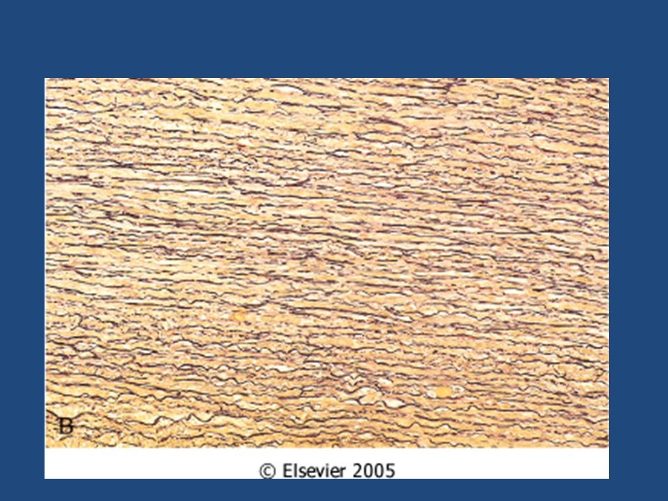
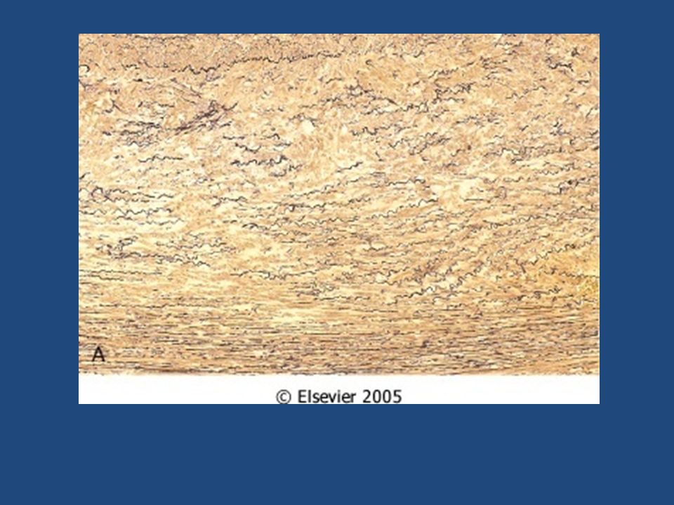
The obliterative endarteritis of the the vasa vasorum of the thoracic aorta can lead to aneurysmal dilation that can include the aortic annulus Ascending aorta and arch May cause aortic valve ring dilation -> valvular insufficiency -> ventricular wall hypertrophy, sometimes to 1000 gm "cor bovinum" (cow's heart) Tertiary stage of syphilis Causes ischemic injury of the aortic media, with patchy loss of the medial elastic fibres and muscle cells followed by inflammation and scarring. With destruction of the media, the aorta loses its elastic recoil and may become dilated, producing a syphilitic aneurysm. Contraction of fibrous scars may lead to wrinkling of intervening segments of aortic intima, noted grossly as "tree-barking."
 Tertiary stage of syphilis. Causes ischemic injury of the aortic media, with patchy loss of the medial elastic fibres and muscle cells followed by inflammation and scarring. With destruction of the media, the aorta loses its elastic recoil and may become dilated, producing a syphilitic aneurysm. Contraction of fibrous scars may lead to wrinkling of intervening segments of aortic intima, noted grossly as tree-barking.")
23
SYPHILITIC (LUETIC) ANEURYSMS
Widening of the commissures between the cusps, and turbulence-induced thickening and rolling of the free margins. Talk about complications of compression, cough, SOB, Swallowing, bone…, rupture Most patients with syphilitic aneurysms die of heart failure induced by aortic valvular incompetence

25
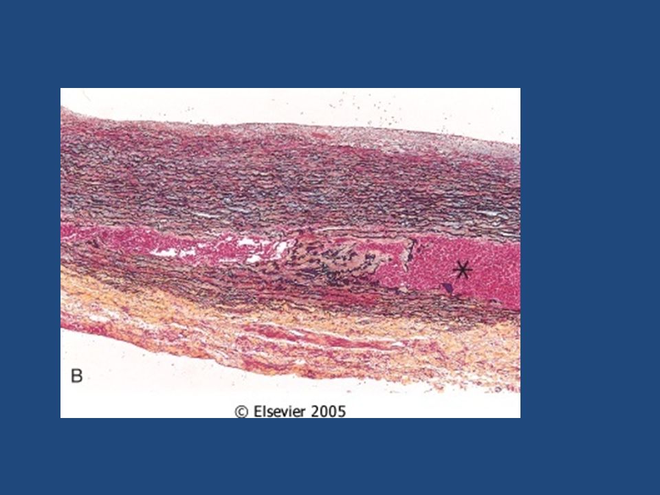
Dissecting hematoma defective fibrillin production in Marfan disease affects elastic tissue synthesis;

30
Dissecting hematoma Causes Hypertension Connective tissue defects
Cannulation or other trauma Preganancy Hypertension years Rarely, for unknown reasons, dissection of the aorta or other branches, including the coronary arteries, occurs during or after pregnancy. unusual in the presence of substantial atherosclerosis or other causes of medial scarring,

31
Types Type A is associated with serious complications
Type A COMMON and Dangerous Type B distal to subclavian

32
Dissecting hematoma Clinical picture
sudden onset of excruciating pain: usually beginning in the anterior chest radiating to the back between the scapulae moving downward as the dissection progresses Previously, aortic dissection was typically fatal, but the prognosis has markedly improved. Rapid diagnosis and institution of intensive antihypertensive therapy, coupled with surgical procedures involving plication of the aorta permits survival of 65% to 75% of patients.

33
Dissecting hematoma Clinical picture
cardiac tamponade aortic insufficiency myocardial infarction extension of the dissection into the great arteries of the neck or into the coronary, renal, mesenteric, or iliac arteries causing critical vascular obstruction; compression of spinal arteries may cause transverse myelitis

Similar presentations


, awareness of heartbeat.>")


>")


