Download presentation
Presentation is loading. Please wait.

1
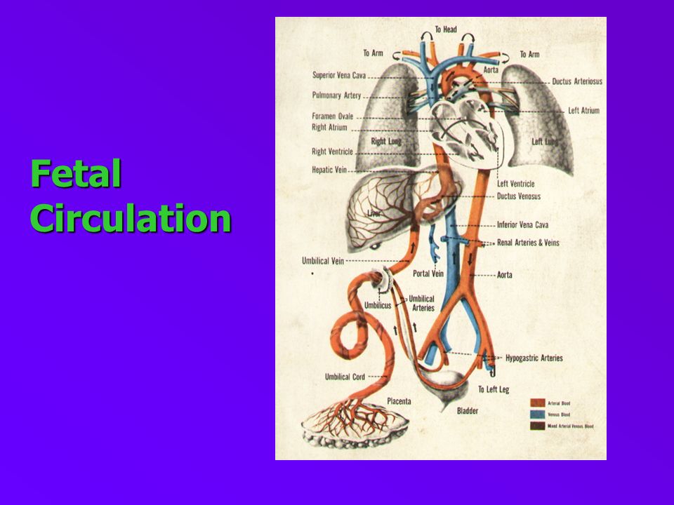
Fetal Circulation

3
Salient Features of Fetal Growth Placenta is very active.Placenta is very active. Liver and lungs are passive.Liver and lungs are passive. Head is growing faster.Head is growing faster.

4
Hemodynamics of Fetal Circulation Venous and arterial blood mix up in atria Venous and arterial blood mix up in atria The venacava carries mixed blood to the right atrium bypassing liver and lungs. The venacava carries mixed blood to the right atrium bypassing liver and lungs. Aorta carries mixed blood back to placenta Aorta carries mixed blood back to placenta Blood circulation in the fetus is never as saturated with oxygen as that blood circulating through the adult. That is, difference in oxygen tension between blood of the umbilical arteries is much less than that of adult venous and arterial blood. Blood circulation in the fetus is never as saturated with oxygen as that blood circulating through the adult. That is, difference in oxygen tension between blood of the umbilical arteries is much less than that of adult venous and arterial blood.

5
Fetal Circulation

6
The arterial blood flows to the fetus through the umbilical vein. The arterial blood flows to the fetus through the umbilical vein. It passes to the undersurface of liver and separates in to two branches. It passes to the undersurface of liver and separates in to two branches. Lager is joined by the portal vein and enters the right lobe of the liver. Lager is joined by the portal vein and enters the right lobe of the liver. The other continued upwards as ductus venosus and joins the inferior venacava. The other continued upwards as ductus venosus and joins the inferior venacava. In the inferior venacava blood carried by the ductus venosus and hepatic veins becomes mixed with blood returning from the lower extremities, so it is a mixture of oxygenated and de oxygenated blood In the inferior venacava blood carried by the ductus venosus and hepatic veins becomes mixed with blood returning from the lower extremities, so it is a mixture of oxygenated and de oxygenated blood Course of Fetal Circulation Pre-delivery phase

7
It enters the right atrium and is directed through the foramen ovale into the left atrium. In the left atrium, it mixes with a small quantity of blood returning from the lungs through the pulmonary veins. It enters the right atrium and is directed through the foramen ovale into the left atrium. In the left atrium, it mixes with a small quantity of blood returning from the lungs through the pulmonary veins. From the left atrium, blood passes into the left ventricle, and from the left ventricle into the aorta. From the aortal it is distributed almost entirely to the head and upper extremities – a small quantity of blood continuing into the descending aorta. From the left atrium, blood passes into the left ventricle, and from the left ventricle into the aorta. From the aortal it is distributed almost entirely to the head and upper extremities – a small quantity of blood continuing into the descending aorta. Continued

8
After circulation in the head and upper extremities, blood is returned by the superior vena cava to the right atrium, where it mixes with a small portion of that blood entering from the inferior vena cava. After circulation in the head and upper extremities, blood is returned by the superior vena cava to the right atrium, where it mixes with a small portion of that blood entering from the inferior vena cava. From the right atrium it descends into the right ventricle and passes into the main pulmonary artery. From the right atrium it descends into the right ventricle and passes into the main pulmonary artery. From the pulmonary artery, most of the blood passes trough the ductus arterioss into the aorta, where it mixes with blood from the left ventricle. From the pulmonary artery, most of the blood passes trough the ductus arterioss into the aorta, where it mixes with blood from the left ventricle. Continued

9
Some of the blood with main pulmonary artery is distributed to the lungs by the right and left pulmonary arteries. The pulmonary veins returns this blood the the left atrium. Some of the blood with main pulmonary artery is distributed to the lungs by the right and left pulmonary arteries. The pulmonary veins returns this blood the the left atrium. The blood passing from the main pulmonary artery through the ductus arteries descends through the aorta and is in part distributed to the lower extremities and viscera of the abdomen and pelvis. The greater amount is conveyed by the hypogastric arteries to the umbilical arteries The umbilical arteries return the blood to the placenta. The blood passing from the main pulmonary artery through the ductus arteries descends through the aorta and is in part distributed to the lower extremities and viscera of the abdomen and pelvis. The greater amount is conveyed by the hypogastric arteries to the umbilical arteries The umbilical arteries return the blood to the placenta. Continued

10
Intermediate Phase Immediately after delivery, the baby begins to breathe, and pulmonary circulation changes, An increased quantity of blood is pumped into the pulmonary arteries by the right ventricle, and a smaller quantity passes through the ductus arteriosus. The ductus arteriosus begins to atrophy, eventually becoming the ligamentum arteriosum Immediately after delivery, the baby begins to breathe, and pulmonary circulation changes, An increased quantity of blood is pumped into the pulmonary arteries by the right ventricle, and a smaller quantity passes through the ductus arteriosus. The ductus arteriosus begins to atrophy, eventually becoming the ligamentum arteriosum. Pulmonary circulation increases, and more blood is returned from the lungs to the left atrium. Pressure rises in the left atrium, causing the foramen ovale to close. Placental circulation ceases to function as soon as the umbilical cord is tied. The ends of the hypogastric arteries atrophy, and these vessels become the hypo-gastric ligaments. Pulmonary circulation increases, and more blood is returned from the lungs to the left atrium. Pressure rises in the left atrium, causing the foramen ovale to close. Placental circulation ceases to function as soon as the umbilical cord is tied. The ends of the hypogastric arteries atrophy, and these vessels become the hypo-gastric ligaments.

11
Adult Phase Closure of ductus arterosus in early postnatal period due to reflex action secondary to increased oxygen tension and the interaction of prosta glandin Closure of ductus arterosus in early postnatal period due to reflex action secondary to increased oxygen tension and the interaction of prosta glandin PDA machinery murmur Obliteration of foramen ovale – 6 to 8 weeks Obliteration of foramen ovale – 6 to 8 weeks Patent foramen ovale with few or no symptoms Occluded umblical vein – ligamentum teres Occluded umblical vein – ligamentum teres Obliterated ductus venosus – ligamentum venosum Obliterated ductus venosus – ligamentum venosum

12
Hemodynamics of Normal Adult Venous and arterial blood no longer mix in the atria Venous and arterial blood no longer mix in the atria The venacava carries only deoxygenated blood into the right atrium and right ventricle which is pumped to the pulmonary arteries and finally to the pulmonary capillaries The venacava carries only deoxygenated blood into the right atrium and right ventricle which is pumped to the pulmonary arteries and finally to the pulmonary capillaries Aorta carries only oxygenated blood from the left heart via the pulmonary veins for distribution to the rest of the body. Aorta carries only oxygenated blood from the left heart via the pulmonary veins for distribution to the rest of the body.

13
Thank You

Similar presentations

RBC 2)WBC 3)Platelet.>")


 starts at the beginning of the third week. Blood vessels first start to develop in the.>")
