Download presentation
Presentation is loading. Please wait.

1
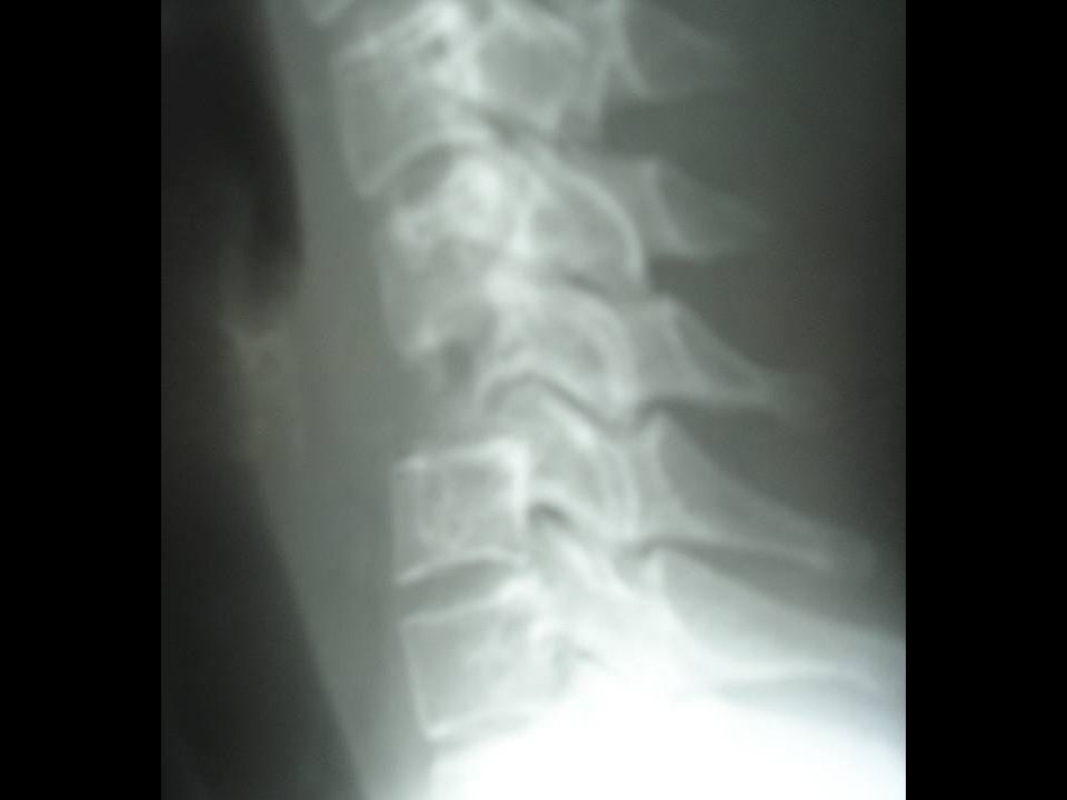
Osteosarcoma Clinical and Imaging 5% of primary malignant spinal tumours 4th decade (older than long bone) Associated with Paget’s, DXRT Mixed lytic / sclerotic appearance Aggressive, soft tissue extension Osteoid mineralisation T1WT2W T1W CT Differential diagnosis Ewings Chondrosarcoma Metastasis Osteoblastoma Lymphoma
 Associated with Paget’s, DXRT Mixed lytic / sclerotic appearance Aggressive, soft tissue extension Osteoid mineralisation T1WT2W T1W CT Differential diagnosis Ewings Chondrosarcoma Metastasis Osteoblastoma Lymphoma")
2
SBTR 4346, AB 22M 7/12 Hx LBP 2/12 Hx right leg / foot pain and weakness more recently urinary problems

3
4346, Ant Bishop, 22M

4
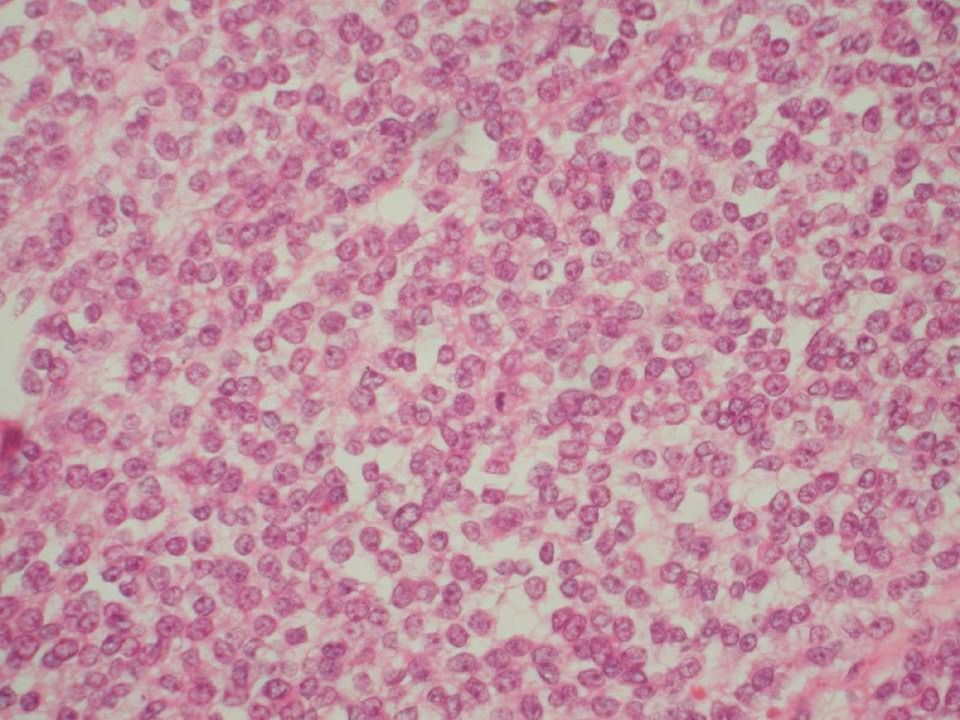
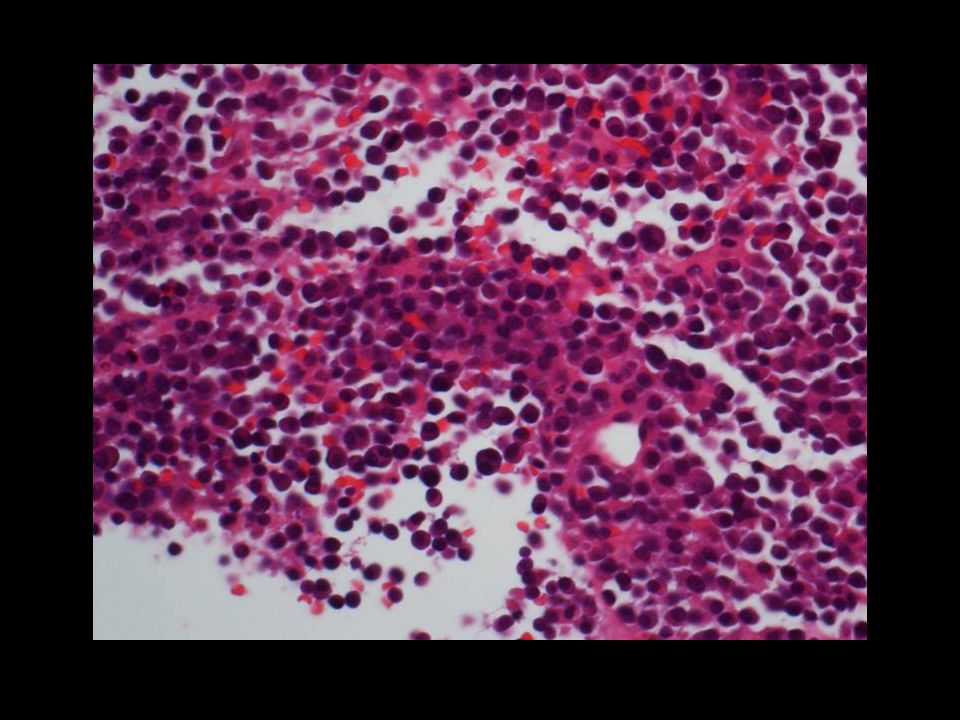
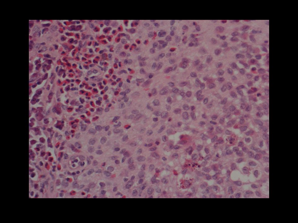
Ewing’s sarcoma Bone or soft tissue Now accepted to be primitive neurectodermal tumour Includes Ewing, PNET, peripheral neuroepithelioma, Askin tumour t (11;22)(q24;q12) and other rearrangements
(q24;q12) and other rearrangements")
6
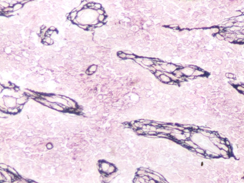
PAS

10
Chimaeric protein Control sequence Control sequence Overexpression of target gene Fusion gene Translocation

11
ES Cytogenetics t(11,22)(q24;q12) (95%)EWS/FLI1 t(21,22)(q22;q12) (5%)EWS/ERG t(7,22)(p22;q12)EWS/ETV1 t(17,22)(q21;q12)EWS/EIAF t(2,22)(q33;q12)EWS/FEV inv(22)(q12;q12)EWS/ZSG t(16,21)(p11;q22)FUS/ERG
(q24;q12) (95%)EWS/FLI1 t(21,22)(q22;q12) (5%)EWS/ERG t(7,22)(p22;q12)EWS/ETV1 t(17,22)(q21;q12)EWS/EIAF t(2,22)(q33;q12)EWS/FEV inv(22)(q12;q12)EWS/ZSG t(16,21)(p11;q22)FUS/ERG")
12
Ewing’s sarcoma Clinical and Imaging Usually 10 – 30yrs >50% metastases at presentation Metastatic involvement more common than primary Permeative pattern of bone destruction Large soft tissue mass and infiltration No matrix mineralisation but reactive sclerosis MR – inhomogeneous, haemorrhage & necrosis T1W T2WGRE Differential diagnosis Osteosarcoma Osteoblastoma Lymphoma Langerhans

13
30F, LBP

14
T1W T2W

26
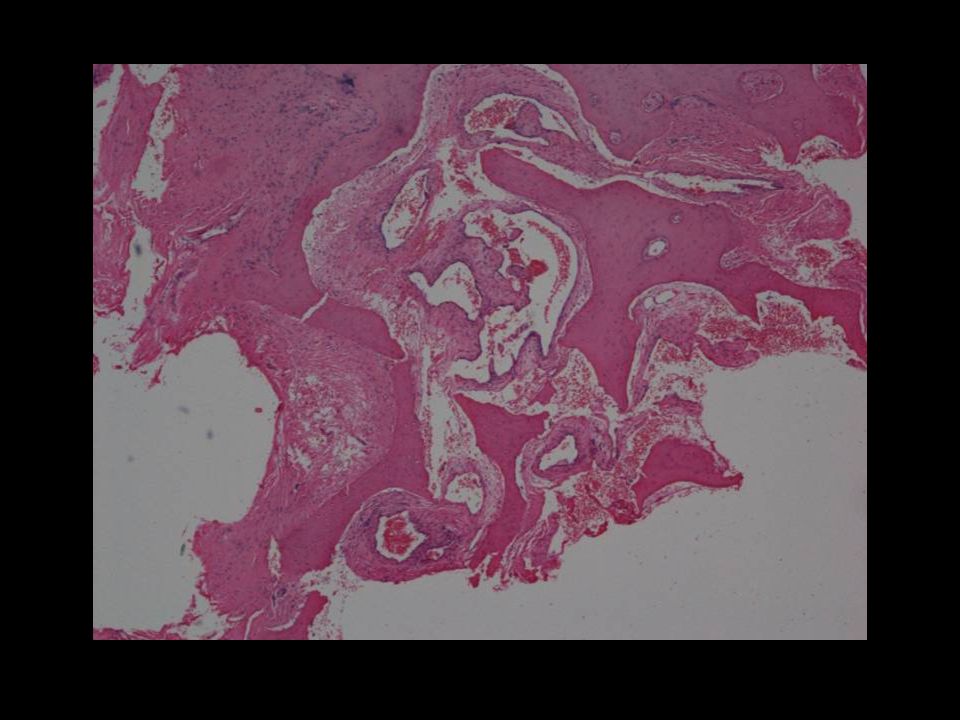
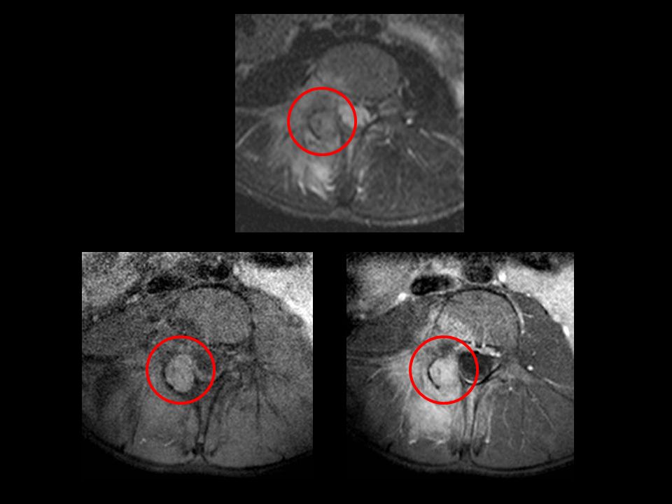
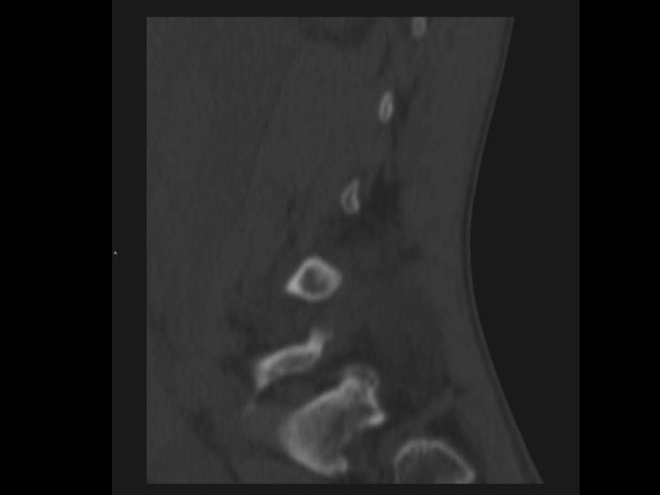
Giant Cell Tumour Clinical and Imaging >60% of primary benign sacral tumours 3rd – 5 th decades, 2F:1M Locally aggressive, 12-50% recurrence rate Lytic, expansile, absent matrix, +/- cortical breakthrough CT / MR may show fluid-fluid levels MR – low signal on T2W due to collagen, high cellularity and haemosiderin from haemorrhage Can undergo sarcomatous degeneration Differential diagnosis Expansile metastasis Chordoma Myeloma Aneurysmal bone cyst Osteoblastoma Brown tumour of hyperparathyoidism

27
DH, SBTR 4513 Mid thoracic pain

30
Myeloma Clinical Proliferation of malignant plasma cells Most common primary bone malignancy Most common in 6 th and 7 th decades Plasmacytoma usually precedes myeloma Differential diagnosis Metastases Lymphoma Sarcoma Chordoma GCT

31
Patterns on T1W - Myeloma Baur-Melnyk. Role of MRI in multiple myeloma. EJR 2005; 55:56 <20% plasma cells 20 – 50% plasma cells >50% plasma cells NormalInfiltrationReplacement

32
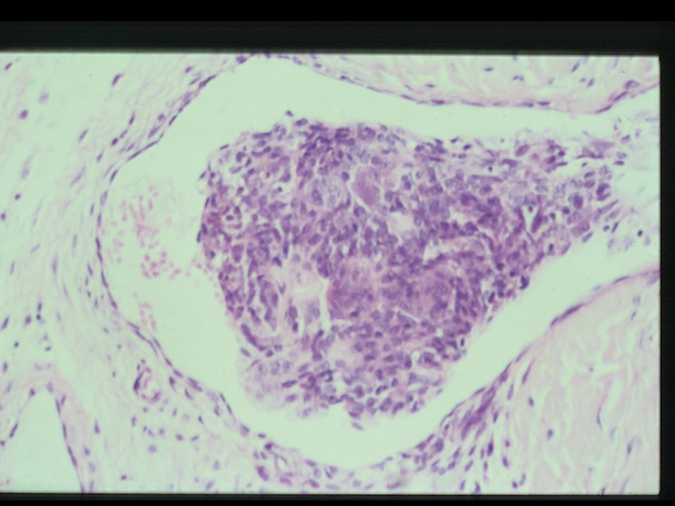
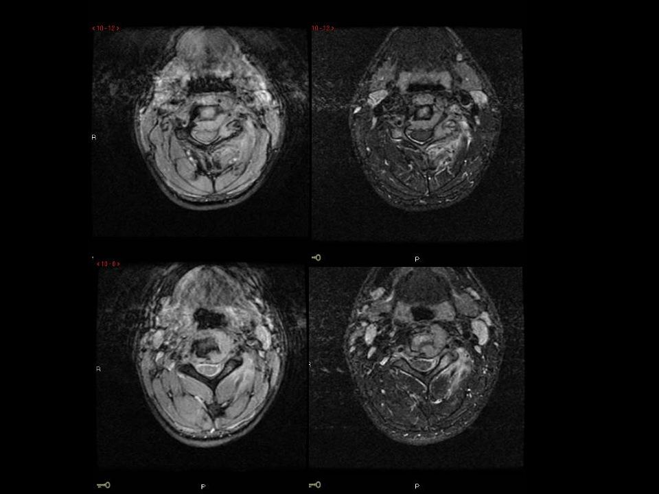
MC, 1907806113 30F Left neck pain Prominent ECA / pulsatile mass ?carotid body tumour

38
Eosinophilic Granuloma 5 – 10yrs 15% axial skeleton Can be asymptomatic Vertebral body Multifocal in 10% Differential Ewing’s Metastatic neuroblastoma ABC

39
60F, LBP Haemangioma

42
56F, right L5 sciatica

43
Pagets – later phase

44
Paget’s disease of bone increased bone turnover osteoclastic and osteoblastic activity age > 40 M>F 3% of routine autopsies aetiology unknown ? viral racial predilection monostotic or polyostotic raised alkaline phosphatase complications fracture deformity and sarcoma

47
Lolge S. Isolated solitary vertebral body tuberculosis. Clin Rad 2003 22F, Indian Differential diagnosis Sarcoma Eosinophilic granuloma GCT

48
Summary: Imaging In children / adults, metastases most frequent bone tumour. Patient age, location and relative frequency important In sacrum, Paget’s, chordoma and GCT Scintigraphy mainly for staging metastatic disease CT - optimal imaging technique for osteoid osteoma CT - bony detail, mineralisation, cortical shell & sequestra CT - often complementary to MRI. MRI - imaging method of choice.

51
SBTR 4319 Wal Miller chondrosarcoma grade 3

52
GJ, SBTR 4530 LBP, Osteoblastoma

Similar presentations


 – A NEW APPROCH AND OUR EXPERIENCE Kamenetsky Natalya (1), Rachmilewitz Eliezer.>")


>")


 - Genetic factors may play a role (p53 and RB mutations) - Bone infarcts, trauma, osteomyelitis,>")


 normal cell of origin Most are classified.>")

 October 29, 2008.>")


