Download presentation
Presentation is loading. Please wait.

1
Xray Rounds - A Hole in the Bone Robbie N Drummond October 31, 2002

2
Overview Hole found on xray incidental vs presenting symptom metastases, benign lesions, malignancies : some basic criteria the impending fracture

3
What do we do when a test we order brings up an incidental finding Diagnosis made my combination of primary care physician, radiologist, interventional radiologist, histopathologist, oncologist and orthopod no definitive pathognomonic findings for specific lesions our role to initiate diagnosis and slant treatment

4
Is the Lesion infective or neoplastic? Is the Lesion benign or malignant? Is it a primary or secondary lesion? Is the tumour creating or destroying bone? Is the cortex of the bone intact, broken or eroded

6
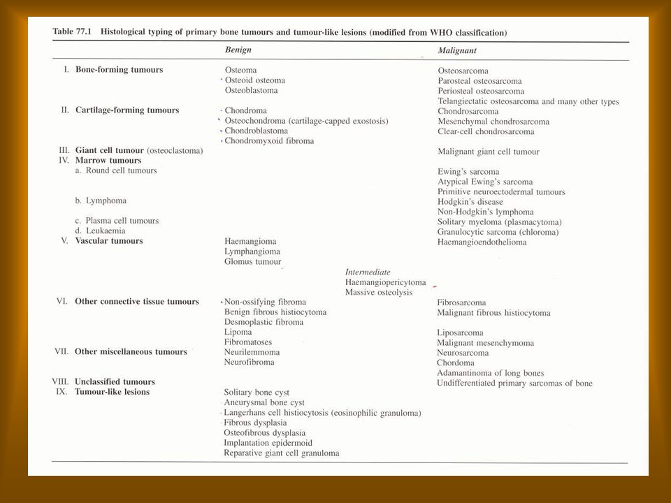
Five basic presentations of hole in bone 1 benign bone tumour 2 malignant bone tumour 3 metasastases 4 non-tumour 5 infection

7
Age Osteosarcoma and most malignant tumours are tumours of child hood Any invasive lesion < 40 Sarcoma any invasive lesion > 40 Metastasis

8
Location of Common Tumours

9
Benign Tumours Intact cortex usually solitary enlarges by expansion and pressure... Slowly the margin is sharp geographic narrow zone of transition if part of lesion looks benign whole lesion usually is periosteum not affected

10
Benign Tumour - Chondromyxoid Fibroma

11
Chondromyxoid Fibroma Uncommon benign tumour found in proximal tibial metaphyses sharply marginated lytic zone of destruction sclerotic rim of bone

12
Malignant Tumours Moth -eaten leading to permeative pattern wide zone of transition ill-defined lucencies small ill-defined lesions periosteum involved often soft-tissue involvement

13
OsteoSarcoma

14
Third most common malignancy found in children 2,500 new cases a year in USA metaphyses usually in femur proximal tibia can develop in any bone at any age mixed sclerotic and lytic lesion periosteal and soft tissue changes almost always solitary

15
X-ray Findings Sclerosis visible as a cloudy density variable pattern permeative moth-eaten pattern often periosteal involvement as in onion-skin change of Ewings Tumour

16
Osteomyelitis

17
Metastases 2,000,000 new cancers a year in USA half metastasize to bone only 8,000 new cases of primary bone cancer a year often metastasis is first presentation of cancer 50 % of bone gone before found on xray hallmark multiple bony lesions (found on bone scan)
")
18
Thyroid Metastasis to Femur (note Codman’s triangle)
")
19
X-ray Appearance Metastasis shows poor margination aggressive looking variable pattern with soft tissue extension periosteal reaction can be lytic, blastic or combined

20
Mets from the Breast

21
Tumours With Predilection for Spread to Bone Prostate 32% blastic goes to pelvis Breast 22% lytic prone to fractures long bones Kidney 16% lytic aggressive long bones Lung lytic can go to hands and feet Thyroid usually solitary and lytic

22
Bone Metastases from breast

23
Bone Cysts Implies hollow often filled with fluid tissue circumferential thinned and slightly expanded cortex no periosteal involvement most are asymptomatic 2/3 found after pathological fracture children, boys more than girls proximal humerus and femur 90% calcaneus and ileum in adults multiple cysts rare

24
Xray Appearance Arise centrally in bone thinning of overlying cortex ovoid, symmetrical most in metaphysis parallel to axis of bone geographic and sclerotic margins

25
Treatment Curretage insertion of bone chips methylprednisolone usually never recur

26
Expanding Aneurysmal Bone Cyst

27
Bone Cyst With Fallen Fragment

28
Benign Bone Cyst

29
The Impending Fracture Osteolytic more prone than osteoblastic or mixed areas of high stress - femur humerus site of endosteal or periosteal resorption with cortical thinning extending more than 50 -75% of original thickness interruption in longitudinal or coronal plane > 50% diameter lesions > 2.5 cm in femur persistent pain on weight-bearing despite treatment can be prevented by change in activity, prophylactic pinning, radiation therapy

30
The Impending Fracture

32
Mirels Risk Score pathological # RISK SCORE VARIABLE 1 2 3 Site upper limb lower limb peritrochanter Pain mild moderate severe Lesion Blastic mixed lytic Size 2/3 (diameter) fracture likely > 10 unlikely < 7
 fracture likely > 10 unlikely < 7")
33
Conclusions We as primary care physicians should be able to initiate the process of diagnosis in lesions found in bone. Should be able to differentiate between benign and malignant lesions, primary and secondary lesions and should have some knowledge of non tumourous lesions should be able to start to advise the patient on the severity of their disease with the help of the pathological fracture scale decide which patient can benefit from prophylactic surgery

Similar presentations


>")


 normal cell of origin Most are classified.>")


. Painful Bone metastases.>")


