Download presentation
Presentation is loading. Please wait.

1
Chapter 13 Pages 339-373 19

2
1. Angio- = vessel 2. Auri- = ear 3. Cardio-, cor- = heart 4. -emia = in the blood 5. Endo- = within 6. Epi = on top of 7. Ische- = to obstruct 8. Myo = muscle 9. Papill- = nipple 10.Peri = around 11.Pulmon- = lung 12.Tend- = tendon 13.Ventr- =underside

3
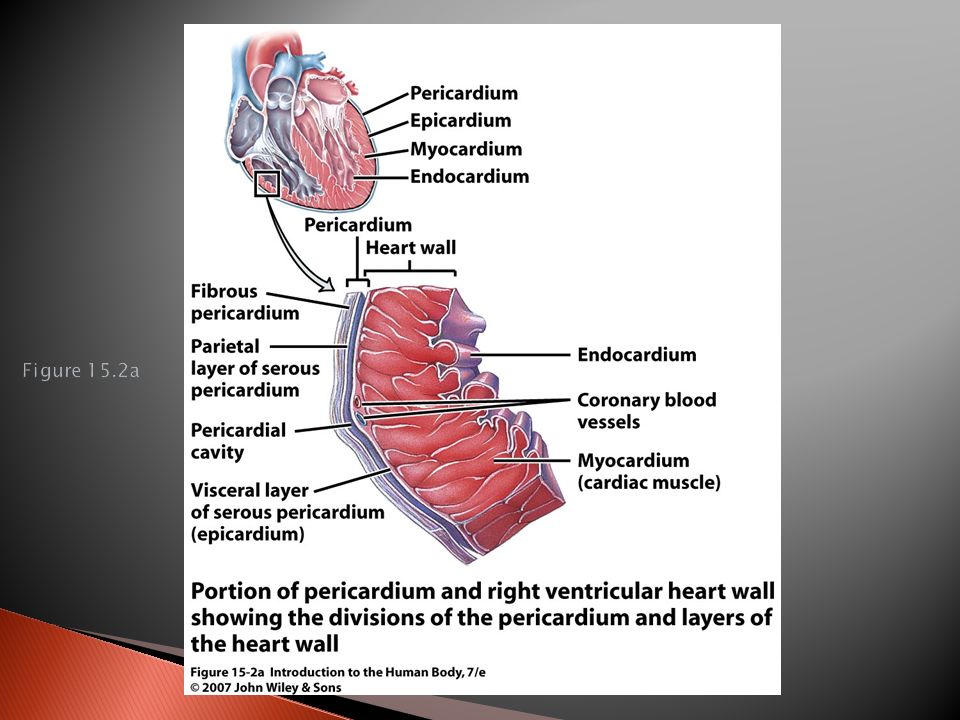
About the size of a fist; weighs less than a pound Found in thoracic cavity between two lungs = mediastinum (medial cavity of thorax) ◦ ~2/3 to left of midline Surrounded by pericardium: 1. Fibrous pericardium- ◦ Inelastic; protects and anchors heart in place 2. Inside is serous pericardium- double layer around heart ◦ Parietal layer fused to fibrous pericardium ◦ Inner visceral layer adheres tightly to heart ◦ Filled with pericardial fluid- reduces friction during beat.

4
Pericarditis inflammation of pericardium –Creates a creaking sound heard by a stethoscope –Can compress fluid and limit heart’s ability to pump blood

5
p. 682

6
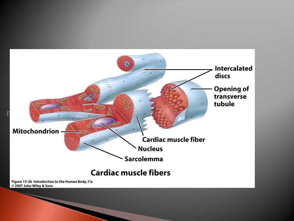
1. Epicardium- outer layer (contains fat) 2. Myocardium- cardiac muscle = bulk of heart; what contracts ◦ Two separate networks via gap junctions in intercalated discs- atrial & ventricular ◦ Networks- contract as a unit 3. Endocardium- Squamous epithelium ◦ Lines inside of myocardium

10
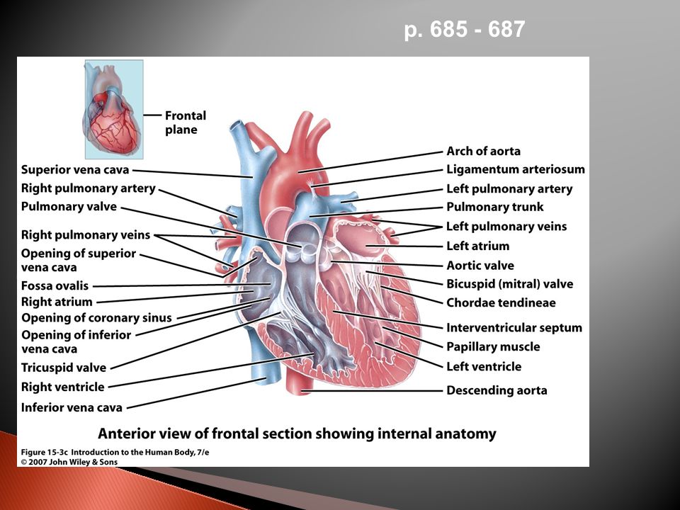
2 superior chambers= atria (receiving chambers) ◦ Contains auricles small, wrinkled, protruding appendages ◦ 2 Parts: ◦ 1.smooth-walled posterior ◦ 2.anterior = ridged by bundles of muscles ◦ Between is interatrial septum ◦ Contains fossa ovalis- remnant of foramen ovalis (opening in fetal heart)
 ◦ Contains auricles small, wrinkled, protruding appendages ◦ 2 Parts: ◦ 1.smooth-walled posterior ◦ 2.anterior = ridged by bundles of muscles ◦ Between is interatrial septum ◦ Contains fossa ovalis- remnant of foramen ovalis (opening in fetal heart)")
11
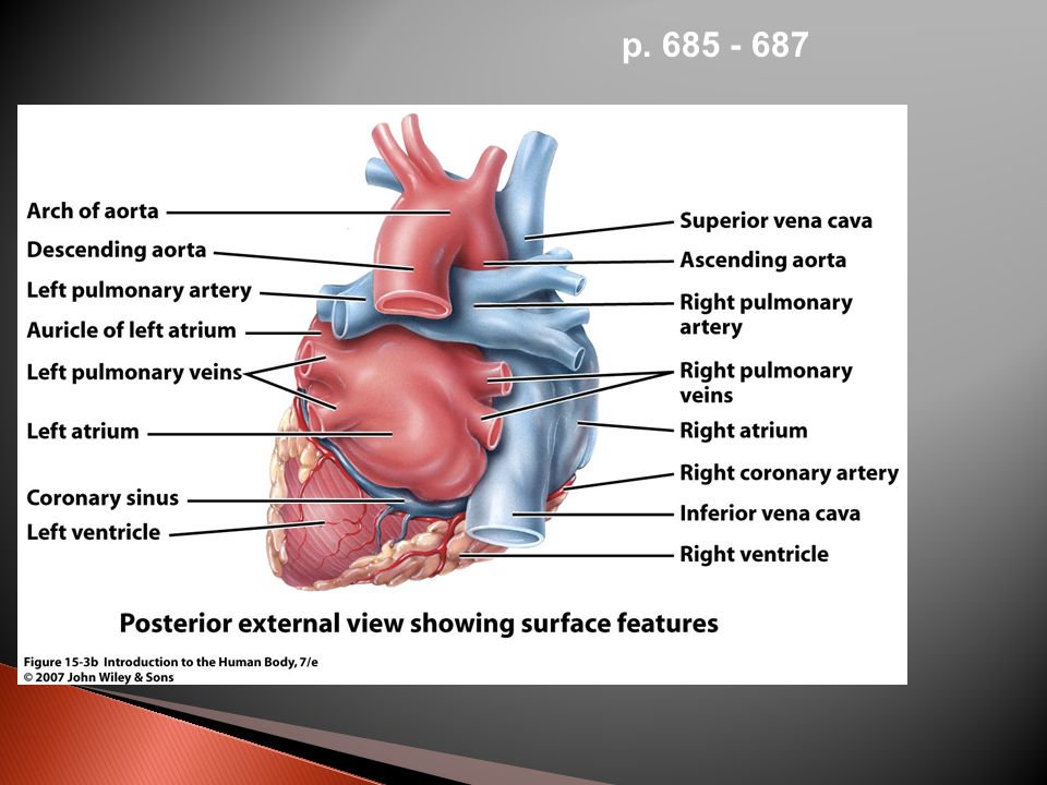
Blood enters right atria via 3 veins: 1.Superior vena cava (from body regions superior to diaphragm) 2.Inferior vena cava (from body areas below diaphragm) 3.Coronary sinus (collects blood from myocardium)
 2.Inferior vena cava (from body areas below diaphragm) 3.Coronary sinus (collects blood from myocardium)")
12
4 pulmonary veins enter the left atrium ◦ Transport blood from the lungs back to the heart

13
2 inferior chambers = ventricles (discharging chambers = pumps) ◦ Make up most of the mass of the heart ◦ Between is interventricular septum ◦ Right ventricle pumps blood into the pulmonary trunk (to lungs for gas exchange) ◦ Left ventricle pumps blood into the aorta = largest artery in the body Wall thickness depends on work load ◦ Atria thinnest ◦ Right ventricle pumps to lungs & thinner than left
 ◦ Make up most of the mass of the heart ◦ Between is interventricular septum ◦ Right ventricle pumps blood into the pulmonary trunk (to lungs for gas exchange) ◦ Left ventricle pumps blood into the aorta = largest artery in the body Wall thickness depends on work load ◦ Atria thinnest ◦ Right ventricle pumps to lungs & thinner than left")
14
Superior & inferior Vena Cavae ◦ Delivers deoxygenated blood to R. atrium from body ◦ Coronary sinus drains heart muscle veins R. Atrium R. Ventricle pumps through Pulmonary Trunk R & L pulmonary arteries lungs

15
Pulmonary Veins from lungs ◦ oxygenated blood L. atrium Left ventricle ascending aorta body Between pulmonary trunk & aortic arch is ligamentum arteriosum fetal ductus arteriosum remnant

16
p. 685 - 687

19
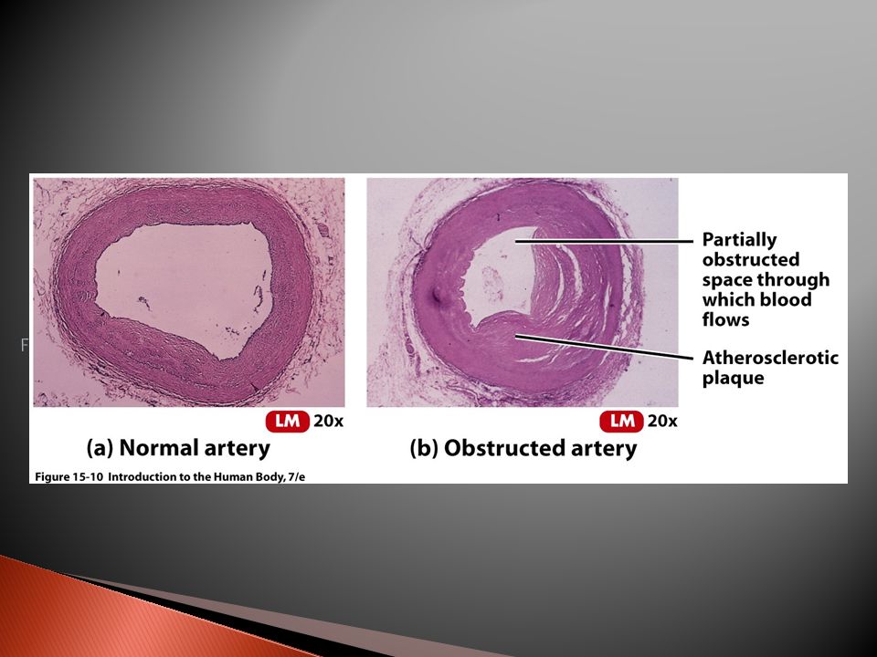
Blood flow through vessels in myocardium = coronary circulation L. & R. coronary arteries ◦ branch from aorta ◦ branch to carry blood throughout muscle Deoxygenated blood collected by Coronary Sinus (posterior) Empties into R. Atrium
 Empties into R. Atrium.")
21
p. 688

22
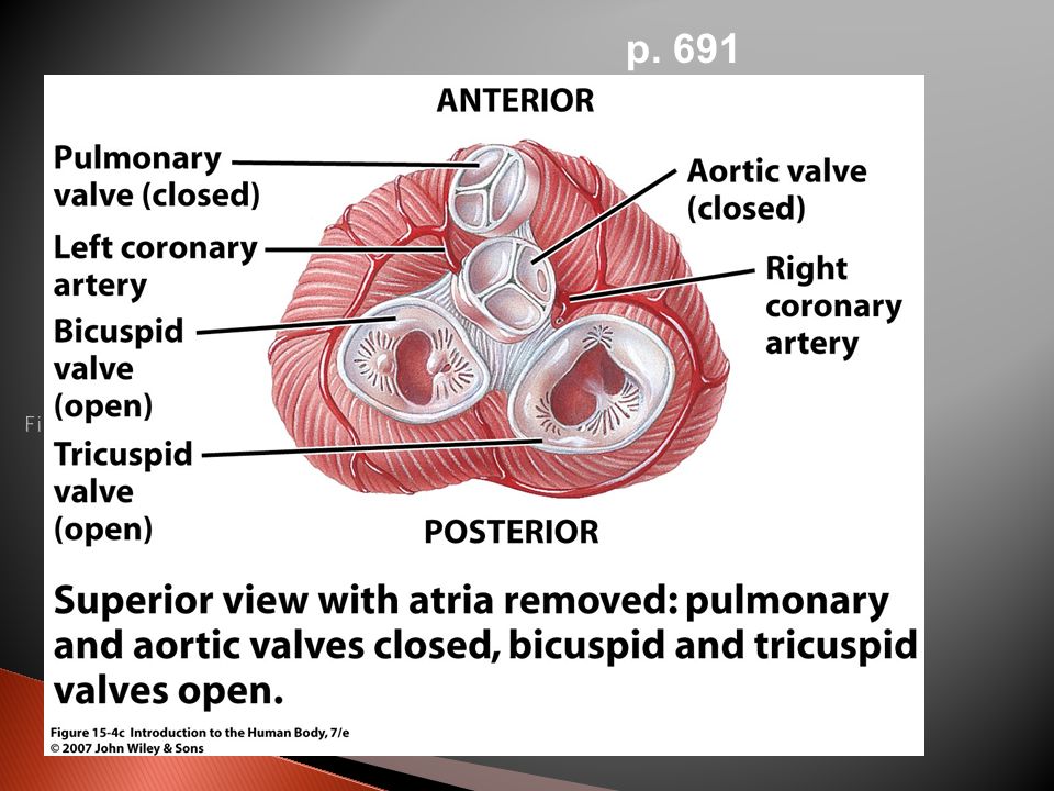
Designed to prevent back flow in response to pressure changes Atrioventricular (AV) valves ◦ Between atria and ventricles ◦ Right = tricuspid valve (3 cusps) ◦ Left = bicuspid or “mitral” valve ◦ Chordae tendineae – “heart strings” = anchor cusps to muscles Semilunar valves near origin of aorta & pulmonary trunk ◦ Aortic & pulmonary (semilunar) valves respectively
 valves ◦ Between atria and ventricles ◦ Right = tricuspid valve (3 cusps) ◦ Left = bicuspid or mitral valve ◦ Chordae tendineae – heart strings = anchor cusps to muscles Semilunar valves near origin of aorta & pulmonary trunk ◦ Aortic & pulmonary (semilunar) valves respectively")
23
p. 691

26
Striated Contracts by sliding Short, fat, branched, interconnected Contains 1 or 2 centrally located nuclei Large mitochondria = high resistance to fatigue

27
1% of cardiac muscle generate action potentials= Pacemaker & Conduction system Normally begins at sinoatrial (SA) node Atria contracts AV node -slows AV bundle (Bundle of His) bundle branches Purkinje fibers apex and up- then ventricles contract
 node Atria contracts AV node -slows AV bundle (Bundle of His) bundle branches Purkinje fibers apex and up- then ventricles contract")
28
Depolarize spontaneously sinoatrial node ~100times /min also AV node ~40-60 times/min in ventricle ~20-35 /min Fastest one run runs the heart = pacemaker Normally the sinoatrial node

30
Recording of currents from cardiac conduction on skin = electrocardiogram (EKG or ECG) P wave= atrial depolarization ◦ Contraction begins right after peak ◦ Repolarization is masked in QRS QRS complex= Ventricular depolarization ◦ Contraction of ventricle T-wave = ventricular repolarization ◦ Just after ventricles relax
 P wave= atrial depolarization ◦ Contraction begins right after peak ◦ Repolarization is masked in QRS QRS complex= Ventricular depolarization ◦ Contraction of ventricle T-wave = ventricular repolarization ◦ Just after ventricles relax")
32
after T-wave ventricular diastole ◦ Ventricular pressure drops below atrial & AV valves open ventricular filling occurs After P-wave atrial systole ◦ Finishes filling ventricle (`25%) After QRS ventricular systole ◦ Pressure pushes AV valves closed ◦ Pushes semilunar valves open and ejection occurs ◦ Ejection until ventricle relaxes enough for arterial pressure to close semilunar valves
 After QRS ventricular systole ◦ Pressure pushes AV valves closed ◦ Pushes semilunar valves open and ejection occurs ◦ Ejection until ventricle relaxes enough for arterial pressure to close semilunar valves")
33
Review muscle Heart has addition of External Ca 2+ creates a plateau prolonged depolarized period. Can not go into tetanus.

35
Cardiac Output (CO) = liters/min pumped Heart Rate (HR) = beats/minute (bpm) Stroke volume (SV) = volume/beat CO = HR x SV
 = liters/min pumped Heart Rate (HR) = beats/minute (bpm) Stroke volume (SV) = volume/beat CO = HR x SV")
36
Degree of stretch = Frank-Starling law ◦ Increase diastolic Volume increases strength of contraction increased S.V. ◦ Increased venous return increased S.V. increased sympathetic activity High back pressure in artery decreased S.V. ◦ Slows semilunar valve opening

37
Pacemaker adjusted by nerves ◦ Cardiovascular center in Medulla parasympathetic- ACh slows ◦ Via vagus nerve Sympathetic - norepinephrine speeds Sensory input for control: ◦ baroreceptors (aortic arch & carotid sinus)- B.P. ◦ Chemoreceptors- O 2, CO 2, pH

38
Hormones: ◦ Epinephrine & norepinephrine increase H.R. ◦ Thyroid hormones stimulate H.R. ◦ Called tachycardia Ions ◦ Increased Na + or K + decrease H.R. & contraction force ◦ Increased Ca 2+ increases H.R. & contraction force

40
Aerobic exercise (longer than 20 min) strengthens cardiovascular system Well trained athlete doubles maximum C.O. Resting C.O. about the same but resting H.R. decreased

Similar presentations


 in thorax, in inferior mediastinum>")


