Download presentation
Presentation is loading. Please wait.

1
Block 1 review

3
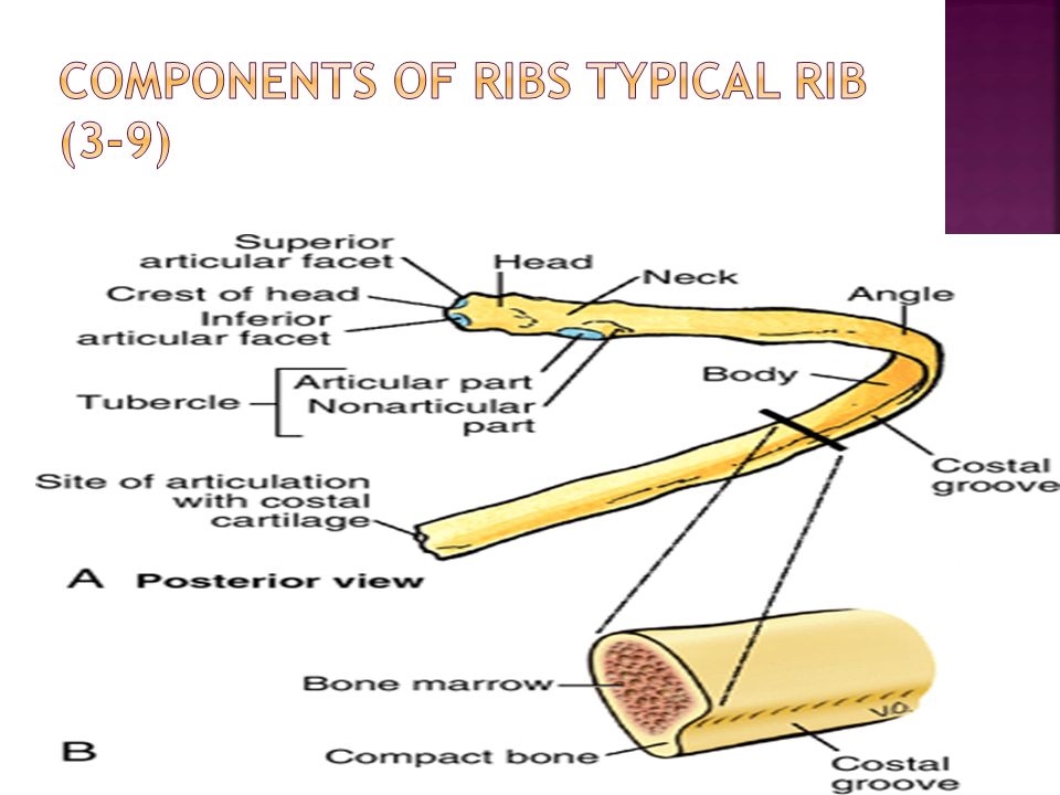
The thoracic wall consists of skeletal elements and muscles 1. Posteriorly, it is made up of twelve thoracic vertebrae and their intervening intervertebral discs 2. Laterally, the wall is formed by ribs (twelve on each side) and three layers of flat muscles. 3. Anteriorly, the sternum, which consists of the manubrium of sternum, body of sternum, and xiphoid process.
 and three layers of flat muscles. 3. Anteriorly, the sternum, which consists of the manubrium of sternum, body of sternum, and xiphoid process..")
6
Relations of Breast Blood vessels Lymphatic drainage

8
Breast Quadrants

10
Carcinoma of the Breast

11
Blood vessels A. Arteries B.Veins Intercostal nerves

15
Different parts of pleura Pleural reflection Recess:Costodiaphragmatic recess Thoracentesis Nerve supply

17
1. Costomediastinal recesses 2. Costodiaphragmati c recesses

18
The parietal pleura is sensitive to pain, temperature, touch, and pressure and is supplied as follows: The costal pleura is segmentally supplied by the intercostal nerves. The mediastinal pleura is supplied by the phrenic nerve. The diaphragmatic pleura is supplied over the domes by the phrenic nerve and around the periphery intercostal nerves.

19
Relations of lungs Impressions on medial surface of lungs Bronchopulmonary segment Trachea Blood vessels Nerve supply

20
1.Pulmonary artery 2. Two pulmonary veins 3. Main bronchus 4. Bronchial vessels 5. Nerves and lymphatics.

21
1. Heart 2.Inferior vena cava 3.Superior vena cava 4.Azygos vein 5.Esophagus

22
1.Heart 2.Aortic arch 3.Thoracic aorta 4. Esophagus

23
Has its own Bronchus Has its own Pulmonary artery (Blue) Drains to multiple pulmonary veins (Red) between segments So, each segment has its own bronchus and artery but not its own vein
 Drains to multiple pulmonary veins (Red) between segments So, each segment has its own bronchus and artery but not its own vein")
24
2 sets of Blood Supply 1.Pulmonary Vessels: for Gas Exchange 2. Bronchial Vessels: for blood supply to lung substance like any other organ

25
Superior mediastinum Inferior mediastinum A. Anterior B. Middle C. Posterior

26
superior mediastinum: [Green] Inferior Mediastinum: Below the plane passing from Sternal Angle/Angle Luise Inferior mediastinum has 3 parts: Purple: anterior mediastinum; Yellow: middle mediastinum; Blue: posterior mediastinum
![ superior mediastinum: [Green] Inferior Mediastinum: Below the plane passing from Sternal Angle/Angle Luise Inferior mediastinum has 3 parts: Purple: anterior mediastinum; Yellow: middle mediastinum; Blue: posterior mediastinum](http://images.slideplayer.com/33/8175176/slides/slide_26.jpg " superior mediastinum: [Green] Inferior Mediastinum: Below the plane passing from Sternal Angle/Angle Luise Inferior mediastinum has 3 parts: Purple: anterior mediastinum; Yellow: middle mediastinum; Blue: posterior mediastinum")
27
The middle mediastinum is centrally located in the thoracic cavity. It contains the pericardium, heart, origins of the great vessels, various nerves, and smaller vessels.

28
Lies between 1.Great vessels anteriorly [aorta and pulmonary trunk] 2.SVC posteriorly During Open heart surgery, clamp is passed here to block the blood flow while patient is put on artificial heart-lung machine.
![Lies between 1.Great vessels anteriorly [aorta and pulmonary trunk] 2.SVC posteriorly During Open heart surgery, clamp is passed here to block the blood flow while patient is put on artificial heart-lung machine.](http://images.slideplayer.com/33/8175176/slides/slide_28.jpg "Lies between 1.Great vessels anteriorly [aorta and pulmonary trunk] 2.SVC posteriorly During Open heart surgery, clamp is passed here to block the blood flow while patient is put on artificial heart-lung machine.")
29
1. Diaphragmatic (inferior) surface on which the pyramid rests 2. Anterior (sternocostal) surface oriented anteriorly 3.Right pulmonary surface 4.Left pulmonary surface.
 surface oriented anteriorly 3.Right pulmonary surface 4.Left pulmonary surface..")
31
1. SA nodal: 60% Pulmonary trunk and SA node 2. Right marginal:Right ventricle and apex of heart 3. Posterior interventricular:67% Right and left ventricles and posterior third of IVS 4. AV nodal

33
Diaphragmatic or Inferior wall infarct True Posterior wall infarct Anterior wall infarct Antero-lateral infract

34
The coronary sinus receives four major tributaries: 1.Great:anterior IV branch of the LCA 2.Middle: accompanies the posterior interventricular branch 3. Small: accompanies the right marginal branch of the RCA 4. Posterior cardiac veins 5. Oblique vein of the left atrium (of Marshall):The oblique vein is the remnant of the embryonic left SVC
:The oblique vein is the remnant of the embryonic left SVC.")
36
X ray Ct Scan

44
1)Right Atrium. 2)Left Atrium. 3)Right Ventricle. 4)Left Ventricle. 5)Descending Aorta. 6)Transverse Process of T7. 7)Right Bronchus. 8)Left Bronchus
Left Ventricle. 5)Descending Aorta. 6)Transverse Process of T7. 7)Right Bronchus. 8)Left Bronchus.")
45
A 37-year-old patient with palpitation was examined by her physician, and one of the diagnostic records included a posterior-anterior chest x-ray film. Which of the following comprises the largest portion of the sternocostal surface of the heart seen on the radiograph? (A) Left atrium (B) Right atrium (C) Left ventricle (D) Right ventricle (E) Base of the heart
 Left atrium (B) Right atrium (C) Left ventricle (D) Right ventricle (E) Base of the heart.")
46
A 19-year-old man came to the emergency department, and his angiogram exhibited that he was bleeding from the vein that is accompanied by the posterior interventricular artery. Which of the following veins is most likely to be ruptured? (A) Great cardiac vein (B) Middle cardiac vein (C) Anterior cardiac vein (D) Small cardiac vein (E) Oblique veins of the left atrium
 Great cardiac vein (B) Middle cardiac vein (C) Anterior cardiac vein (D) Small cardiac vein (E) Oblique veins of the left atrium.")
47
A 27-year-old cardiac patient with an irregular heartbeat visits her doctor's office for examination.Where should the physician place the stethoscope to listen to the sound of the mitral valve? (A) Over the medial end of the second left intercostal space (B) Over the medial end of the second right intercostal space (C) In the left fourth intercostal space at the midclavicular line (D) In the left fifth intercostal space at the midclavicular line (E) Over the right half of the lower end of the body of the sternum
 Over the medial end of the second left intercostal space (B) Over the medial end of the second right intercostal space (C) In the left fourth intercostal space at the midclavicular line (D) In the left fifth intercostal space at the midclavicular line (E) Over the right half of the lower end of the body of the sternum.")
48
A thoracentesis is performed to aspirate an abnormal accumulation of fluid in a 37-year-old patient with pleural effusion. A needle should be inserted at the midaxillary line between which of the following two ribs so as to avoid puncturing the lung? (A) Ribs 1 and 3 (B) Ribs 3 and 5 (C) Ribs 5 and 7 (D) Ribs 7 and 9 (E) Ribs 9 and 11
 Ribs 1 and 3 (B) Ribs 3 and 5 (C) Ribs 5 and 7 (D) Ribs 7 and 9 (E) Ribs 9 and 11.")
Similar presentations


 in thorax, in inferior mediastinum>")


 in the diaphragm. Give the.>")


