Download presentation
Presentation is loading. Please wait.

1
Dr. ANAND SRINIVASAN

2
Able to : Describe, identify and draw the histological features of : Blood vessels

3
Histologically 5 main types of blood vessels : ARTERIES ▪ Large A. (Elastic A.) ▪ Medium sized A. (Muscular A.) ARTERIOLES CAPILLARIES ▪ Continuous capillary ▪ Fenestrated capillary ▪ Sinusoidal capillary VENULES VEINS ▪ Medium sized V. ▪ Large V.
 ▪ Medium sized A. (Muscular A.) ARTERIOLES CAPILLARIES ▪ Continuous capillary ▪ Fenestrated capillary ▪ Sinusoidal capillary VENULES VEINS ▪ Medium sized V. ▪ Large V..")
4
All blood vessel have same basic structure TUNICA INTIMA ▪ Innermost lining endothelium (simple squamous) and subendothelial connective tissue TUNICA MEDIA ▪ Smooth muscle and connective tissue TUNICA ADVENTITIA ▪ Fibroelastic connective tissue
 and subendothelial connective tissue TUNICA MEDIA ▪ Smooth muscle and connective tissue TUNICA ADVENTITIA ▪ Fibroelastic connective tissue")
5
Thick wall E.g. Aorta and its branches Tunica intima Endothelium and subendothelial connective tissue Subendothelial cells contain mactophages and smooth muscle like cells “Myointimal cells” Separated from Tunica media by poorly defined internal elastic lamina.

6
Tunica media Mainly made of 40 – 70 layers of fenestrated elastic laminae arranged circularly. Between elastic laminae there are smooth muscle cells and collagen fibers Outermost / External elastic lamina is thick. Tunica adventitia Fibroelastic connective tissue carrying small blood vessels and sympathetic nerves

8
Thickening of tunica intima – due to migration and proliferation of smooth muscle cells from tunica media Accumulation of lipid in myointimal cells and macrophages Formation of fibrofatty plaques in tunica intima (Atheroma) Calcification of tunica media (Arteriosclerosis)
 Calcification of tunica media (Arteriosclerosis)")
9
Age related changes (Atheroma ± arteriosclerosis) Not only in large A. but also coronary and cerebral A. Can lead to ischemia / infarction

10
Atrophy of tunica media – loss of elasticity of artery

11
E.g. branches of external carotid A., radial A. etc. Distributes blood to various parts of body Tunica intima : Endothelium and internal elastic lamina No subendothelium Internal elastic lamina clearly seen and thrown into folds due to contraction of smooth muscle in media

12
Tunica media Smooth muscle cells (~40 layers) arranged circularly Also contains elastic and few collagen fibers Tunica adventitia Presence of external elastic lamina Inner part of adventitia contains more elastic Middle part contains more collagen Outer part contains loose connective tissue
 arranged circularly Also contains elastic and few collagen fibers Tunica adventitia Presence of external elastic lamina Inner part of adventitia contains more elastic Middle part contains more collagen Outer part contains loose connective tissue")
14
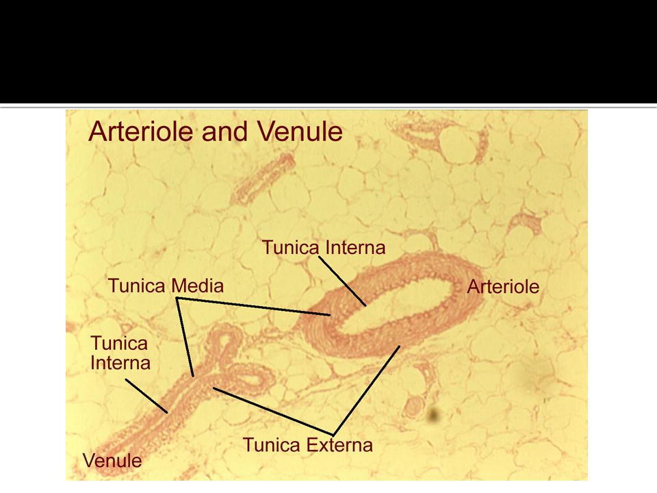
Small artery having diamater less than 0.5 mm. Meta arterioles – terminal branches of arterioles acting like sphincter Tunica intima Only endothelial lining No subendothelium or internal elastic lamina Tunica media 1 – 5 layer of circular layer of smooth muscle Tunica adventitia Thin and poorly developed & contain sympathetic

16
CONTINUOUS Commonest type present in tissue, mucle, brain FENESTRATED Found in kidney, intestines SINUSOIDAL Found in liver, spleen and bone marrow

18
Larger diameter than arterioles (0.5 – 1 mm) Tunica intima Endothelium Tunica media 1 – 2 layer of smooth muscle cells Tunica adventitia Thick made of collagen fibers
 Tunica intima Endothelium Tunica media 1 – 2 layer of smooth muscle cells Tunica adventitia Thick made of collagen fibers")
20
Tunica intima Endothelium & subendothelium No internal elastic lamina Tunica media Few circular smooth muscle, mostly collagen Tunica adventitia Loose fibroelastic connective tissue Compared to Medium sized A. Collapsed lumen Thin wall of tunica media with few smooth muscle & elastic fibers No internal elastic lamina Presence of valves

22
E.g. Superior vena cava Tunica intima Well developed endothelium and subendothelium Tunica media Thin / absent Tunica adventitia Well developed and thickest coat Longitudinally arranged smooth muscle

24
Wheater’s Functional Histology Cell Biology & Histology – Board Review Series Microanatomy workbook – RAKMHSU

Similar presentations


 Inner layer of arteries and veins Endothelium made of simple squamous epithelial cells.>")


, (10x obj.) TI TM TA elastic fibers.>")


