Download presentation
Presentation is loading. Please wait.

1
Histology of the Circulatory System Heart - pump the blood Arteries - efferent vessels Capillaries - anastomosing thin tubules where interchange between blood and tissue takes place Veins - convergence of the capillaries into a system of larger channels to the heart Lymphatic vascular system

2
Basic structure of vessels Tunica intima Endothelial layer Subendothelial layer Internal elastic lamina (arteries) Tunica media Concentric layers of helically arranged smooth muscle cells Varying amounts of elastic fibers, reticular fibers, and proteoglycans External elastic membrane-(larger arteries) Tunica Adventitia - longitudinally oriented collagen (type I)
 Tunica media Concentric layers of helically arranged smooth muscle cells Varying amounts of elastic fibers, reticular fibers, and proteoglycans External elastic membrane-(larger arteries) Tunica Adventitia - longitudinally oriented collagen (type I)")
3
Classic muscular artery- elastic stain

4
Small muscular artery

5
Vasa Vasorum In larger vessels Small vessels branch profusely in the adventitia and outer part of the media Nourish the media

6
Vaso vasorum Media of large art.

7
Innervation Most vessels have a profuse network of sympathetic fibers Sympathetic transmitter cause vasoconstriction - epinephine Arteries in skeletal muscle also have cholinergic vasodilatior nerve supply Sensory nerve endings - baroreceptors in the carotid sinuses and arch of aorta and chemoreceptors in the carotid and aortic bodies

9
Capillaries Single layer of endothelial cells Diameter 7 to 9 m Gap junctions are present Held together by zonula occludens Pericytes - partly surround the endothelial cells- they have myosin, actin, & tropomyosin--contractile function Most endothelial cells are continuous Fenestrated - kidney, intestine, endocrine Discontinuous - liver, spleen, bone marrow

10
Capillary

12
Capillary(b) and venule(a)
 and venule(a)")
13
Capillary in heart(B)
")
14
Capillary - pericyte

15
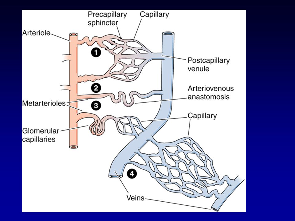
Fenestrated capillary (kidney)
")
16
Arteries -resist changes in blood pressure and regulate blood flow Arterioles - < 0.5mm in dia., endothelial cells, thin subendothelial layer, most have no internal elastic layer, 1 or 2 layers of smooth muscle, no external elastic lamina

17
Small artery & Vein (arrow) artery
 artery")
18
Arteriole & venule

19
Arteriole --small artery

20
Muscular Arteries Most arteries in the body Thin intimal layer Well developed internal elastic lamina Muscle layer up to 40 layers Varying intermingled elastic fibers adventitia consists of nerves, vessels, collagen, elastic fibers, fibroblasts and adipose cells

21
Muscular artery- very thin intima, thick internal elastic lamina

22
Small muscular artery

23
Large Elastic Arteries Aorta and its large branches Thick intima (subintima) Internal elastic lamina Media concentrically perforated elastic lamina with smooth muscle and ground substance in between Poorly formed external layer
 Internal elastic lamina Media concentrically perforated elastic lamina with smooth muscle and ground substance in between Poorly formed external layer")
24
Elastic artery Aorta

25
Elastic artery- note thin intima and wavy elastic fibers

26
Elastic artery- elastic stain

27
Adventitia of large artery

28
Carotid Bodies Small structures near the bifurcation of the carotid artery Chemoreceptors(low 02 tension, high CO2 and low blood pressure) Composed of glomus cells surrounded by fenestrated capillaries and many afferent nerve fibers Glomus cells have numerous dense core vesicles that store dopamine, norepinephrine and seratonin
 Composed of glomus cells surrounded by fenestrated capillaries and many afferent nerve fibers Glomus cells have numerous dense core vesicles that store dopamine, norepinephrine and seratonin")
29
Carotid Sinuses Slight dilation of internal carotid artery Contain baroreceptors that detect changes in blood pressure

30
Arteriovenous anastomoses Direct communication between arterial and venous circulation Plays a role in regulation of blood pressure, flow and temperature in various areas Glomus - complex anastomoses in fingerpads, fingernail beds, and ears

31
Arteryvein

32
Veins Capacitance vessels - 70% of blood volume Venules - thin walls - only contractile pericytes Small and medium sized veins - thin intimia, thin media, valves

33
Muscular artery (a) and vein(b)
 and vein(b)")
34
Muscular vein

35
Muscular vein with valve

36
Vein with valve

37
Arteriole (b) and venule (a)
 and venule (a)")
38
lymphatic

39
Lymphatic with valve

40
Heart Pump Produces atrial natriuretic factor Fibrous skeleton - base for the valves and site of origin and insertion of muscle cells

41
Tunics of the Heart Endocardium Endothelial layer Subendothelial layer - veins, nerves and Purkinje cells Myocardium Cardiac muscle cells Epicardium Visceral layer of the pericardium - mesothelium Subepicardial layer of loose connective tissues - veins, nerves and ganglia

42
Layers of the heart

43
Heart - * endocardium

44
Heart - epicardium

45
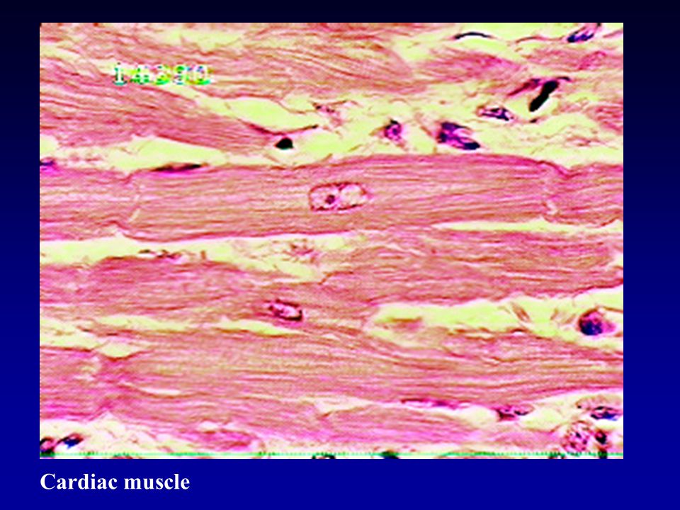
Cardiac muscle

47
Cardiac muscle - intercalated disks

48
Conduction System(Purkinji fibers) Subendocardial area Cells larger, pale cytoplasm, few myofibrils, rich in glycogen but no T tubule system Junctions via desmosomes and gap junctions rather than intercalated disks
 Subendocardial area Cells larger, pale cytoplasm, few myofibrils, rich in glycogen but no T tubule system Junctions via desmosomes and gap junctions rather than intercalated disks")
49
Sinoatrial node - pacemaker of the heart Close to the entrance of the superior vena cava into the right atrium Modified cardiac muscle cells Fewer myofibrils

50
Atrioventricular node In wall of membranous septum Atrioventricular bundle of this formed by Purkinje cells Divides into Rt and Lt bundle branches

51
AV node

52
Bundle of His Bundle

53
Innervation of the Heart Both parasympathetic and sympathetic Ganglionic nerve cells and nerves are present near both nodes

Similar presentations


 Dept.of Histology and Embryology.>")


, (10x obj.) TI TM TA elastic fibers.>")


 Arteries; b) Veins; c) Microcirculatory bed 4. Lymphatics 5. Heart.>")

