Download presentation
Presentation is loading. Please wait.

8
H EMOPTYSIS... O R T HAT LARGE TESTICULAR MASS I’ VE BEEN IGNORING Morning Report Christine Williams, MD April 28, 2010

9
EPIDEMIOLOGY Most common malignancy in men 15-35y 1% of cancers of men overall Cryptoorchidism causes 3 – 17x higher risk Kleinfelter’s syndrome have increased risk, particularly of mediastinal germ-cell tumors Germ cell tumors 95%, often mixed histology Teratoma, choriocarcinoma, yolk sac, carcinoma Seminomas are very radiosensitive Mixed seminomas with nonseminomatous tissue should be treated as nonseminomatous tumors (chemo for metastatic disease)
")
10
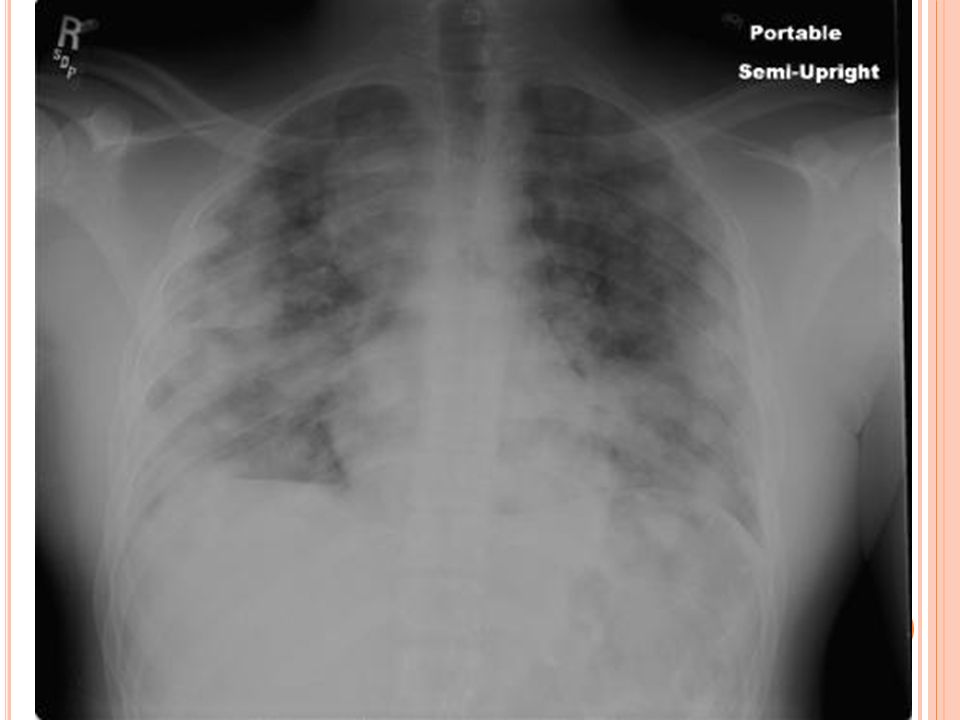
PRESENTATION Painless testicular or scrotal mass (only 10% have pain) Dull ache or heavy sensation in the lower abdomen Symptoms often the result of metastatic dz: Supraclavicular LN Cough or dyspnea Anorexia, n/v, GI hemorrhage Low back pain or bone pain Neuropathy Unilateral or bilateral lower extremity swelling Gynecomastia Paraneoplastic hyperthyroidism (due to Bhcg) or limbic encephalitis (anti-Ma2 antibodies)
 Dull ache or heavy sensation in the lower abdomen Symptoms often the result of metastatic dz: Supraclavicular LN Cough or dyspnea Anorexia, n/v, GI hemorrhage Low back pain or bone pain Neuropathy Unilateral or bilateral lower extremity swelling Gynecomastia Paraneoplastic hyperthyroidism (due to Bhcg) or limbic encephalitis (anti-Ma2 antibodies)")
11
DIFFERENTIAL DIAGNOSIS Hydrocele Varicocele Epididymitis Epididymorchitis Testicular torsion Hernia Hematoma Spermatocele Gumma

12
EVALUATION Physical exam w/particular focus on: Testes, abdominal wall & organs, supraclavicular LN, chest Scrotal ultrasound can help distinguish hydrocele or epididymitis from mass but is only about 70% correct in diagnosis of tumor type Cystic or fluid-filled lesions unlikely neoplastic Seminomas are well-defined hypoechoic lesions Nonseminomatous germ cell tumors (NSGCTs) inhomogeneous with calcifications, cystic areas, and indistinct margins
 inhomogeneous with calcifications, cystic areas, and indistinct margins")
13
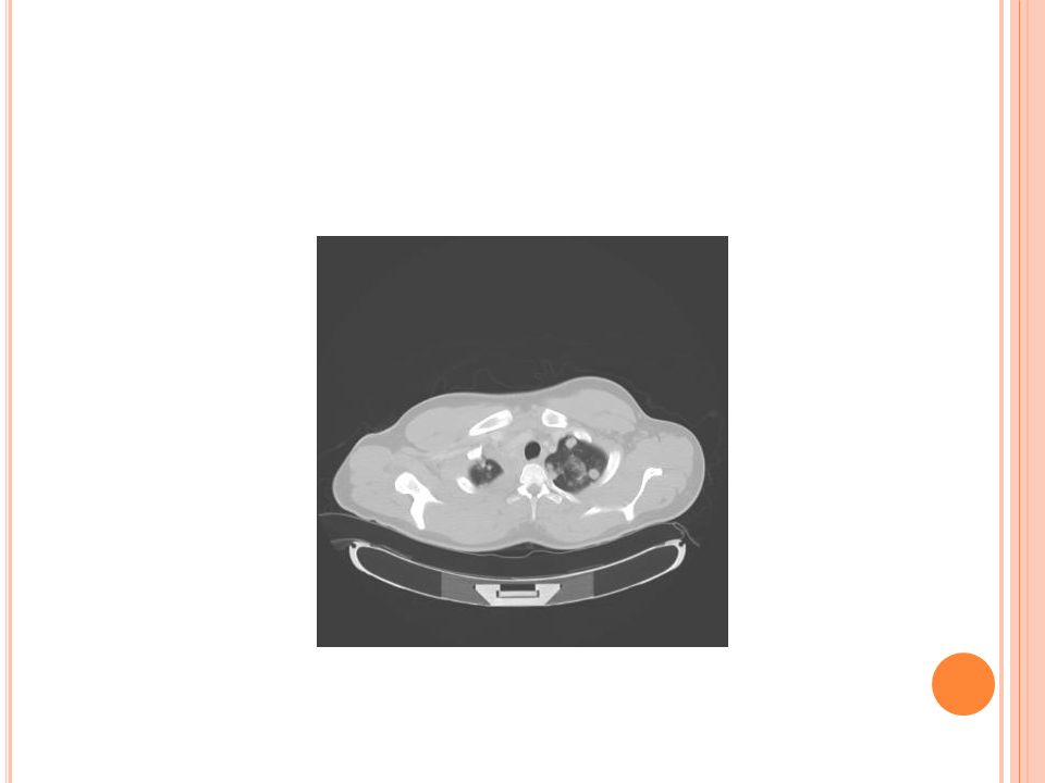
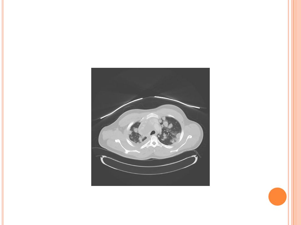
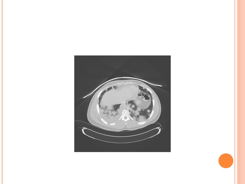
DIAGNOSIS/STAGING Radical inguinal orchiectomy – curative in stage I disease CXR then CT Chest if abnormal CT Abdomen & Pelvis Occult micrometastasis in retroperitoneal LN is common and may be missed by CT (up to 44% false negative depending on LAD classification) Brain MRI if mets suspected PET scan often false negative (but high PPV) for initial staging but can be used to evaluate post-therapy residual masses Retroperitoneal LN dissection may be considered Immunohistochemical, cytogenetic studies and molecular gene profiling may be required for poorly differentiated tumors Isochromosome 12p is suggestive of a germ cell tumor if present
 Brain MRI if mets suspected PET scan often false negative (but high PPV) for initial staging but can be used to evaluate post-therapy residual masses Retroperitoneal LN dissection may be considered Immunohistochemical, cytogenetic studies and molecular gene profiling may be required for poorly differentiated tumors Isochromosome 12p is suggestive of a germ cell tumor if present")
14
SERUM BIOMARKERS Alpha fetoprotein (AFP) – also elevated in HCC, half-life is 5-7 days Β-HCG – also elevated in trophoblastic differentiation of lung or gastric cancer, pregnancy or trophoblastic disease in women. Half life is 24-36 hours Lactate dehydrogenase (LDH) Other biomarkers like CEA, CA19-9 others can be used for following response to therapy but are not diagnostic BiomarkerNSGCTSeminoma AFPHigh (85%)Normal B-hcgHigh (85%)Normal or High (20%)
 Other biomarkers like CEA, CA19-9 others can be used for following response to therapy but are not diagnostic BiomarkerNSGCTSeminoma AFPHigh (85%)Normal B-hcgHigh (85%)Normal or High (20%).")
15
TREATMENT Overall 5 yr survival rate is 95% BEP: bleomycin, etoposide, cisplatin PVB: cisplatin, vinblastine, bleomycin Taxane & platinum based therapy also used Radiation for intracranial mets For advanced testicular cancer Overall response rate 62% Complete response rate 26%

16
MKSAP #1 A 30yo M is evaluated for an enlarged, painless, L testicular mass. PMH is noncontributory and FH is unremarkable. Exam is otherwise normal. Labs show B- hCG 28mU/mL (nl <5) and AFP of 35ng/mL. LDH is normal. Chest, A/P CT are normal. Scrotal ultrasound shows solid hypoechoic 4cm diameter mass. Radical orchiectomy is performed and tumor confirmed as embryona carcinoma w/chorionic elements. A week after surgery the B-hcg is 1.8 mU/ML and AFP Is 16.3ng/mL Which of the following is the most appropriate next step in management? A. Chemotherapy B. Repeat B-hCG and AFP in 1 week C. Abdominal radiation therapy D. Biopsy of the opposite testicle
 and AFP of 35ng/mL. LDH is normal. Chest, A/P CT are normal. Scrotal ultrasound shows solid hypoechoic 4cm diameter mass. Radical orchiectomy is performed and tumor confirmed as embryona carcinoma w/chorionic elements. A week after surgery the B-hcg is 1.8 mU/ML and AFP Is 16.3ng/mL Which of the following is the most appropriate next step in management. A. Chemotherapy B. Repeat B-hCG and AFP in 1 week C. Abdominal radiation therapy D. Biopsy of the opposite testicle.")
17
MKSAP #1 - A NSWER B- repeat AFP in 1 week of 14 days after surgery Half life of AFP is 1 week wheras Bhcg is 24 hrs Serum hCG has normalized and AFP concentration has decreased at expected rate for removal of all cancer Stage I Nonseminomatous testicular cancer confined to the testis likely cured by initial surgery. Chemotherapy would be overly aggressive Synchronous testicular tumors are rare <1% Abdominal radiation is inappropriate, chemo is standard of care for metastatic disease

18
MKSAP #2 A 28 yo M is evaluated for a 1-month history of a sensation of scrotal heaviness and an enlarging testicle. His PMH is otherwise noncontributory and FH unremarkable. On exam he has a large, firm, right testicular mass. Remainder of exam is normal. Preoperative serum B-hCG is 20 mU/mL (nl <5) and AFP and LDH are normal. Chest CT is normal but abdominal CT shows three enlarged retroperitoneal LN each 2-3cm in diameter. High inguinal radial orchiectomy reveals a tumor that is 95% seminomatous but with 5% nonseminomatous elements. A week after surgery, serum tumor markers are normal. Which of the following is the most appropriate next step in management A. Repeated CT of the abdomen B. Abdominal radiation therapy C. Chemotherapy D. Abdominal radiation therapy plus chemotherapy
 and AFP and LDH are normal. Chest CT is normal but abdominal CT shows three enlarged retroperitoneal LN each 2-3cm in diameter. High inguinal radial orchiectomy reveals a tumor that is 95% seminomatous but with 5% nonseminomatous elements. A week after surgery, serum tumor markers are normal. Which of the following is the most appropriate next step in management A. Repeated CT of the abdomen B. Abdominal radiation therapy C. Chemotherapy D. Abdominal radiation therapy plus chemotherapy.")
19
MKSAP #2 A NSWER C. Chemotherapy Stage II testicular cancer metastatic to the retroperitoneal LN. Chemotherapy is highly effective and curative for the seminomatous and nonseminomatous elements of testicular cancer and should be administered immediately. Normalized concentrations of tumor markers after orchiectomy do not change the clinical disease stage. Abdominal radiation therapy, alone or in combination with chemo is not appropriate because mixed tumors should be managed as NSGCT. Radiation therapy alone is inadequate to cure nonseminomatous tumors. The addition of radiation therapy to chemo is unnecessary for cure, would result in more acute toxicity and confer the additional risk of radiation-induced cancer. Results of a repeated CT scan of the abdomen would be unlikely to have changed and would not change management.

20
MKSAP #3 A 27yo M is evaluated for increasing abdominal girth w/intermittent midabdominal pain radiating to the back and a 4.5kg weight loss over the past 3 months. He has no symptoms of reflux. PMH and FH are unremarkable. Exam is normal. Abdomen is nontender but there is a sense of fulllness in the midabdomen. Testes are descended w/no palpable abnormalities. CT demonstrates a 10cm retroperitoneal mass. Serum B-hCG is 212mU/mL (nl <5) and serum AFP of 478ng/mL. Testicular ultrasound is normal. A needle biopsy of the mass reveals a poorly differentiated carcinoma, with molecular genetic analysis detecting isochromosome 12p. Which of the following is the most appropriate next step in management? A. Testicular biopsy B. Cisplatin-based chemotherapy C. Upper Endoscopy D. Radiation Therpay
 and serum AFP of 478ng/mL. Testicular ultrasound is normal. A needle biopsy of the mass reveals a poorly differentiated carcinoma, with molecular genetic analysis detecting isochromosome 12p. Which of the following is the most appropriate next step in management. A. Testicular biopsy B. Cisplatin-based chemotherapy C. Upper Endoscopy D. Radiation Therpay.")
21
MKSAP #3 - A NSWER B. Chemotherapy The patient has a poorly differentiated tumor that is primarily in the midline, expresses germ cell cancer markers, and contains isochromosome 12p. This type of tumor is likely to respond to cisplatin-based chemotherapy and may occasionally be cured. Although isochromosome 12p is not diagnostic of a germ cell malignancy, this clinical pattern is highly suggestive of an extragonadal germ cell cancer. Performing a biopsy is unlikely to be helpful because ultrasound was normal. The abdominal pain is likely due to his large midline mass; he is unlikely to derive any additional benefit from an upper endoscopy. The systemic illness present in this patient is potentially curable with cisplatin-based chemotherapy, which is the typical treatement approach for patients with germ cell cancer. Although radiation therapy has the potential to diminish the size of the patient’s abnormal abdominal mass, it is not curative and therefore not warranted.

22
MKSAP #4 A 35 yo M is evaluated after a recent diagnosis of metastatic nonseminomatous testicular cancer. His treatement plan includes retroperitoneal LN dissection with possible chemotherapy, depending on histologic results and extent of disease. The remainder of his medical history is noncontributory and FH is unremarkable. Exam is normal. The patient is concerned about the risks associated with his cancer treatment. Which of the following treatment-related risks is most likely in this patient? A. Decreased fertility B. Anorgasmia C. Retrograde ejaculation D. Erectile dysfunction E. Congenital malformations in offspring

23
MKSAP #4 - A NSWER A. Decreased fertility Approximately 25% of patients have decreased sperm counts or sperm motility and dx suggesting an underlying fertility problem associated with this disease; however, patients almost invariable become azoospermic shortly after chemotherapy is initiated. Over a period of 2 years, may patients’ sperm counts return to normal, and these men are ab le to produce offspring but many remain infertile or subferile. Consequently, sperm storage should be offered to men with testicular cancer before they undergo chemotherapy. Neither RPLND nor chemo has been shown to decrease the ability of men to have an orgasm once the acute side effects of therapy have resolved. The traditional method for RPLND has routinely led to retrograde ejaculation but not sexual impotence; however, newer limited techniques for RPLND have eliminated the problem of retrograde ejaculation wtihout increasing the risk for disease recurrence in the abdomen or diminishing cute rates. Although they are not large, f/u studies of chemo in patients with testicular germ cell cancer have not shown a higher-than-expected risk for congenital malformations in offspring.

24
MKSAP #5 A 40 yo M is evaluated during routine exam after being discharged from care by his medical oncologist. PMH is significant for metastatic testicular germ cell cancer for which he was treated with bleomycin – etoposide – cisplatin therapy 5 ½ years ago and has been disease free ever since. The remainder of PMH and FH is unremarkable and PE is normal. Which of the following is the most appropriate management strategy for this patient at this time? A. Biopsy of the remaining testicle B. Semiannual testicular US of remaining testicle C. Periodic audiometry D. Annual serum creatinine and BUN measurement E. Periodic review of symptoms

25
MKSAP #5 - A NSWER E. Periodic ROS Most testicular cancer relapses occur within 2 years after the completion of definitive therapy; relapse after that point is rare. Many expers advocate f/u examinations including annual CT scans or laboratory studies focused only on new symptoms rather than an arbitrary schedule of workups. Contralateral testicular tumors occur rarely and neither routine testicular biopsy or US is known to be helpful in establishing early dx or changing outcomes. False-positive US results can be problematic and performing a blind bx of the testicle is invasive na dsubject to sampling error. Cisplatin can cause decreased high-frequency hearing, it affects sound frequencies in the conversational range infrequently and does not require routine audiometry. Likewise, although cisplatin can cause ong-term subclinical deficits in renal function, renal failure after the usual curative BEP regimen is rare; therefor, routine cr and BUN measurement is unnecessary.

Similar presentations


; FRCS (Ireland); MMed (Wits); FCS (SA) Urology 38 th BMA CONGRESS.>")


