Download presentation
Presentation is loading. Please wait.

1
UNIT SIX CHAPTERS 10 AND 11

2
CELL DIVISION CHAPTER 10

3
THE MAIN GOAL OF CELLULAR DIVISION, IN BOTH EUKARYOTIC AND PROKARYOTIC CELLS, IS WHAT?

4
BINARY FISSION Single circular chromosome located in the nucleoid region Compaction of the chromosome is completed by SMC proteins (structural maintenance of chromosome) DNA must be replicated prior to division
 DNA must be replicated prior to division")
5
BACTERIAL CHROMOSOME DETAILS ORI, origin of replication is where the chromosomal replication begins Copying proceeds in both directions, speeds up the process Replication ends at a specific site of termination

6
CHROMOSOME SEPARATION The replicated chromosomes need to move to opposite ends of the cell Last event in replication is decatenation, the untangling of the chromosomes The new pieces are segregated by the formation of the septum through the process of septation and use of the FtsZ proteins, which is a protein similar to tubulin

8
EUKARYOTIC CHROMOSOMES Discovered by Walther Flemming in 1879 while looking at salamander larvae He witnessed cellular division and called it mitosis, from the Greek “mitos”, meaning thread Normal human chromosome number is 46, 23 from mom and 23 from dad Missing a chromosome is monosomy Having an extra chromosome is trisomy Organisms have specific chromosome numbers

9
CHROMOSOME NUMBER Diploid = 2n Haploid =1n

10
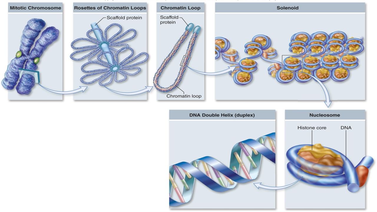
WHAT IS A CHROMOSOME MADE OF? Made of chromatin, a mix of DNA and protein, 40% and 60% respectively One human chromosome contains about 140 million nucleotides and would be about 5cm long if stretched out How does all the information fit in a cell?

12
KARYOTYPES Chromosomes vary in many ways Size Staining properties Location of centromere Relative length of arms Visualize chromosomes with a karyotype

13
HOMOLOGOUS CHROMOSOMES

14
WE KNOW WHY CELLS NEED TO DIVIDE, WE UNDERSTAND MORE ABOUT THE CHROMOSOMES AND THEIR PACKAGING, SO, HOW DO CELLS DIVIDE? WHAT IS THE PROCESS?

15
INTERPHASE G1, S, and G2 After S phase the sister chromatids share a common centromere, in actuality, there are two complete DNA molecules They are held together with a protein, cohesion Each chromatid has its own set of kinetochore proteins The G2 phase is when the chromosomes begin to condense using motor proteins Centrioles form in G2

16
PROPHASE The condensed chromosomes can be seen Centrioles move apart and spindle begins to form (no centrioles in plant cells) In animal cells a spindle aster forms
 In animal cells a spindle aster forms")
17
PROMETAPHASE Chromosomes attach to spindle at the kinetochores Critical step, if something is attached inappropriately the chromsomes may not separate correctly later on, resulting in one cell with an extra chromosome and once cell with one less chromosome (nondisjunction) Chromosomes begin to align at the center of the cell Assembly and disassembly of microtubules Motor proteins Most likely a combination of the two processes
 Chromosomes begin to align at the center of the cell Assembly and disassembly of microtubules Motor proteins Most likely a combination of the two processes")
18
METAPHASE Alignment of the chromosomes at the metaphase plate

19
ANAPHASE Shortest phase of mitosis Chromatids separate at the centromere, the cohesion proteins are broken down Anaphase A Kinetochores are pulled toward the poles Shortening process, tubulin subunits are removed at the ends of the kinetochore fibers Anaphase B Poles move apart Cell becomes elongated

20
TELOPHASE Spindle disassembles Nuclear envelope reforms Chromosomes uncoil, genes begin being expressed again, rRNA genes especially, so, nucleolus reappears Cell division is still not complete

21
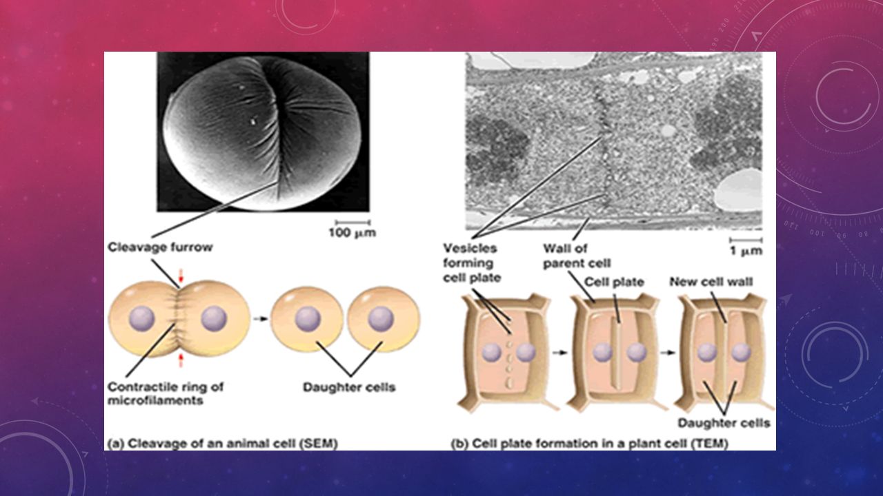
CYTOKINESIS Animal cells Constricting belt of actin pinches in the membrane Cleavage furrow Plant cells A membrane partition called a cell plate forms Plate grows until it reaches the cell membrane and fuses with it Cellulose is laid down on the new membrane, the space between the cells becomes impregnated with pectins, and forms the middle lamella

23
CONTROLLING THE CELL CYCLE Two irreversible points Replication of genetic material Separation of sister chromatids Cell can halt specific functions at checkpoints

24
CONTROL FACTORS MPF M phase-promoting factor Low levels during G 2 and peaking in mitosis Activity of MPF involves phosphorylation of proteins Cyclins Produced in synchrony with the cell cycle Two forms Peaks at G 1 /S Peaks at G 2 /M

25
MPF AND CYCLIN ACTIVITY

26
GENETIC ANALYSIS OF THE CELL CYCLE Yeast were the model system Studies indicated that there are two critical control points Commitment to DNA synthesis, termed START Commitment to mitosis One particular gene was found to be key to both processes, cdc2 gene

27
WHAT DOES THE CDC2 GENE DO? Cdc2 gene was determined to be a gene for a protein kinase Purification of MPF showed that it is composed of a cyclin and the cdc2 protein kinase The cdc2 protein was termed a Cdk (cyclin dependent kinase) Cdk enzymes drive the cell cycle
 Cdk enzymes drive the cell cycle.")
28
THE THREE CHECKPOINTS

29
HOW DO CDKS WORK?

30
YEAST CELL CYCLE Accumulation of G 1 cyclins seems to be the trigger Causes enzymes to be made for DNA replication

31
ANAPHASE-PROMOTING COMPLEX Sensing system of the spindle checkpoint (APC), also known as a cyclosome The whole purpose of the APC/C complex is to trigger anaphase APC does not directly act on cohesion, it marks a protein, securin for destruction Securin is an inhibitor for separase, which is a protein specific to a component of the cohesion complex Separase destroys cohesion allowing the chromatids to separate Also destroys mitotic cyclins to drive the cell cycle out of mitosis by marking proteins for destruction, they proteins are marked with a protein called ubiquitin
, also known as a cyclosome The whole purpose of the APC/C complex is to trigger anaphase APC does not directly act on cohesion, it marks a protein, securin for destruction Securin is an inhibitor for separase, which is a protein specific to a component of the cohesion complex Separase destroys cohesion allowing the chromatids to separate Also destroys mitotic cyclins to drive the cell cycle out of mitosis by marking proteins for destruction, they proteins are marked with a protein called ubiquitin")
32
HOW DO THE CELLS OF A MULTICELLULAR ORGANISM KNOW WHEN TO STOP DIVIDING? Contact inhibition When cells come into contact with one another receptor proteins in the membrane activate a signal transduction pathway to inhibit Cdk action, preventing the cell cycle from proceeding

33
MAMMALIAN CELL CYCLE More Cdks controlling the mammal cell cycle Greater opportunity for input, both internal and external

34
YEAST VERSUS MAMMAL

35
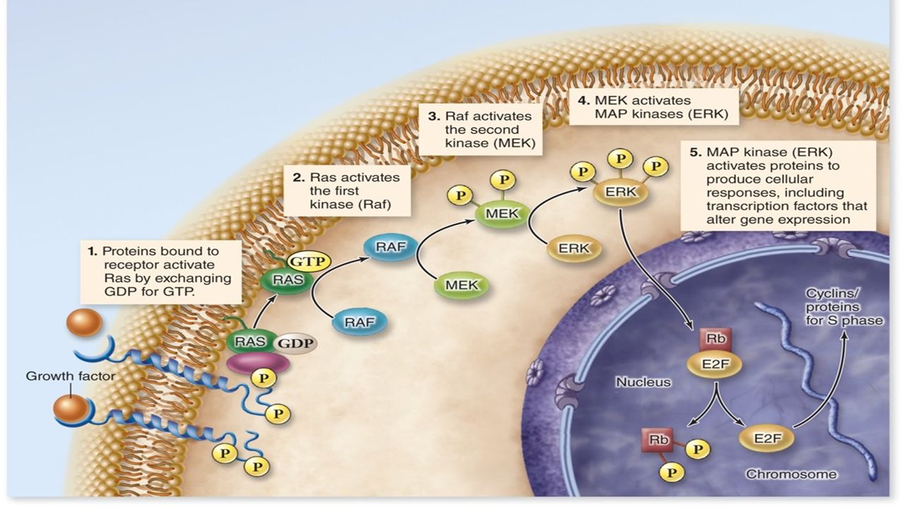
GROWTH FACTORS Function is to trigger intracellular signaling systems One of the first identified was platelet-derived growth factor (PDGF) It is an RTK that initiates a MAP kinase response to stimulate cell division To date, over 50 different growth have been identified Many trigger MAP kinase cascades to activate transcription factors through phosphorylation, which in turn stimulate G 1 cyclin production
 It is an RTK that initiates a MAP kinase response to stimulate cell division To date, over 50 different growth have been identified Many trigger MAP kinase cascades to activate transcription factors through phosphorylation, which in turn stimulate G 1 cyclin production")
37
CANCER: A CORRUPTED CELL CYCLE Cancer is nothing more than unrestrained or uncontrolled cell growth Something has caused the cell cycle to not stop at the appropriate checkpoints The p53 gene is considered one of the most important genes in regulating the cell cycle, if it becomes corrupted cancer is the result

38
P53, A TUMOR SUPPRESSOR GENE Monitors integrity of DNA during the G 1 checkpoint

39
LOSS OF CELL CYCLE CONTROL Oncogenes Genes that when introduced to a cell can cause cancer Proto-oncogenes Normal genes that if they become mutated act as oncogenes PDGF EGF (epidermal growth factor) If one copy of the proto-oncogenes goes bad cancer can result, acts in a genetically dominant fashion
 If one copy of the proto-oncogenes goes bad cancer can result, acts in a genetically dominant fashion")
40
TUMOR SUPPRESSOR GENES P53 acts as a tumor suppressor Both copies of this gene must be bad for a cancer to develop Acts in a genetically recessive fashion Retinoblastoma susceptibility gene (Rb)
")
41
SEXUAL REPRODUCTION AND MEIOSIS CHAPTER 11

42
WHAT IS SEXUAL REPRODUCTION?

43
DISCOVERY OF MEIOSIS Edouard van Beneden Discovered different numbers of chromosomes in Ascaris gametes Somatic cells had 4 chromosomes Gametes only had 2 chromosomes He determined that the gametes must contain half the number because when they fuse it would restore the normal number He called the fused cell a zygote The fusion of the gametes is fertilization or syngamy

44
MEIOSIS IS A REDUCTION DIVISION

45
SEXUAL REPRODUCTION INVOLVES AN ALTERNATION OF GENERATIONS Part of the time life is diploid Part of the time life is haploid Another way of saying it is that meiosis and fertilization alternate

46
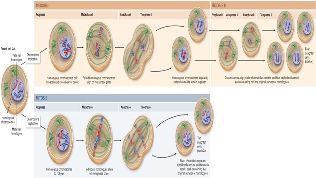
WHAT HAPPENS IN MEIOSIS Two rounds of division containing prophase, metaphase, anaphase, and telophase Homologous chromosomes pair up in a process called synapsis A synaptonemal complex will form Genetic recombination or crossing over of chromosomal material occurs

47
WHAT THE CHROMOSOMES ARE DOING Synaptonemal complex Crossing over

48
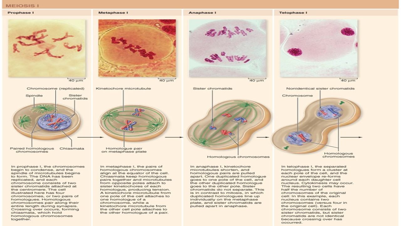
PROPHASE I DNA coils together and the chromosomes can begin to be seen The homologous chromosomes find each other; synapsis Crossing over occurs with the help of recombination nodules that are thought to contain the enzymatic machinery to power the process After crossing over the synaptonemal complex breaks down, but the chromosomes stay attached at the chiasmata The four chromatids are held together: Sister chromatids held together with cohesion proteins Exchange of genetic material locks all four chromatids together

49
METAPHASE I The paired homologous chromosomes line up on the metaphase plate Microtubules from opposite poles attach to the kinetochores

51
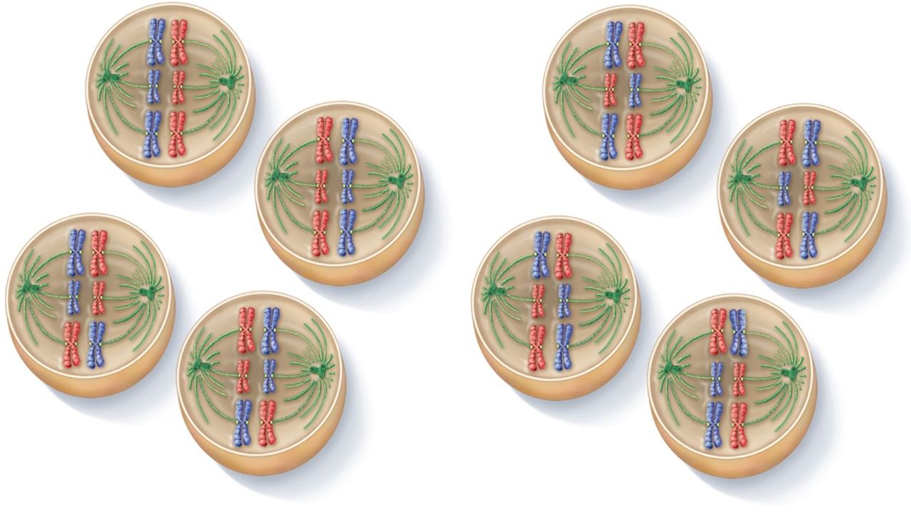
ANAPHASE I Centromeres stay in tact, but the homologous chromosomes release from one another Homologs are pulled to opposite poles by the kinetochore microtubules The random orientation of the homologous chromosomes on the metaphase plate means the chromosomes can sort independently regardless of the maternal or paternal origin, known as independent assortment

52
TELOPHASE I Nuclear membranes reform Sister chromatids are no longer identical due to crossing over, so genetic variability for potential offspring is increased dramatically

54
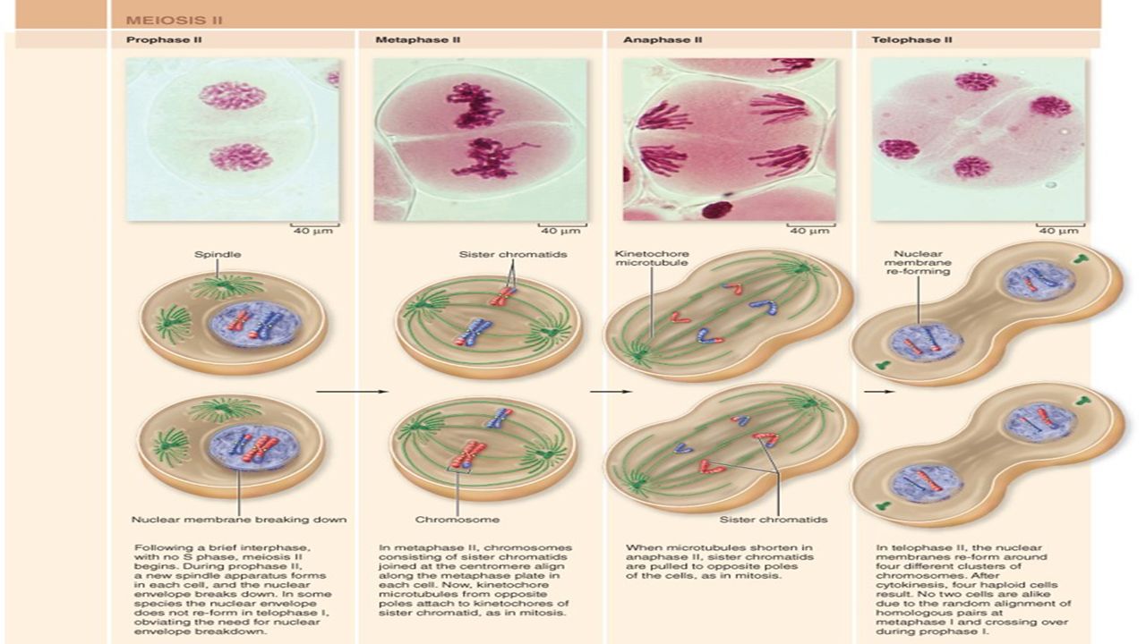
MEIOSIS II All the phases are the same as meiosis I, but there is DNA replication prior to meiosis II No replication of DNA means the number of chromosomes in the final product is reduced

56
MEIOSIS MISTAKES Nondisjunction is the failure of a chromosome to move to the opposite pole Results in a cell that has one extra chromosome and a cell with one less These gametes with improper chromosome number are called aneuploidy gametes and typically result in spontaneous abortion

57
MEIOSIS SUMMARY Crossing over during meiosis I Sister chromatids remain connected at the centromere and segregate during anaphase I Kinetochores of sister chromatids are attached to the same pole in meiosis I and to opposite poles in mitosis DNA replication does not occur between meiosis I and meiosis II

Similar presentations


 a progression from primitive.>")


