Download presentation
Presentation is loading. Please wait.

1
Biology 210 Chapter 13 The Central Nervous System
Edited by John McGill Supplemental Notes by Beth Wyatt Original PowerPoint By: Jack Bagwell Last Updated: March 28, 2017

2
DIVISIONS OF THE NERVOUS SYSTEM
There Are Several Ways That the Nervous System Can Be Organized BASED ON LOCATION OF ORGANS CENTRAL NERVOUS SYSTEM (CNS) PERIPHERAL NERVOUS SYSTEM (PNS)
 PERIPHERAL NERVOUS SYSTEM (PNS)")
3
DIVISIONS OF THE NERVOUS SYSTEM
Organs centrally located CENTRAL NERVOUS SYSTEM (CNS) BRAIN SPINAL CORD
 BRAIN. SPINAL CORD.")
4
DIVISIONS: Peripheral Nervous System
Organs (Nerves) Peripherally Located 2 Kinds of Nerves CRANIAL NERVES (Originate From Brain) SPINAL NERVES (Originate From Spinal Cord)
 Peripherally Located. 2 Kinds of Nerves. CRANIAL NERVES (Originate From Brain) SPINAL NERVES (Originate From Spinal Cord)")
5
Classification Based on Pathways
BASED ON DIRECTION IN WHICH PATHWAYS CARRY INFORMATION NEURONS CONDUCT NERVE IMPULSES) AFFERENT (aa ferent) DIVISION: All Afferent Neurons EFFERENT (ee ferent) DIVISION: All Efferent Neurons
 AFFERENT (aa ferent) DIVISION: All Afferent Neurons. EFFERENT (ee ferent) DIVISION: All Efferent Neurons.")
6
Classification Based on Effectors
BASED ON EFFECTORS INNERVATED/REGULATED SOMATIC NERVOUS SYSTEM (SNS) AUTONOMIC NERVOUS SYSTEM (ANS)
 AUTONOMIC NERVOUS SYSTEM (ANS)")
7
Somatic: Voluntary Effectors
SOMATIC NERVOUS SYSTEM (SNS) Effectors: Skeletal Muscles Voluntary Somatic Effectors
 Effectors: Skeletal Muscles. Voluntary. Somatic Effectors.")
8
Autonomic: Involuntary Effectors
BASED AUTONOMIC NERVOUS SYSTEM (ANS) Effectors: Cardiac and Smooth Muscle,Glands Involuntary Autonomic/Visceral Effectors)
 Effectors: Cardiac and Smooth Muscle,Glands. Involuntary. Autonomic/Visceral Effectors)")
9
Autonomic: 2 Divisions SYMPATHETIC DIVISION-Response of Autonomic Effectors During Stress PARASYMPATHETIC DIVISION-Response of Autonomic Effectors During Nonstress

10
COVERINGS OF THE BRAIN AND SPINAL CORD
Coverings Provide Protection 2 Kinds OUTER COVERING - BONE CRANIAL BONES: BRAIN VERTEBRAE: SPINAL CORD INNER COVERING – MEMBRANES (MENINGES) LAYERS OF MENINGES DURA MATER ARACHNOID MEMBRANE PIA MATER
 LAYERS OF MENINGES. DURA MATER. ARACHNOID MEMBRANE. PIA MATER.")
11
BONE COVERS THE BRAIN AND SPINAL CORD
CRANIAL BONES: BRAIN VERTEBRAE: SPINAL CORD

12
Vertebra & Spinal Cord

13
INNER COVERING MEMBRANES (MENINGES)
LAYERS OF MENINGES DURA MATER ARACHNOID MEMBRANE PIA MATER

14
LAYERS OF MENINGES DURA MATER Outermost Layer of Meninges Tough, White
An Extension of Periosteum

15
Dura Mater of Inferior Sheep Brain
Pituitary Optic Chiasma

16
LAYERS OF MENINGES ARACHNOID MEMBRANE Middle Layer of Meninges
Delicate

17
LAYERS OF MENINGES PIA MATER Innermost Layer of Meninges Thin
Transparent Adheres to CNS Contains Blood Vessels

18
Pia Mater of Superior Sheep Brain

19
SPACES BETWEEN MENINGES
EPIDURAL SPACE SUBDURAL SPACE SUBARACHNOID SPACE

20
EPIDURAL SPACE “Space Above Dura” (Between Bone & Dura Mater)
Contains Connective Tissue (Adipose)
")
21
Epidural Space

22
SUBDURAL SPACE “Space Below Dura” (Between Dura Mater & Arachnoid)
Contains a Serous Fluid That Provides Lubrication

23
SUBARACHNOID SPACE SPACES “Space Below the Arachnoid” (Between Arachnoid & Pia Mater) Contains Cerebrospinal Fluid
 Contains Cerebrospinal Fluid.")
24
CEREBROSPINAL FLUID The Fluid That Circulates In and Around the CNS
FUNCTION Protection (A Cushion of Fluid) FLUID SPACES The Spaces Where Cerebrospinal Fluid Circulates
 FLUID SPACES. The Spaces Where Cerebrospinal Fluid Circulates.")
25
CEREBROSPINAL FLUID: FLUID SPACES
WITHIN BRAIN VENTRICLES 4 Fluid Spaces Within the Brain LATERAL (FIRST AND SECOND) VENTRICLES THIRD VENTRICLE FOURTH VENTRICLE CEREBRAL AQUEDUCT WITHIN SPINAL CORD CENTRAL CANAL
 VENTRICLES. THIRD VENTRICLE. FOURTH VENTRICLE. CEREBRAL AQUEDUCT. WITHIN SPINAL CORD. CENTRAL CANAL.")
26
CEREBROSPINAL FLUID within the BRAIN
VENTRICLES: 4 Fluid Spaces Within the Brain LATERAL (FIRST AND SECOND) VENTRICLES 2, Located in Each Cerebral Hemisphere The Largest of the Ventricles
 VENTRICLES. 2, Located in Each Cerebral Hemisphere. The Largest of the Ventricles.")
27
CEREBROSPINAL FLUID within the BRAIN
VENTRICLES: 4 Fluid Spaces Within the Brain THIRD VENTRICLE Lies Inferior and Medial to the Lateral Ventricles

28
CEREBROSPINAL FLUID within the BRAIN
VENTRICLES: 4 Fluid Spaces Within the Brain FOURTH VENTRICLE Diamond Shaped Space, Located Between the Brainstem and the Cerebellum

29
CEREBROSPINAL FLUID within the BRAIN
CEREBRAL AQUEDUCT Canal That Connects the Third and Fourth Ventricles

30
CEREBROSPINAL FLUID within the SPINAL CORD
CENTRAL CANAL Passageway Within the Spinal Cord Continuous With the Fourth Ventricle

31
CEREBROSPINAL FLUID Circulation
AROUND BRAIN AND SPINAL CORD SUBARACHNOID SPACE

32
CEREBROSPINAL FLUID Formation
FORMATION FROM BLOOD Choroid Plexuses: Capillary Networks Located in Each of the Ventricles As Blood Flows Through Choroid Plexuses, Some of the Fluid from Blood Filters Through the Plexuses and into the, Ventricles, The Fluid is Now Known as CSF

33
Choroid Plexuses

34
CEREBROSPINAL FLUID Circulation
FROM LATERAL VENTRICLES THIRD VENTRICLE CEREBRAL AQUEDUCT FOURTH VENTRICLE* CENTRAL CANAL SUBARACHNOID SPACE BACK TO BLOOD (ARACHNOID VILLI)
")
35
CEREBROSPINAL FLUID Circulation 2nd Pathway
*NOTE: ADDITIONAL PATHWAY: ONCE IN FOURTH VENTRICLE SUBARACHNOID SPACE BACK TO BLOOD (ARACHNOID VILLI) Arachnoid Villi: Fingerlike Extensions of Arachnoid That Project into Blood Vessels
 Arachnoid Villi: Fingerlike Extensions of Arachnoid That Project into Blood Vessels.")
36
SPINAL CORD STRUCTURE LOCATION, EXTENT, SHAPE Spinal Cavity
Extends from Foramen Magnum to Lower Border of the First Lumbar Vertebra (approx. 18 inches) Oval Cylinder
 Oval Cylinder.")
37
SPINAL CORD STRUCTURE: Grooves
ANTERIOR MEDIAN FISSURE Deep Groove in the Anterior Midline POSTERIOR MEDIAN SULCUS Groove in the Posterior Midline

38
Anterior Median Fissure
Olive Pyramids Cerebellum

39
Posterior Median Sulcus: 400X+

40
Posterior Median Sulcus & Others

41
SPINAL CORD: NERVE ROOTS
NERVE ROOTS/SPINAL NERVES Spinal Nerves Attached to Spinal Cord by Nerve Roots (example nerves) DORSAL NERVE ROOT: NF OF AFFERENT NEURONS VENTRAL NERVE ROOT: NF OF EFFERENT NEURONS
 DORSAL NERVE ROOT: NF OF AFFERENT NEURONS. VENTRAL NERVE ROOT: NF OF EFFERENT NEURONS.")
42
DORSAL NERVE ROOT: NF OF AFFERENT NEURONS
Unipolar Receptors/Dendrites in Sense Organs Axons (Peripheral Portion) in Spinal Nerve Cell Bodies in Dorsal Root Ganglion Axons (Central Portion) in Dorsal Nerve Root
 in Spinal Nerve. Cell Bodies in Dorsal Root Ganglion Axons (Central Portion) in Dorsal Nerve Root.")
43
VENTRAL NERVE ROOT: NF OF EFFERENT NEURONS
Efferent Neurons: Multipolar Dendrites/Cell Bodies in Gray Matter of Spinal Cord Axons in Ventral Nerve Root and S. Nerve

44
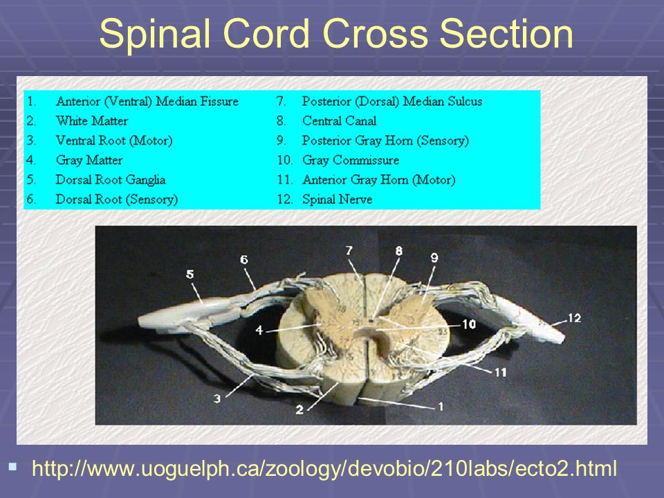
Spinal Cord Cross Section
45
Spinal Cord: Afferent (unipolar) & Efferent (multipolar) Neurons
 & Efferent (multipolar) Neurons")
46
Spinal Cord: Gray and White Matter
The spinal cord consists of Gray Matter: Consists of cell bodies of interneurons and motor neurons White Matter Consists of axons of neurons originating in the spinal cord and brain.

47
SPINAL CORD: Gray Matter
In Spinal Cord, Gray Matter Primarily Consists of Cell Bodies of Interneurons and Motor Neurons

48
SPINAL CORD: Gray Matter
LOCATION: Central Portion of Spinal Cord, Resembles Letter “H” DIVISIONS: GRAY HORNS (COLUMNS), ANTERIOR, POSTERIOR, & LATERAL
, ANTERIOR, POSTERIOR, & LATERAL.")
49
SPINAL CORD: White Matter
Consists of axons of neurons originating in the spinal cord and brain.

50
SPINAL CORD: White Matter
LOCATION: Peripheral to Gray Matter DIVISIONS WHITE COLUMNS: ANTERIOR, POSTERIOR, LATERAL TRACTS White Columns Further Subdivided into Tracts Names of Tracts Often Indicate 2 Things About Tract: Where Tract Begins/Ends White Column in Which Tract Located

51
SPINAL CORD: White Matter
WHITE COLUMNS ANTERIOR POSTERIOR LATERAL

52
Gray and White Matter

53
SPINAL CORD: White Matter
FUNCTIONS PROVIDES CONDUCTION ROUTES (2 WAY) FOR NERVE IMPULSES TRAVELING TO AND FROM THE BRAIN (WHITE MATTER) White Matter of the Spinal Cord (Tracts) Conducts Impulses Toward and Away from the Brain Ascending tracts conduct impulses up the SC toward the brain Descending tracts conduct impulses down the SC away from the brain
 FOR NERVE IMPULSES TRAVELING TO AND FROM THE BRAIN (WHITE MATTER) White Matter of the Spinal Cord (Tracts) Conducts Impulses Toward and Away from the Brain. Ascending tracts conduct impulses up the SC toward the brain. Descending tracts conduct impulses down the SC away from the brain.")
54
SPINAL CORD: ASCENDING TRACTS
Conduct Impulses Up the SC Toward the Brain FUNCTION Ascending Tracts Have a Sensory Function (Carry Impulses Related To General Sensations: Hot, Cold, Pain, Pressure, Touch & Kinesthesia) IMPORTANT ASCENDING TRACTS SPINOTHALAMIC TRACTS FASCICULI GRACILIS AND CUNEATUS SPINOCEREBELLAR TRACTS
 IMPORTANT ASCENDING TRACTS. SPINOTHALAMIC TRACTS. FASCICULI GRACILIS AND CUNEATUS. SPINOCEREBELLAR TRACTS.")
55
SPINAL CORD: DESCENDING TRACTS
DESCENDING TRACTS Elaboration Conduct Impulses Down the SC Away from the Brain FUNCTION Descending Tracts Have a Motor Function (Carry Impulses that Will Result in Voluntary Movement of Skeletal Muscles) IMPORTANT DESCENDING TRACTS CORTICOSPINAL TRACTS RETICULOSPINAL TRACTS RUBROSPINAL TRACTS
 IMPORTANT DESCENDING TRACTS. CORTICOSPINAL TRACTS. RETICULOSPINAL TRACTS. RUBROSPINAL TRACTS.")
56
Spinal Cord Tracts

57
Ascending Tracts Spinocerebellar tracts Spinothalamic tracts
Impulses from stretch receptors are carried to the spinal cord. These tracts originate in the spinal cord and transmit signals to the cerebellum Involved in regulation of muscle tone without reaching consciousness. Spinothalamic tracts Fibers concerned with pain, thermal sense, and light touch Originate in the spinal cord. Convey sensory impulses to the thalamus. Dorsal column: Fasciculi Gracilis and Cuneatus Tracts Arise from spinal ganglion cells The fasciculi terminate upon large nuclear masses (the nuclei gracilis and cuneatus) in the medulla. Conveys signals associated with tactile, pressure, and kinesthetic (or positional) sense to sensory areas of the cerebral cortex.
 in the medulla. Conveys signals associated with tactile, pressure, and kinesthetic (or positional) sense to sensory areas of the cerebral cortex.")
58
Descending Tracts Corticospinal tract Rubrospinal tract
Concerned with skilled voluntary activity, the corticospinal tract originates from premotor, primary motor, and primary sensory cortex. Synapse with interneurons and motor neurons. Rubrospinal tract Arises from cells in the midbrain. Fibers of this tract descend the spinal cord and terminate on interneurons. Cells of the midbrain receive input from the motor cortex and from the cerebellum (via the superior cerebellar peduncle). The rubrospinal tract brings flexor muscle tone under the control of these two regions of the brain. Reticulospinal tract Arises from the reticular formation of the pons and medulla Fibers of this tract influence voluntary movements, muscle tone, and a variety of spinal reflexes. Fibers terminate at all spinal levels. Receive input from regions of the motor cortex.
. The rubrospinal tract brings flexor muscle tone under the control of these two regions of the brain. Reticulospinal tract. Arises from the reticular formation of the pons and medulla. Fibers of this tract influence voluntary movements, muscle tone, and a variety of spinal reflexes. Fibers terminate at all spinal levels. Receive input from regions of the motor cortex.")
60
Spinal Cord Tracts

61
SPINAL CORD : Reflexes REFLEX CENTER FOR ALL SPINAL REFLEXES (GRAY MATTER) Gray Matter of the Spinal Cord Functions as Reflex Centers for Spinal Reflexes Reflex Center: Center of a Reflex Arc Spinal Reflexes: Reflexes Whose Arcs Pass Through the SC

62
SPINAL CORD : Reflexes

63
SPINAL CORD : Reflexes

64
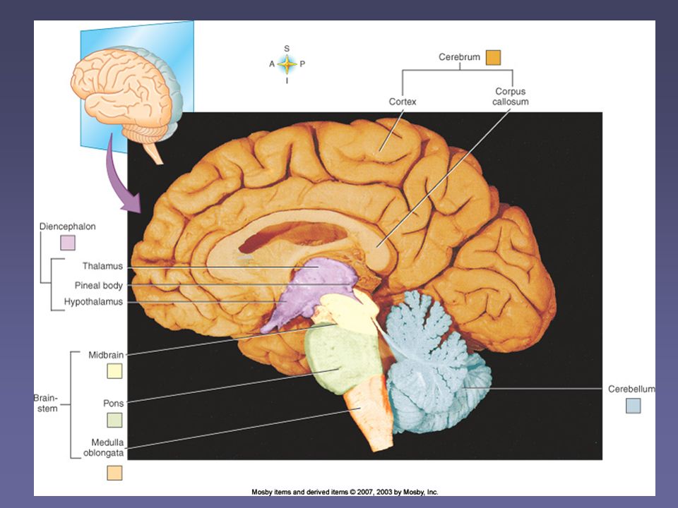
THE BRAIN DIVISIONS SIZE BRAINSTEM CEREBELLUM DIENCEPHALON CEREBRUM
MEDULLA OBLONGATA PONS MIDBRAIN CEREBELLUM DIENCEPHALON CEREBRUM SIZE In Adults, Weighs Approx. 3 Pounds

66
THE BRAIN: Brainstem BRAINSTEM STRUCTURE
Extension of SC into the Cranial Cavity STRUCTURE MEDULLA OBLONGATA Lowermost Division PONS Middle, Swollen Division MIDBRAIN Uppermost Division

67
THE BRAIN: Brainstem MIDBRAIN: Uppermost Division; Has 2 Important External Landmarks CEREBRAL PEDUNCLES: Form Floor of Midbrain CORPORA QUADRIGEMINIA

68
Cerebral Peduncles

69
Midbrain: Copora Quadrigemina
CORPORA QUADRIGEMINIA Forms Roof of Midbrain CQ Consists of 4 Rounded Structures SUPERIOR COLLICULI (2): Visual Centers INFERIOR COLLICULI (2): Auditory Centers
: Visual Centers. INFERIOR COLLICULI (2): Auditory Centers.")
70
CORPORA QUADRIGEMINIA

71
THE BRAIN: Reticular Formation
LOCATIONS OF GRAY AND WHITE MATTER IN BRAINSTEM WHITE MATTER –outside RETICULAR FORMATION – inside Reticular Formation = Mixture of Gray and White Matter Located in the Core of the Brainstem

72
Reticular Formation http://www. medinfo. ufl

73
Reticular Formation http://www. medinfo. ufl

74
THE BRAIN: Brainstem Tracts (white matter)
FUNCTIONS The Functions of the Brainstem are Similar to the Functions of the SC PROVIDES CONDUCTION ROUTES (2 WAY) FOR NERVE IMPULSES TRAVELING BETWEEN SPINAL CORD AND BRAIN (WHITE MATTER) The White Matter of the Brainstem is Organized into Tracts (Ascending & Descending Which are an Extension of The Tracts of the Spinal Cord
 FOR NERVE IMPULSES TRAVELING BETWEEN SPINAL CORD AND BRAIN (WHITE MATTER) The White Matter of the Brainstem is Organized into Tracts (Ascending & Descending Which are an Extension of The Tracts of the Spinal Cord.")
75
THE BRAIN: Reticular Formation
REFLEX CENTER FOR BRAIN REFLEXES (GRAY MATTER) Portions of Gray Matter Located Within the Reticular Formation Functions as Reflex Centers for Brain Reflexes (Brain Reflexes: Reflexes Whose Arcs Pass Through the Brainstem)
 Portions of Gray Matter Located Within the Reticular Formation Functions as Reflex Centers for Brain Reflexes. (Brain Reflexes: Reflexes Whose Arcs Pass Through the Brainstem)")
76
THE BRAIN: Brain Reflexes
2 Kinds of Reflex Centers in Brainstem VITAL REFLEX CENTERS (MEDULLA) Reflex Centers for Vital Reflexes Vital Reflexes: Reflexes that are Essential for Survival; Examples: Breathing, Heart rate, BP Names of Vital Reflex Centers: Respiratory Centers, Cardiac Control Centers, Vasomotor Center All of the Vital Reflex Centers are Located in the Medulla (Medulla Associated with Basic Survival)
 Reflex Centers for Vital Reflexes. Vital Reflexes: Reflexes that are Essential for Survival; Examples: Breathing, Heart rate, BP. Names of Vital Reflex Centers: Respiratory Centers, Cardiac Control Centers, Vasomotor Center. All of the Vital Reflex Centers are Located in the Medulla (Medulla Associated with Basic Survival)")
77
THE BRAIN: Brain Reflexes
NONVITAL REFLEX CENTERS (MEDULLA, PONS, MIDBRAIN) Reflex Centers for Nonvital Reflexes (Not Essential for Basic Survival: Vomiting, Coughing, Hiccuping, Swallowing, Sneezing, etc.); Centers Have Same Names as Reflexes Nonvital Reflex Centers Located in All Parts of the Brainstem
 Reflex Centers for Nonvital Reflexes (Not Essential for Basic Survival: Vomiting, Coughing, Hiccuping, Swallowing, Sneezing, etc.); Centers Have Same Names as Reflexes. Nonvital Reflex Centers Located in All Parts of the Brainstem.")
78
Brain Illusions and Information

Similar presentations


>")


![The Nervous System. Divisions of the Nervous System Central Nervous System [CNS] = Spinal Cord Brain Peripheral Nervous System [PNS]= Spinal Nerves.](/16/5239306/big_thumb.jpg "The Nervous System. Divisions of the Nervous System Central Nervous System [CNS] = Spinal Cord Brain Peripheral Nervous System [PNS]= Spinal Nerves.>")

 and Nerves. NERVOUS SYSTEM 1.Collect sensory input 2.Integrate sensory input 3.Motor output Functions of Nervous System.>")


 Bone – Cranium, Vertebrae 2) Meninges – Three connective tissue membranes covering the brain and spinal cord a) Dura Mater – outermost,>")