Download presentation
Presentation is loading. Please wait.

1
The Cardiovascular System: Blood Vessels and Circulation

2
Blood Vessels Arteries- from heart
Elastic => large Muscular => distribution to organs Arterioles => distribution to capillaries- mostly muscle Capillaries- thin walled for diffusion Veins- to heart Venules => from capillaries Veins from tissue to vena cavae to heart Blood Vessels

3
Figure 16.1ab

4
Figure 16.1c

5
Blood Vessel Structure
Three layers Tunica Intima (Interna)- Innermost, endothelial layer Tunica Media- Middle, muscular layer Tunica Externa- Outermost layer Blood Vessel Structure
- Innermost, endothelial layer. Tunica Media- Middle, muscular layer. Tunica Externa- Outermost layer. Blood Vessel Structure.")
6
Blood Vessel Structure
Differences Arteries-> thicker tunica media Elastic tissue and/or muscle As they get smaller-> more muscle Arterioles-> very muscular- control Veins- bigger lumen and thinner walls Veins-> valves to prevent backflow Venules very thin, no valves Blood Vessel Structure

7
Vessel Functions Differences
Muscular arteries & arterioles regulate flow Sympathetic activity to smooth muscle vasoconstriction (narrowing) Decreased sympathetic activity or NO causes relaxation or vasodilation Arterioles adjust flow into capillaries Systemic veins & venules serve as blood reservoirs (~64% total blood volume) Vessel Functions
 Decreased sympathetic activity or NO causes relaxation or vasodilation. Arterioles adjust flow into capillaries. Systemic veins & venules serve as blood reservoirs (~64% total blood volume) Vessel Functions.")
8
Capillary Details Capillaries only have endothelium
Very thin cells & cell nuclei protrude into lumen- easy diffusion Connected from arterioles to venules in networks Sometimes direct route from arteriole to venule Filling controlled by small arterioles & precapillary sphincters Capillary Details

9
Figure 16.2a

10
Figure 16.2b

11
Capillary Exchange Slow flow through capillaries Blood pressure
Allows time for exchange through wall Blood pressure filtration of fluid out of capillary Mostly in first ½ of vessel length Osmosis (protein concentration) Reabsorption of fluid from outside to inside Mostly in last ½ of vessel length Balance determines fluid in circulation Excess fluid returned via lymphatic system Local signals can adjust capillary flow Capillary Exchange
 Reabsorption of fluid from outside to inside. Mostly in last ½ of vessel length. Balance determines fluid in circulation. Excess fluid returned via lymphatic system. Local signals can adjust capillary flow. Capillary Exchange.")
12
Figure 16.3

13
Venous Return Blood enters veins at very low pressure.
Needs more pumping to get back to heart = action of heart; muscle pumps; respiratory pump Some pressure from heart action Not enough to overcome gravity Venous Return

14
Muscle & Respiratory Pumps
Contracting skeletal muscles squeeze veins emptying them Venous valves flow is toward heart Respiratory pump has similar action Inhalation decreased thoracic pressure & increased abdominal pressure Blood flows toward heart Exhalation allows refilling of abdominal veins Muscle & Respiratory Pumps

15
Figure 16.4

16
From high pressure area to lower pressure area, i. e
From high pressure area to lower pressure area, i.e. down pressure gradient Greater gradient greater flow Ventricular contraction blood pressure (BP) Highest in aorta and declines as flows through vessels mmHg in aorta ~16 mmHg at venules 0 at R. Atrium Resistance= opposition to flow Blood Flow
 Highest in aorta and declines as flows through vessels mmHg in aorta ~16 mmHg at venules 0 at R. Atrium. Resistance= opposition to flow. Blood Flow.")
17
Resistance Depends on: Vessel lumen diameter
Smaller lumen greater resistance Blood viscosity (thickness) Higher viscosity greater resistance Viscosity of blood depends on Hct Total vessel length Longer the length of flow the more friction with wall Total body resistance increases with growth and addition of tissue Resistance
 Higher viscosity greater resistance. Viscosity of blood depends on Hct. Total vessel length. Longer the length of flow the more friction with wall. Total body resistance increases with growth and addition of tissue. Resistance.")
18
Figure 16.5

19
Regulation of Blood Pressure & Flow
Fast responses: e.g. standing up Slower responses: e.g. blood volume Distribution: e.g. to working muscles Balance of CO (cardiac output) with flow to body Interacts with many other control systems Cardiovascular (CV) Center major regulator Regulation of Blood Pressure & Flow
 with flow to body. Interacts with many other control systems. Cardiovascular (CV) Center major regulator. Regulation of Blood Pressure & Flow.")
20
Inputs Higher centers: Sensory receptor input: Cerebral cortex,
Limbic system Hypothalamus Flow adjusted for body temperature Sensory receptor input: Proprioceptors Baroreceptors Chemoreceptors Inputs

21
Inputs Proprioceptors: Baroreceptors: in aorta & carotid
Start HR change as activity starts Baroreceptors: in aorta & carotid pressure parasympathetic & sympathetic stimulation CO Chemoreceptors: in aorta & carotid Low O2, high H+, CO2 vasoconstriction BP Inputs

22
Figure 16.6

23
Output ANS to heart Sympathetic HR & force of contraction
Parasympathetic HR Vasomotor To arterioles vasomotor tone (vasoconstriction) To veins move blood to heart BP Output
 To veins move blood to heart BP. Output.")
24
Hormone regulation Renin-Angiotensin system
Angiotensin II vasoconstriction+ thirst aldosterone Na+ & water loss in urine on Epinephrine & Norepinephrine CO ADH = Vasopressin constriction BP Thirst & water retention in kidney BP ANP- from cells in atria Vasodilation & loss of salt & water in urine BP Hormone regulation

25
Figure 16.7

26
Checking Circulation- Pulse
Pulse in arteries = HR (Heart Rate) Use radial artery at wrist carotid artery- neck brachial artery- arm Tachycardia = rapid rest rate (>100 bpm) Bradycardia= slow rest rate (<50 bpm) Checking Circulation- Pulse
 Use radial artery at wrist. carotid artery- neck. brachial artery- arm. Tachycardia = rapid rest rate (>100 bpm) Bradycardia= slow rest rate (<50 bpm) Checking Circulation- Pulse.")
27
Blood Pressure Arterial Blood Pressure Systolic pressure
Peak arterial pressure during ventricular systole Diastolic pressure Minimum arterial pressure during diastole Blood Pressure

28
Blood Pressure Use sphygmomanometer Raise pressure above systolic-
Usually on brachial artery Raise pressure above systolic- stop flow Lower pressure in cuff until flow just starts first sound Systolic Pressure Lower until sound suddenly gets faint Diastolic pressure Blood Pressure

29
Blood Pressure Normal = 120/80 Hypertension Hypotension
Abnormally high blood pressure Greater than 140/90 Hypotension Abnormally low blood pressure Less than 90/60 Blood Pressure

30
Aging Stiffening of aortae Loss of cardiac muscle strength
Reduced CO & increased systolic pressure Coronary artery disease Congestive heart failure Atherosclerosis Aging

31
Anatomy of Blood Vessels

32
Blood flow through Vessels
From Heart Arteries Arterioles Capillaries Venules Veins To Heart Blood flow through Vessels

33
Figure 16.1ab

34
Figure 16.1c

35
Blood Vessel Structure
Three layers Tunica Intima (Interna)- Innermost, endothelial layer Tunica Media- Middle, muscular layer Tunica Externa- Outermost layer Blood Vessel Structure
- Innermost, endothelial layer. Tunica Media- Middle, muscular layer. Tunica Externa- Outermost layer. Blood Vessel Structure.")
36
Blood Vessel Structure
Differences Arteries-> thicker tunica media Veins- bigger lumen and thinner walls, valves to prevent backflow Capillaries only have endothelium Blood Vessel Structure

37
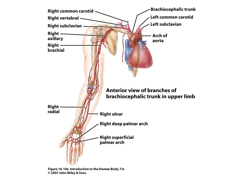
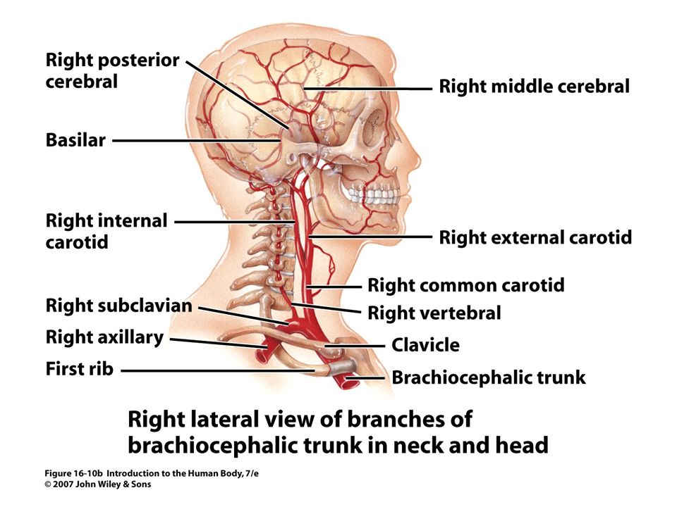
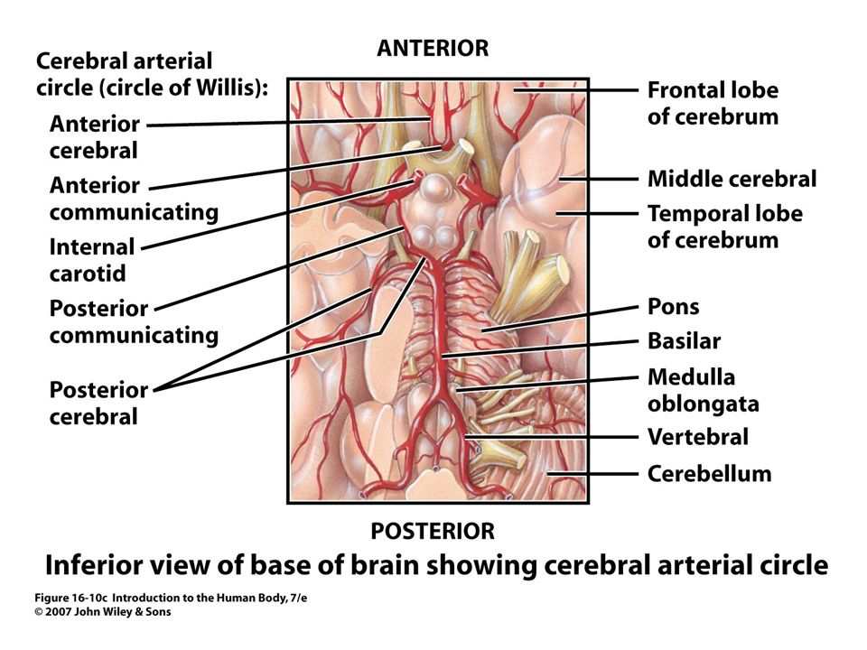
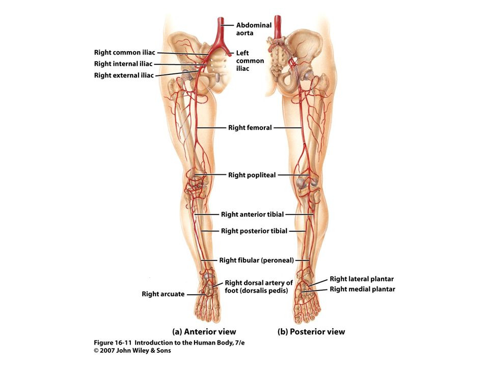
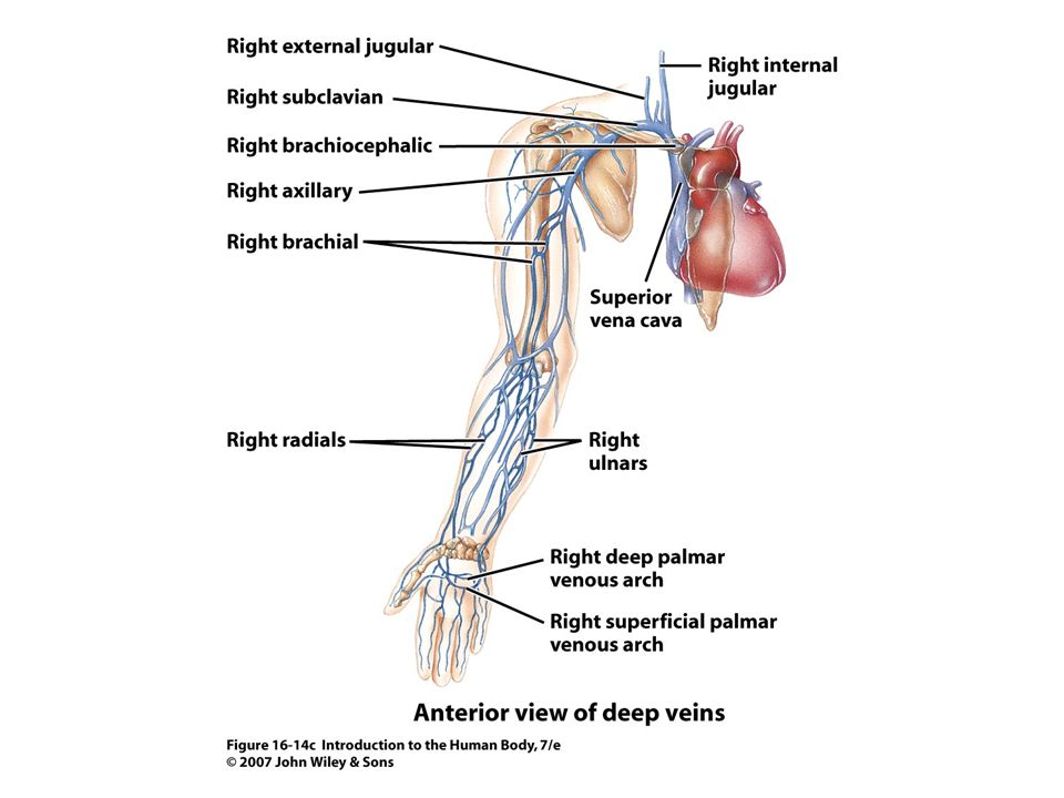
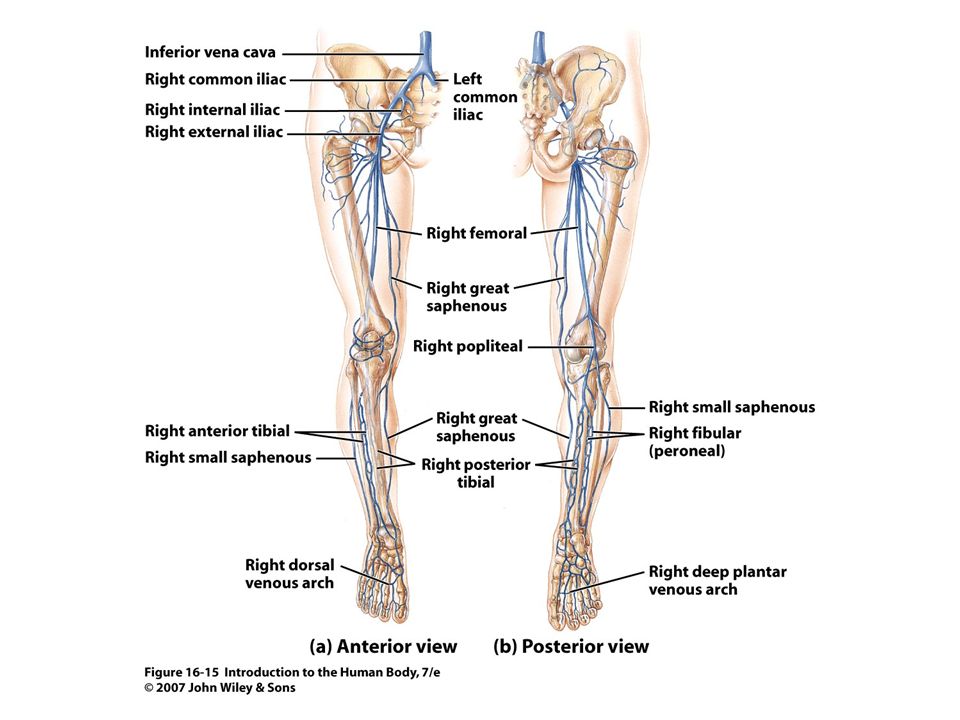
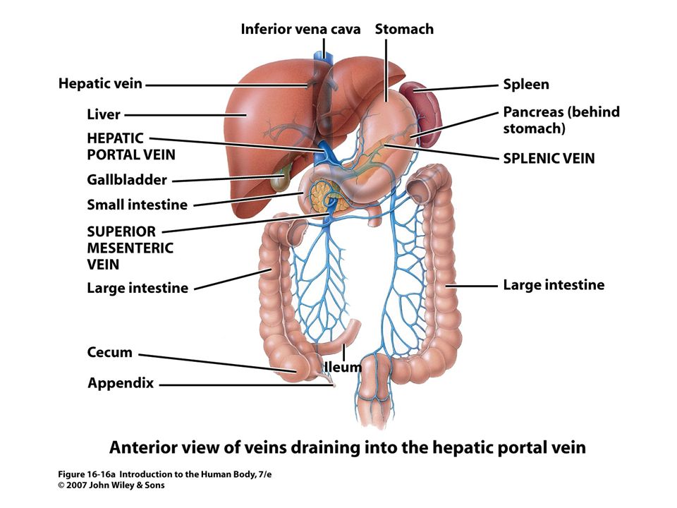
Circulatory Routes Two parts: Systemic & Pulmonary
Systemic circulation- throughout body Oxygenated blood deoxygenated as it goes All systemic arteries branch from aorta All systemic veins empty into Superior Vena Cava, Inferior Vena Cava or the Coronary Sinus Carry deoxygenated blood to heart Circulatory Routes

38
Pulmonary Circulation
From right ventricle pulmonary trunk R. & L. pulmonary arteries Carry deoxygenated blood R. & L. lungs Gas exchange occurs 2 R. & 2 L. pulmonary veins Carry oxygenated blood L. atrium Pulmonary Circulation

40
Pulmonary circuit (veins) Systemic circuit (arteries)
Brain Upper limbs Pulmonary circuit (veins) Lungs LA Systemic circuit (arteries) Left ventricle Kidneys Spleen Digestive organs Liver Gonads Lower limbs 40
 Lungs. LA. Systemic circuit (arteries) Left ventricle. Kidneys. Spleen. Digestive organs. Liver. Gonads. Lower limbs. 40.")
41
Pulmonary circuit (arteries) Systemic circuit (veins)
Brain Upper limbs Pulmonary circuit (arteries) Lungs RA Right ventricle Systemic circuit (veins) Kidneys Digestive organs Liver Gonads Lower limbs 41
 Lungs. RA. Right ventricle. Systemic circuit (veins) Kidneys. Digestive organs. Liver. Gonads. Lower limbs. 41.")
Similar presentations