Download presentation
Presentation is loading. Please wait.

1
Medical Imaging: the Glass Patient Prof.dr.ir. Bart M. ter Haar Romeny Technische Universiteit Eindhoven Dept. of Biomedical Engineering

2
Image Acquisition Techniques Classical X-Ray Classical X-Ray Computed Tomography Computed Tomography Nuclear Medicine Nuclear Medicine Ultrasound Ultrasound Magnetic Resonance Imaging Magnetic Resonance Imaging

3
28 December 1895 Prof. Röntgen presenting his invention at Würzburg, 23 January 1896

4
The first X-ray ever: the hand of Röntgen’s wife, end 1895. One of the first medical examples: a shot of hail in a hand, 1896

5
Anode connection + kV Filament connection High Voltage supply + - Principle of the X-ray tube: The kinetic energy of the electrons is released by the collision at the anode. The tube is vacuum. vacuum Tungsten anode cathode X-rays output

6
Classical X-ray images

8
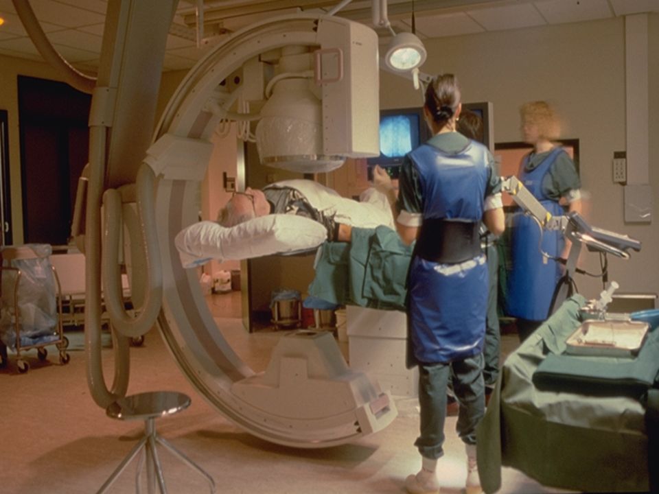
Fluoroscopy with the image intensifier during angioplasty: Real-time visualization of catheters and vessels. Image intensifier X-ray tube High voltage generator

9
DSA = Digital Subtraction Angiography = Röntgen X-ray with contrast in vessels Dotter procedure: Blow up balloon in obstructed vessel

11
Tomoscan AV EasyVision CT = Computed Tomography = Röntgen X-ray slices 3D Greek = to cut, to slice

12
CT: solve for 512x512 pixels by 512x512 equations CT: solve for 512x512 pixels by 512x512 equations Result: a slice Result: a slice

13
Examples CT

14
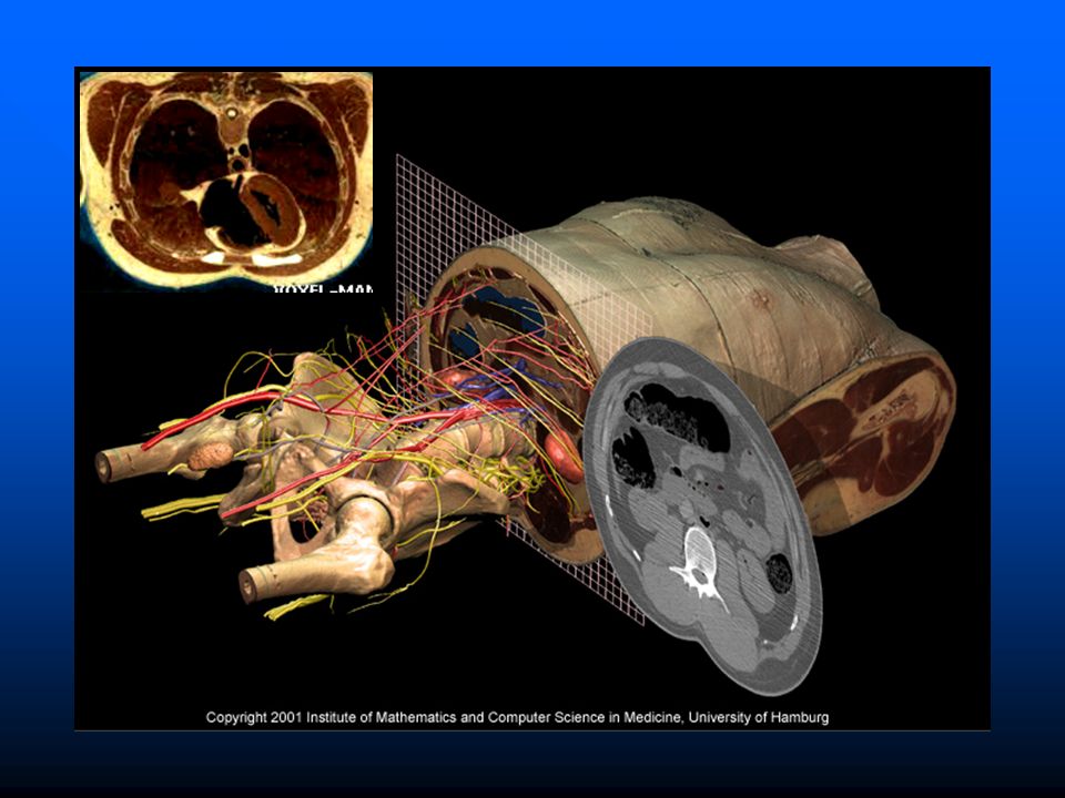
3D visualization Simulation of the physics of light reflection (ray casting/tracing) “2.5D” image
 2.5D image")
16
Nuclear Medicine Principle: Instable radioactive isotopes are made, and build in a pharmacon Instable radioactive isotopes are made, and build in a pharmacon Patient gets contrast medium injected, which specifically stores in tissue Patient gets contrast medium injected, which specifically stores in tissue Signal position is measured with a gamma-camera Signal position is measured with a gamma-camera

17
Ionizing radiation: GAMMA When the nucleus gets too large, the “strong force” is not strong enough to compensate the repulsive force of the protons Alpha radiation: He nuclei (come only microns far in tissue) Beta radiation: electrons (come only cm far in tissue) Gamma radiation: high energy photons (easily go through tissue) GAMMA photon(s)
 Beta radiation: electrons (come only cm far in tissue) Gamma radiation: high energy photons (easily go through tissue) GAMMA photon(s)")
18
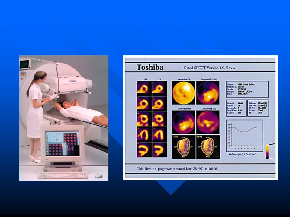
Nuclear Imaging Camera 3-rotating-head SPECT scanner SPECT = Single Photon Emission Computed Tomography

20
PET = Positron Emission Tomography No taskDuring task Positron = anti-electron When it meets an electron → annihilation (explosion) Two photons go in opposite direction, ring coincidence detector
 Two photons go in opposite direction, ring coincidence detector")
21
Molecular Imaging Nano-vesicles: - antibody bindings - 90.000 Gadolinium atoms - container for pharmaca - break by US shockwave - less side effects - chemotherapy on target Highly specific tracer biomolecules

22
Ultrasound Kretz Medicor 530D

23
F 0 F 1 skin vessel (red) bloodcells F d = F 0 - F 1 = 2 x V x cos c V F d = Doppler (‘difference’) frequency transducer Doppler
 bloodcells F d = F 0 - F 1 = 2 x V x cos c V F d = Doppler (‘difference’) frequency transducer Doppler")
24
3D ultrasound

25
Magnetic Resonance Imaging (MRI) X Y Z ( ) Receiver Coil
 X Y Z ( ) Receiver Coil")
26
Philips Medical Systems 1000 x 1000 pixels = 1 million measurements

27
Superconducting Magnet

28
MR Angiography Excitation only of a thin slice Non excited blood flows in the slice Readout of little ‘zero-signal’ areas For all slices → angiogram

29
Why so many imaging modalities? Choice modality: Tissues have different properties for different physical interactions Choice modality: Tissues have different properties for different physical interactions Contrast: Tissue types differ in one or more of these properties Contrast: Tissue types differ in one or more of these properties Anatomical imaging versus functional imaging Anatomical imaging versus functional imaging CT MR

30
Anna Vilanova, Vienna TU / TUE - BMT Univ. of Dusseldorf Philips Medical Systems A new 3D technique: Virtual endoscopy

31
New Eyes are assisting the Radiologist The overwhelming amount of data calls for condensed presentation and analysis Philips Medical Systems Vital Images Groeller - TU Vienna

32
Image Guided Surgery

33
Bev Doolittle: The forest has eyes Physics everywhere Image Acquisition Pattern recognition Computer aided diagnosis Biomedical research New researchers Strong benefit for the patient

Similar presentations


 – Introduction of medical imaging and MRI – Basic.>")


 describe the nature of X-rays Stowmarket Physics X-rays - nature Forms of electromagnetic radiation Short wavelength High frequency.>")


