Download presentation
Presentation is loading. Please wait.

1
Medical Imaging and Anatomy Mike Houston, Anthony Sherbondy, Ruwen Hess

2
Where do the pictures come from? The imagination Dissection X-ray, CT, MRI, Cryosection, PET/SPECT, etc.

3
X-Ray Transmission Imaging Shoot x-rays through the patient onto detector film. Different tissues absord and deflect x-rays to different degrees. The film is exposed less when x- rays encounter higher density material like bone. Low resolution. Hard to distinguish between blood vessals and tissue without an injection of iodine or barium

4
Computed Tomography (CT) Sort of a 3D x-ray. An x-ray emitter is rotated around the patient and a receiver measures the intensity of the transmitted rays from different angles Uses an electronic receiver instead of film. Became generally available in mid 1970's and have gotten MUCH better in resolution and accuracy. Still have problems with metal in the body...

5
Magnetic Resonance Imaging (MRI) Subject body to strong magnetic field (0.08-4T) causing the nuclei of magnetic isotopes to align their orientation. This causes the nuclei to absorb energy and enter a higher energy state. When magnetic field is turned off, nuclei return to equilibrium state emitting energy. Each element has a unique energy signature that can then be measured. Getting more common and very cheap. Can now get a full body MRI scan for ~$500

6
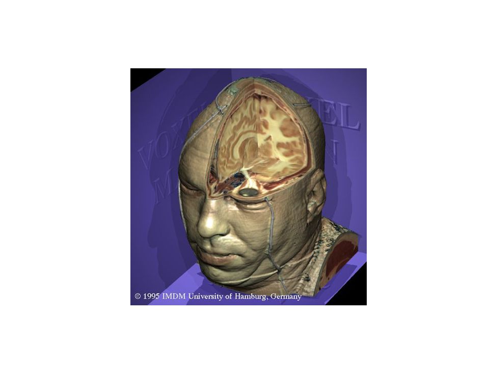
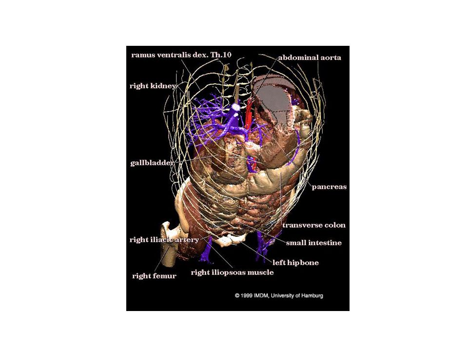
Cryosection Freeze specimen or embed in a plastic polymer Cut specimen into as many slices as possible along an axis Photograph each slice Example: Visible Male and Visible Female

7
Positron Emission Tomography (PET) Inject a metabolically active tracer into subject with an affinity to a certain molecule. 18 F will accumulate in the brain where glucose is used as primary energy source. The radioactive nuclei decay by positron emmission which collides with a free electron resulting in a gamma ray Detectors pick up the events and the a reconstruction is computed

8
Medical Imaging for Education

9
Exploring the human body The "Ancients" Gray's Anatomy Visible Human Project

15
The Visible Human Project Create a digital image dataset of complete human male and female cadavers in MRI, CT and anatomical modes. MR 256x256 resolution at 4mm intervals, 12-bit/pixel CT 512x512 resolution at 1mm intervals, 12-bits/pixel Cryosection – Low res: 2048x1216 at 1mm intervals, 24- bits/pixel – High res: 4096x2700 at 1mm intervals, 24- bits/pixel 1871 slices per mode Note: specs are for visible male

16
So what? We now have a digitized model of an "average" male and female using the current major medical imaging techniques We can now get views of the body that were previously difficult if not impossible But, we know have LOTS of data and have to figure out how to visualize it effectively

17
Lots of examples

Similar presentations


 quantum property of protons energy absorbed when precession frequency.>")


 CT scanning or (CAT scanning) is using X-rays to create a 3D image of the inside of an object. CT stands for computed tomography.>")

, was made possible in part by NIH NLM contract# HHSN276201000580P,>")


 –X-rays –CT (Computer Tomography) –MRI (Magnetic Resonance Imaging)>")


