Download presentation
Presentation is loading. Please wait.

1
Fluid and Electrolytes Jan Bazner-Chandler CPNP, CNS, MSN, RN

2
Alteration in Fluid and Electrolyte Status Normal routes of fluid excretion in infants and children. Lungs Skin Urine & feces Ball & Bender

3
Regulatory Mechanisms Kidneys Gastrointestinal tract Thermoregulatory mechanism Thirst mechanism

4
Kidneys Regulate fluid by their ability to concentrate and dilute urine. When serum sodium levels are high, ADH is secreted and increases permeability of kidney’s distal tubules and ducts. Angiotensin-renin system along with aldosterone assists in regulating fluids and electrolytes homeostasis.

5
Gastrointestinal Tract In GI tract – water and sodium are reabsorbed and potassium is secreted. Fluid is replaced through oral intake. Due to large surface area of GI tract – changed in fluid and electrolyte balance can occur rapidly.

6
Thermoregulatory Mechanism Insensible loss – passive water loss through skin and lungs No electrolytes are lost

7
Thirst Mechanism Thirst center is located in the hypothalamus Thirst is stimulated by decrease in intravascular volume

8
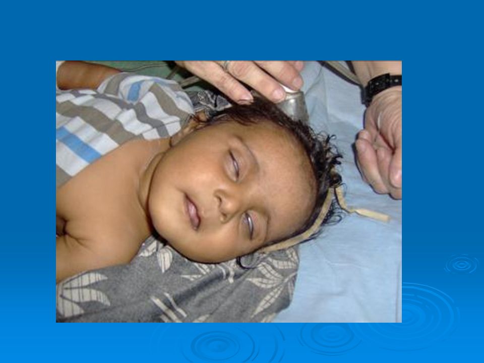
Developmental and Biological Variances Infants younger than 6 weeks do not produce tears. In an infant a sunken fontanel may indicate dehydration. Infants are dependant on others to meet their fluid needs. Infants have limited ability to dilute and concentrate urine.

9
Developmental and Biological The smaller the child, the greater the proportion of body water to weight and proportion of extracellular fluid to intracellular fluid. Infants have a larger proportional surface are of the GI tract than adults. Infants have a higher metabolic rate than adults. (increased HR and RR)
.")
10
Developmental and Biologic Because of immature kidney function, children lack ability to adjust to major changes in sodium and other electrolytes. Normal urine output is 1 mL / kg / hr. More prone than adults to conditions that affect fluid and electrolyte status (diarrhea, vomiting, high fever).
..")
11
Increased Water Needs Fever Vomiting and Diarrhea Diabetes insipidus Burns Shock (hypovolemic) Tachypnea
 Tachypnea")
12
Decreased Water Needs Congestive Heart Failure Mechanical Ventilation Renal failure Head trauma / meningitis

13
Focused Health History Recent fluid intake including type of fluid ingested How many voids in past 12 to 24 hours. Recent weight loss or gain

14
Focused Physical Assessment How does the child look? Skin: Skin: TemperatureTemperature Dry skin and mucous membranesDry skin and mucous membranes Poor turgor, tenting, dough-like feelPoor turgor, tenting, dough-like feel Sunken eyeballs; no tearsSunken eyeballs; no tears Pale, ashen, cyanotic nail beds or mucous membranes.Pale, ashen, cyanotic nail beds or mucous membranes. Delayed capillary refill > 2-3 secondsDelayed capillary refill > 2-3 seconds

16
Loss of Skin Elasticity Loss of skin elasticity Due to dehydration.

17
Cardiovascular Pulse rate change: Tachycardia #1 sign that something is wrong Tachycardia #1 sign that something is wrong Note rate and quality: rapid, weak, or thready Note rate and quality: rapid, weak, or thready Bounding or arrhythmias Bounding or arrhythmias Increased HR may be first subtle sign of hypovolemia Increased HR may be first subtle sign of hypovolemia

18
Blood Pressure Note increase or decrease (remember it takes a 25% decrease in fluid or blood volume for change to occur)
")
19
Respiratory Change in rate or quality Dehydration or hypovolemia Tachypnea Tachypnea Apnea Apnea Deep shallow respirations Deep shallow respirations Fluid overload Moist breath sounds Moist breath sounds Cough Cough

20
Weight Weigh the child and compare with previous recent weights if available. Substantial fluid loss or gain will be reflected in weight changes. Most accurate indicator of fluid status. In the hospitalized child daily weight may be ordered.

21
Diagnostic Tests Highly recommended: sodium, potassium, chloride, BUN, creatinine Recommended: calcium, glucose, hemoglobin and hematocrit, serum osmolarity Optional: urinalysis, urine sodium, urine osmolarity

22
Kidney Function Urine output Urine specific gravity Blood Urea Nitrogen BUN > 100 mg/dl = dehyration BUN > 100 mg/dl = dehyration Albumin Creatinine

23
Hemoglobin and Hematocrit Measures hemoglobin, the main component of erythrocytes, which is the vehicle for transporting oxygen. Hgb and hct will be increased in extracellular fluid volume loss. Hgb and hct will be increased in extracellular fluid volume loss. Hgb and hct will be decreased in extracellular fluid volume excess. Hgb and hct will be decreased in extracellular fluid volume excess.

24
Urine Specific Gravity Normal values: Neonate: 1.001 to 1.020 Neonate: 1.001 to 1.020 Infant / child: 1.010 to 1.020 (infant) 1.010 to 1.030 in older child / adult Infant / child: 1.010 to 1.020 (infant) 1.010 to 1.030 in older child / adult Low specific gravity = fluid excess or kidney disease High specific gravity = fluid deficit (hypovolemia).
 to in older child / adult Infant / child: to (infant) to in older child / adult Low specific gravity = fluid excess or kidney disease High specific gravity = fluid deficit (hypovolemia).")
25
Electrolytes Electrolytes account for approximately 95% of the solute molecules in body water. Sodium Na+ is the predominant extracellular cation. Potassium K+ is the predominant intracellular cation.

26
Sodium Sodium is the most abundant cation and chief base of the blood. The primary function is to chemically maintain osmotic pressure and acid-base balance and to transmit nerve impulses. Normal values: 135 to 148 mEq / L

27
Hyponatremia Serum sodium levels less than 130 mEq/L.

28
Clinical Manifestations Anorexia, nausea, lethargy and apathy More advanced symptoms: disorientation, agitation, irritability, depressed reflexes, seizures Severe: coma and seizures: sodium concentration less than 120 mEq/L

29
Management IV sodium and fluid replacement Restricting water intake Oral re-hydration commercial fluids Stop diuretic therapy Make sure family is preparing formula correctly – do not over-dilute

30
Hypernatremia Serum sodium levels exceeding 150 mEq/L

31
Primary Sodium Excess Improperly mixed formula or re-hydration solution Ingestion of sea water Hypertonic saline IV High breast milk sodium

32
Clinical Pearl Most infant with severe dehydration have a history of lethargy, listlessness, and decreased responsiveness; those with hypernatremia tend to be irritable with stimulation with high-pitched cry.

33
Clinical Pearl Neonatal hypernatremic dehydration is associated with breast-feeding malnutrition Neonates should re-gain any weight loss within a few days of birth and regain their birth weight by the tenth day of life. First signs of neonatal dehydration: failure to have bowel movements, presence of urine crystals, weight loss (> 10% of birth weight).
..")
34
Management Bring sodium levels down to normal and restore hydration gradually over 48 hours. Check for proper formula preparation – to little water mixed with formula Lactation consultant Do not give boiled skim milk

35
Potassium High or low values can lead to cardiac arrest. With adequate kidney function excess potassium is excreted in the kidneys. If kidneys are not functioning, the potassium will accumulate in the intravascular fluid

36
Potassium Adults: 3.5 to 5.3 mEq /L Child: 3.5 to 5.5 mEq / L Infant: 3.6 to 5.8 mEq / L Panic Values 7.0 mEq / L 7.0 mEq / L

37
Hyperkalemia Defined as potassium level above 5.0 mEq / L Causes: dehydration or renal disease

38
Diagnostic tests: Serum potassium ECG Bradycardia Bradycardia Heart block Heart block Ventricular fibrillation Ventricular fibrillation

39
Interdisciplinary Interventions Calcium gluconate 10% IV to stabilize cell membrane Peritoneal dialysis until kidney function is restored

40
Hypokalemia Potassium level below 3.5 mEq / L Before administering make sure child is producing urine. A child on potassium wasting diuretics is at risk – Lasix

41
Clinical Manifestations: Hypokalemia Neuromuscular manifestations are: neck flop, diminished bowel sounds, truncal weakness, limb weakness, lethargy, and abdominal distention. Neuromuscular manifestations are: neck flop, diminished bowel sounds, truncal weakness, limb weakness, lethargy, and abdominal distention.

42
Causes of Hypokalemia Vomiting / diarrhea Malnutrition / starvation Stress due to trauma from injury or surgery. Gastric suction / intestinal fistula Potassium wasting diuretics Ingestion of large amounts of ASA

43
Nursing Alert Before administering a potassium supplement make sure the child is producing urine.

44
Foods high in potassium Apricots, bananas, oranges, pomegranates, prunes Baked potato with skin, spinach, tomato, lima beans, squash Milk and yogurt Pork, veal and fish

45
Treatment Modalities Peripheral IV with IV house.

46
Intraosseous Therapy Intraosseous needle in place for emergency vascular access.

47
Dehydration Significant depletion of body water. Signs and symptoms include thirst, lethargy, dry mucosa, decreased urine output, and as the degree of dehydration progresses, tachycardia, hypotension, and shock.

48
Cause of Dehydration Most common cause is fluid loss in the GI tract from vomiting, diarrhea or both. Hypovolemic Shock = second most common cause of cardiac arrest in infants / children Loss of Fluids Loss of Fluids Loss of blood volume Loss of blood volume

49
Diarrhea Most common cause of diarrhea in infant / child is Rotovirus WHO recommends immunization against Rotovirus to decrease infant deaths world wide.

50
Dehydration

51
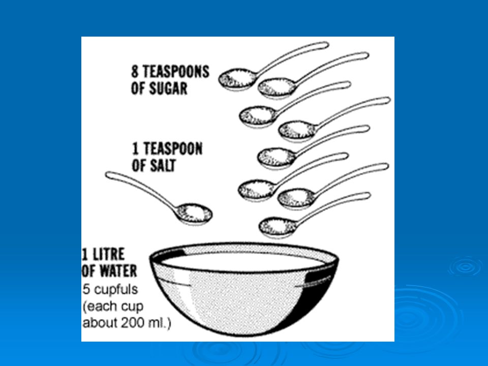
Treatment of Mild to Moderate ORT – oral re-hydration therapy 50 ml / kg every 4 hours 50 ml / kg every 4 hours Increase to 100 ml / kg every 4 hours Increase to 100 ml / kg every 4 hours No carbonated soda, jell-o, fruit juices or tea. No carbonated soda, jell-o, fruit juices or tea. Commercially prepared solutions are the best. Commercially prepared solutions are the best.

52
Re-hydration Therapy Increase po fluids if diarrhea increases. Give po fluids slowly if vomiting. Stop ORT when hydration status is normal Start on BRAT diet Bananas Bananas Rice Rice Applesauce Applesauce Toast Toast

54
Teaching / Parent Instruction Call PMD If diarrhea or vomiting increases No improvement seen in child’s hydration status. Child appears worse. Child will not take fluids. NO URINE OUTPUT

55
Moderate to Severe Dehydration IV Therapy needed

56
Fluid Resuscitation Crystalloid Solution: used for volume resuscitation to expand the interstitial volume rather that the plasma volume. Isotonic Saline is the prototype crystalloid fluid. 0.9% NaCl or normal saline. Isotonic Saline is the prototype crystalloid fluid. 0.9% NaCl or normal saline.

57
Fluid Replacement Standard Orders: Normal Saline or 0.9% NaCl at 20 mL / kg Normal Saline or 0.9% NaCl at 20 mL / kg Followed by Dextrose 5% in 0.45 normal saline Followed by Dextrose 5% in 0.45 normal saline Followed by Dextrose 5% in 0.45 normal saline with 20 mEq KCL per 1000 mL Followed by Dextrose 5% in 0.45 normal saline with 20 mEq KCL per 1000 mL Potassium is only added to the IV when there is documentation of voiding. Potassium is only added to the IV when there is documentation of voiding.

58
Nursing Interventions Assess child’s hydration status Vital signs with temperature and weight most accurate way to monitor fluid levels most accurate way to monitor fluid levels Hourly monitoring of IV rate and site of infusion Intake and output

59
Care Reminder A severely dehydrated child will need more than maintenance to replace lost fluids. 1 ½ to 2 times maintenance. It is the nurses responsibility to check fluid calculations at the beginning of the shift (24 hour fluid needs / hourly IV rate)
.")
60
Over hydration Occurs when child receives more IV fluids that needed for maintenance. In pre-existing conditions such as meningitis, head trauma, kidney shutdown, nephrotic syndrome, congestive heart failure, or pulmonary congestion.

61
Assessment of over-hydration Tachypnea Dyspnea Cough Moist breath sounds Weight gain from edema Jugular vein distention

62
Safety Precautions Use small bags of fluid or buretrol to control fluid volume. Check IV solution infusion against physician orders. Always use infusion pump so that the rate can be programmed and monitored. Calculate 24 hour fluid needs Record IV rate q hour

63
Acid – Base Imbalances Acidosis: Respiratory acidosis is too much carbonic acid in body. Metabolic Acidosis is too much metabolic acid. Alkalosis. Respiratory alkalosis is too little carbonic acid. Metabolic alkalosis is too little metabolic acid.

64
Respiratory Acidosis Carbonic acid excess: CO2 is retained and pH decreases Caused by the accumulation of carbon dioxide in the blood. Acute respiratory acidosis can lead to tachycardia and cardiac arrhythmias.

65
Causes of Respiratory Acidosis Any factor that interferes with the ability of the lungs to excrete carbon dioxide can cause respiratory acidosis. Aspiration, spasm of airway, laryngeal edema, epiglottitis, croup, pulmonary edema, cystic fibrosis, and Bronchopulmonary dysplasia. Sedation overdose, head injury, or sleep apnea.

66
Assessment Respiratory distress CNS depression: disorientation, coma Hypoxia: restlessness, irritability, tachycardia, arrhythmias Muscle weakness

67
Medical Management Correction of underlying cause Bronchodilators: asthma Antibiotics: infection Mechanical ventilation Decreasing sedative use

68
Respiratory Alkalosis Carbonic acid deficit; not enough CO2 is retained, and pH increases. Excess carbon dioxide loss is caused by hyperventilation.

69
Causes of hyperventilation Hypoxemia Hypoxemia Anxiety Anxiety Pain Pain Fever Fever Salicylate poisoning: ASA Salicylate poisoning: ASA Meningitis Meningitis Over-ventilation Over-ventilation

70
Assessment Dizziness Numbness or paresthesias of fingers and toes Tetany Convulsions Unconsciousness

71
Management Stress management if caused by hyperventilation. Pain control. Adjust ventilation rate. Treat underlying disease process. Have child slow respirations, breathe into paper bag

72
Metabolic Acidosis Bicarbonate deficit

73
Causes: Gain in acid: ingestion of acids, oliguria, starvation (anorexia), DKA or diabetic ketoacidosis, tissue hypoxia. Loss of bicarbonate: diarrhea, intestinal or pancreatic fistula, or renal anomaly.

74
Assessment Kussmaul respirations – slow and deep SOB on exertion Weakness Drowsiness to stupor When pH is < 7.2 cardiac contractility is reduced – BP will decrease

75
Management Treat and identify underlying cause. IV sodium bicarbonate in severe cases. Provide low-protein, high-calorie diet Position to facilitate ventilation

76
Metabolic Alkalosis A gain in bicarbonate or a loss of metabolic acid can cause metabolic alkalosis.

77
Causes: Gain in bicarbonate: Ingestion of baking soda or antacids. Loss of acid: Vomiting, nasogastric suctioning, diuretics massive blood transfusion

78
Assessment Signs similar to dehydration Tachycardia Hypoventilation Muscle hypertonicity Confusion, irritability, coma

79
Treatment Administer fluid containing sodium and potassium Avoid antacids Management: Correct the underlying condition

Similar presentations


 Integumentary System (chapters 44- 46)>")


![1 Acid and Base Balance and Imbalance. 2 pH Review pH = - log [H + ] H + is really a proton Range is from 0 - 14 If [H + ] is high, the solution is acidic;](/14/4450368/big_thumb.jpg "1 Acid and Base Balance and Imbalance. 2 pH Review pH = - log [H + ] H + is really a proton Range is from 0 - 14 If [H + ] is high, the solution is acidic;>")


 Assistant Prof. in Pathology Al Maarefa College.>")