Download presentation
Presentation is loading. Please wait.

1
SURGICAL DISEASES DIAGNOSIS HYSTORY CLINICAL EXAMINATION CLINICAL EXAMINATION LAB.TESTS LAB.TESTS IMAGISTIC INVESTIGATIONS

2
TREATMENT MODALITIES SURGERY - CLASIC OR MINIMALLY INVASIVE (LAPAROSCOPIC) MINIMALLY INVASIVE (LAPAROSCOPIC) MEDICAL- COMORBIDITIES, DEFFICITS CORRECTION: SEVERE ANEMIA HYPOVOLEMIA, DISELECTROLYTEMIA, ANTIBIOTICS, ANTICOAGULANTS ADJUVANT, NEOADJUVANT: RADIOTHERAPY, CHEMOTHERAPY RADIOTHERAPY, CHEMOTHERAPY
 MINIMALLY INVASIVE (LAPAROSCOPIC) MEDICAL- COMORBIDITIES, DEFFICITS CORRECTION: SEVERE ANEMIA HYPOVOLEMIA, DISELECTROLYTEMIA, ANTIBIOTICS, ANTICOAGULANTS ADJUVANT, NEOADJUVANT: RADIOTHERAPY, CHEMOTHERAPY RADIOTHERAPY, CHEMOTHERAPY")
3
SURGICAL TREATMENT THE RIGHT OPERATION PERFORMED WELL THE RIGHT OPERATION PERFORMED BADLY THE WRONG OPERATION PERFORMED WELL THE WRONG OPERATION PERFORMED BADLY In only one case the patient will have the best result

4
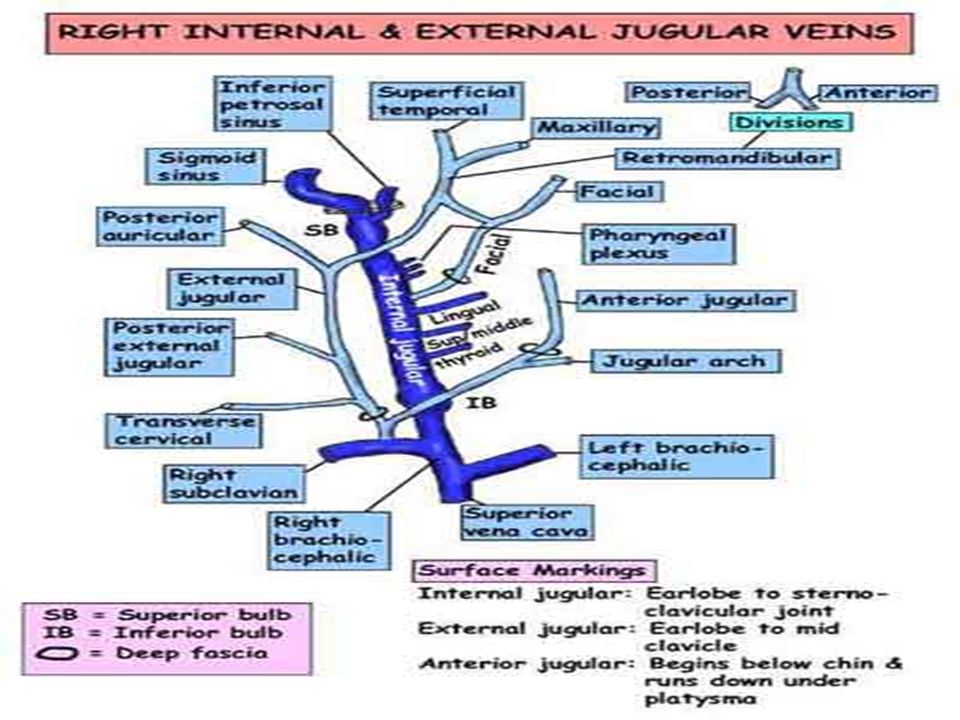
SURGERY OF THE NECK ANATOMY DEFINITIONSTRUCTURES: –SKIN, SUPERFICIAL FASCIA, VEINS, NODES –DEEP CERVICAL FASCIA –MAIN ARTERIES, VEINS, NERVES –VISCERA OF THE NECK –THE ROOT OF THE NECK

14
NECK SURGERY LUMPS IN THE NECK WOUNDS OF THE NECK SURGICAL INFECTIONS IN THE NECK

15
LUMPS IN THE NECK HISTORY AND EXAMINATION HISTORY:- rate of growth of the lump, - symptoms:pain, discharge EXAMINATION- characteristics of the lump- site, shape, size, surface, tenderness, fixation, consistency, fluctuence, pulsatility, associated lymph- adenopathy Clinical diagnosis- benign or malignant -cystic or solid lump -cystic or solid lump

16
LUMPS IN THE NECK CAUSES 1. Lymph node enlargement: -lymphomas, -lymph node metastases, -inflammatory lymphadenopathy from acute or chronic infections in the neck, -AIDS related lymphadenopathy 2. Congenital cysts: thyroglossal, branchial, cystic hygroma 3. Lumps in the skin: lipoma, epidermal cyst 4. Rare tumors: carotid body tumors 5. Thyroid and parathyroid nodules

17
LYMPH NODE ENLARGEMENT CERVICAL TUBERCULOSIS Synonims: tuberculous cervical adenitis :cervical tuberculous lymphadenopathy :mycobacterial lymphadenitis :extrapulmonary tuberculosis

18
CERVICAL TUBERCULOSIS Acquired chronic infection of the nodes Acquired by drinking milk infected cattle The primary infection occurs in the tonsils Secondary involvement of the cervical nodes- enlarged, matted together The incidence of coexisting pulmonary TB is less 5%.

19
CERVICAL TUBERCULOSIS Clinical features Long history of a lump in the neck Medical advise- the lump has become painful Presentation: - just lump in the neck - discharging sinus - discharging sinus - cold abscess - cold abscess - lump adherent to the skin - lump adherent to the skin

20
CERVICAL TUBERCULOSIS Clinical features 90% unilateral 90% involve only one node group The commonest- deep jugular vein - submandibular - submandibular - in the posterior triangle - in the posterior triangle

21
CERVICAL TUBERCULOSIS Diagnosis History Physical examination: characteristics of an inflammatory lump Specific investigations: –Positive tuberculin test –Excisional biopsy of the lump –Culture of Mycobacterium tuberculosis

22
CERVICAL TUBERCULOSIS Treatment Excisional biopsy Antituberculous chemotherapy for 9-12 months Matted nodes attached to the internal jugular vein- functional neck dissection (preservation of the SCM, accessory nerve, jugular vein if possible) *In a kid- remove and examine histologically the tonsils before removing the lymph nodes *Surgery not followed by chemotherapy- a persistent discharging sinus will form and later ugly scar
 *In a kid- remove and examine histologically the tonsils before removing the lymph nodes *Surgery not followed by chemotherapy- a persistent discharging sinus will form and later ugly scar")
23
LUMPS IN THE NECK CAUSES 1.LYMPH NODE ENLARGEMENT: c. ACUTE INFECTIONS d. CHRONIC INFECTIONS

24
CERVICAL LYMPH NODES CLINICAL ENTITIES Lymphomas= neoplastic disorders of lymphoid cells –Solid tumors –Involve the lymphoid tissue –Hodgkin’s disease=malignant neoplasm originating in lymphoid tissue –Characteristic Reed-Sternberg cells

25
CERVICAL LYMPH NODES CLINICAL ENTITIES Cervical lymph nodes metastases –Primary tumors located in the head neck, chest or abdomen –Tumors of the head and neck, send metastases to nodes- submandibular region and upper part of the anterior triangle –Tumors of the chest and abdomen send metastases to nodes- lower part of the posterior triangle (Virchow’s node)
")
26
LUMPS IN THE NECK CAUSES 1.LYMPH NODE ENLARGEMENT: a. PRIMARY TUMORS b. SECONDARY TUMORS a. PRIMARY TUMORS b. SECONDARY TUMORS -lymphomas- -lymph node metastases- -lymphomas- -lymph node metastases-

27
CONGENITAL NECK LUMPS CYSTIC HYGROMA Synonim- cystic lymphangioma Tumor of lymph vessels which forms multilocular cyst- like spaces Painless lump just below the angle of the mandible, soft, fluctuant, transilluminable Surgical excision- the best option in fit patients Incision and drainage when infected

28
LUMPS IN THE NECK CAUSES 2. CONGENITAL CYST: c. CYSTIC HYGROMA c. CYSTIC HYGROMA

29
LUMPS IN THE NECK CAUSES 2. CONGENITAL CYST: a. THYROGLOSSAL CYSTS a. THYROGLOSSAL CYSTS

30
CONGENITAL NECK LUMPS THYROGLOSSAL DUCT CYST The commonest midline neck cyst Remnant of the thyroglossal duct- incomplete regression may result in a cyst It is attached to the base of the tongue and hyoid bone Painless cystic neck lump, moving up with tongue protrusion, mobile, transilluminable Tenderness when infected May drain spontaneouslly with fistula formation Surgical excision with central part of the hyoid bone

31
CONGENITAL NECK LUMPS BRANCHIAL CYST Remnant of the 2 nd pharyngeal pouch Painless lump in the side of the neck, deep to the SCM, 1/3-2/3, anterior triangle Painful lump if infected, soft, fluctuant Complete surgical excision or incision and drainage if infected, preventing injuries to the ICA-ECA. Branchial fistula discharges a glairy mucinous substance

32
LUMPS IN THE NECK CAUSES 2. CONGENITAL CYST: b. BRANCHIAL CYST b. BRANCHIAL CYST

33
LUMPS IN THE NECK CAUSES 3.LUMPS IN THE SKIN: a. LIPOMA a. LIPOMA

34
LUMPS IN THE NECK CAUSES 3.LUMPS IN THE SKIN: b. EPIDERMAL CYST b. EPIDERMAL CYST

35
CAROTID BODY TUMOR Synonim=chemodectomas High incidence in Peru, high altitude Chronic hypoxia at high altitudes leads to carotid body hyperplasia Firm ovoid tumor, firmly adherent to the bifurcation of the CCA Painless palpable lump Seldom grows to more than 4-5 cm.

36
CAROTID BODY TUMOR DIAGNOSIS AND TREATMENT Long history of a lump in the neck Physical examination- characteristics of a solid tumor Investigations- carotid angiogram, doppler echography Surgical removal- risk of morbidity and mortality Indications limited for malignant resectable tumors, interfering swallowing, speaking, breathing Radiotherapy indicated in poor- risc surgical pt. or metastatic diasease

37
LUMPS IN THE NECK CAUSES 4.RARE TUMORS: CAROTID BODY TUMORS CAROTID BODY TUMORS

38
LUMPS IN THE NECK CAUSES Multinodular goitre

43
LUMPS IN THE NECK CAUSES Parathyroid adenoma

44
WOUNDS OF THE NECK SUICIDAL CUT-THROAT HOMICIDAL CUT-THROAT OTHER WOUNDS OF THE NECK

45
CUT-THROAT Is due to attempted suicide Head extended- great vessels escape injury, the air passages are open- repair, tracheostomy Bleeding from thyroid artery- secured IJV- ligated above and below the injury site Pharyngeal injury- suture, septic risk NG tube for feeding

50
HOMICIDAL CUT-THROAT The wound is in the lower part of the neck Great vessels injured- is fatal Treatment: - proper exploration, - debridement of ischemic tissue, - repair of damaged structures, - suture with drainage

51
OTHER WOUNDS OF THE NECK STABS GUNSHOT WOUNDS WHEN VITAL STRUCTURES ESCAPE, REMAINS SEPTIC RISK TREATMENT: - ENLARGEMENT OF THE WOUND - DEBRIDEMENT - REPAIR

52
NECK INJURIES Anatomically, the neck can be divided into 3 major zones, in order to aid in the decision making- diagnostic tests,-timing of surgery Zone I- below the cricoid Zone III- above the angle of the mandible Zone II- the most frequently involved region

53
Neck injuries- signs and symptoms Vascular injury- shock, hematoma, hemorrhage, pulse deficit, neurologic deficit, bruits in neck Laryngo/tracheal injury- subcutaneous emphysema, airway obstruction, sucking wound, hemoptysis, dyspnea, stridor, dysphonia Pharyngo/esophageal injury- subcut.emphysema, dysphagia, odinophagia

54
NECK INJURY- MANAGEMENT Patients with refractory shock, uncontrolable hemorrhage should undergo immediat neck exploration Patients who are stable, should undergo directed explorations with subsequent repair of injured structures

55
ZONE I INJURY- MANAGEMENT Penetrating injuries are potentially fatal Angiography for suspected vascular injury Esophageal examination- a missed esophageal injury- mediastinitis

56
ZONE II INJURY- MANAGEMENT Symptomatic- penetrating injury- neck exploration Asymptomatic- mandatory neck exploration or directed evaluation and serial examinations

57
ZONE III INJURY- MANAGEMENT Potential for injury to major blood vessels and the cranial nerves near the skull base Surgical exposure – difficult Angiogram- interventional radiology

58
INFECTIONS IN THE NECK 1. Retropharyngeal abscess 2. Parapharyngeal abscess 3. Submandibular abscess

59
INFECTIONS IN THE NECK RETROPHARYNGEAL ABSCESS

60
INFECTIONS IN THE NECK PARAPHARYNGEAL ABSCESS

61
INFECTIONS IN THE NECK SUBMANDIBULAR SPACE INFECTIONS (LUDWIGS ANGINA)
")
Similar presentations


 –Zone II injury –haemodynamically stable.>")


