Download presentation
Presentation is loading. Please wait.

1
DNA: The Genetic Material
Chapter 16 The Molecular Basis of Inheritance DNA: The Genetic Material

2
Intro to DNA Video

3
Just a thought… Make a stack of books totaling about 10,000 pages.
That stack of books represents only about one-fiftieth of the information contained in the DNA of every human cell. Correlate this with the amount of information required to code for a human being.

5
Quick Review: 1. What is the structure of a chromosome. 2
Quick Review: 1. What is the structure of a chromosome? 2. Define the term gene. 3. Identify the stage in the cell cycle in which DNA is copied. 4. What are mutations? 5. Summarize Mendel’s theory of heredity.

6
Answers 1. A chromosome consists of two replicated strands of DNA tightly coiled around proteins. The two strands, called chromatids, are attached at a point called a centromere. 2. A gene is a segment of DNA that codes for a protein or RNA molecule. 3. A cell’s DNA is copied during the synthesis (S) phase. 4. When chromosomes break, the broken pieces can detach completely or can reattach in various ways. Therefore, the chromosome is changed, or mutated.
 phase. 4. When chromosomes break, the broken pieces can detach completely or can reattach in various ways. Therefore, the chromosome is changed, or mutated.")
7
5. (a) For each inherited trait, an individual has two copies of the gene, one from each parent.
(b) There may be alternative versions of genes. (c) When two different alleles occur together, one of them may be completely expressed, while the other may have no observable effect on the organism’s appearance. (d) When gametes are formed, the alleles for each gene in an individual separate independently of one another, and when gametes unite during fertilization, each gamete contributes one allele.
 There may be alternative versions of genes. (c) When two different alleles occur together, one of them may be completely expressed, while the other may have no observable effect on the organism’s appearance. (d) When gametes are formed, the alleles for each gene in an individual separate independently of one another, and when gametes unite during fertilization, each gamete contributes one allele.")
8
Vocabulary: 1. DNA /double helix, 2. Nucleosome
3. Semi conservative replication, 4. DNA polymerase, 5. Okazaki fragment

9
Vocabulary vaccine virulent transformation bacteriophage double helix
nucleotide deoxyribose base-pairing rules complementary base pair DNA replication DNA helicase replication fork DNA polymerase

10
1. What are the two chemical components of chromosomes?

11
DNA and protein

12
Chapter: DNA: The Genetic Material
1. Identifying the Genetic Material A. Transformation B. Viral Genes and DNA

13
2. Why did researchers originally think that protein was the genetic material?

14
Until the 1940s, the case for proteins seemed stronger, especially since biochemists had identified them as a class of macromolecules with great heterogeneity (uniformity) and specificity of function, essential requirements for the hereditary material. Also, little was known about nucleic acids, whose physical and chemical properties seemed far too uniform to account for the multitude of specific inherited traits exhibited by every organism.

15
Identifying the Genetic Material
The experiments of Griffith and of Avery yielded results that suggested DNA was the genetic material.

16
Frederick Griffith--Discovery of the Transforming Principle (Video Clip)
")
17
Griffith’s Discovery of Transformation

18
The capsule killed, non capsule did not
The capsule killed, non capsule did not! Heat-killed capsule, did not KILL! BUT A MIX OF THE 2 DID!

19
3. Distinguish between the virulent and non-virulent strains of Streptococcus pneumoniae studied by Frederick Griffith.

20
The virulent strains ( with capsules) are pathogenic (disease-causing), whereas the nonvirulent strains ( no capsule) are nonpathogenic (harmless).
 are pathogenic (disease-causing), whereas the nonvirulent strains ( no capsule) are nonpathogenic (harmless).")
21
4. What was the purpose of Griffith’s studies?

22
Griffith was attempting to develop a vaccine against pneumonia.

23
What happened? 5. Use this figure to summarize the experiment in which Griffith became aware that hereditary information could be transmitted between two organisms in an unusual manner.

24
information could be transmitted between two organisms in an unusual manner.
Griffith studied two strains of the bacterium Streptococcus pneumoniae. Bacteria of the S (smooth) strain can cause pneumonia in mice; they are pathogenic because they have an outer capsule that protects them from an animal’s defense system. Bacteria of the R (rough) strain lack a capsule and are nonpathogenic. To test for the trait of pathogenicity, Griffith injected mice with the two strains. Griffith concluded that the living R bacteria had been transformed into pathogenic S bacteria by an unknown, heritable substance from the dead S cells that allowed the R cells to make capsules.
 strain can cause pneumonia in mice; they are pathogenic because they have an outer capsule that protects them from an animal’s defense system. Bacteria of the R (rough) strain lack a capsule and are nonpathogenic. To test for the trait of pathogenicity, Griffith injected mice with the two strains. Griffith concluded that the living R bacteria had been transformed into pathogenic S bacteria by an unknown, heritable substance from the dead S cells that allowed the R cells to make capsules.")
25
Transformation Something in the heat-killed bacteria that once were able to produce a capsule that caused them to kill the mice was able to “transform” the once non-virulent bacteria into capsule producing, virulent ones! But what was that something????

26
6. Define transformation.
A change in genotype and phenotype due to the assimilation of external DNA by a cell. When the external DNA is from a member of a different species, transformation results in horizontal gene transfer.

27
Griffith 1928 1. What happened to the normally non-virulent rough bacteria when mixed with the virulent smooth heat killed ones? 2. Based on what happened to the bacteria, what was Griffith’s experiment called? 3. At the end of Griffith’s experiment, did scientists know if DNA or proteins caused the changes?

28
Griffith 1928 1. What happened to the normally non-virulent rough bacteria when mixed with the virulent smooth heat killed ones? they became virulent 2. Based on what happened to the bacteria, what was Griffith’s experiment called? the transforming experiment 3. At the end of Griffith’s experiment, did scientists know if DNA or proteins caused the changes? _no

29
Avery’s experiment DNA destroying enzymes helped support the fact that it was the DNA not proteins that were the transforming factors!

30
7. What did Oswald Avery determine to be the transforming factor?
Explain his experimental approach.

31
Only when DNA was allowed to remain active did transformation occur.
Avery broke open the heat-killed pathogenic bacteria and extracted the cellular contents. He treated each of three samples with an agent that inactivated one type of molecule, and then tested the sample for its ability to transform live nonpathogenic bacteria. Only when DNA was allowed to remain active did transformation occur.

32
Avery 1944 4. Avery attempted to do what?
5. Did people accept Avery’s results?

33
Avery 1944 4. Avery attempted to do what? identify if the substance was protein or DNA 5. Did people accept Avery’s results? NO

34
Hershey and Chase used the bacteriophage T2 and radioactive labels to show that viral genes are made of DNA, not protein.

35
Alfred Hershey and Martha Chase--Acceptance within scientific community of DNA as genetic material (Video Clip)
")
36
bacteriophage 8. Sketch a T2 bacteriophage and label its head, tail sheath, tail fiber, and DNA.

37
9. How does a bacteriophage destroy a bacterial cell?

38
The T4 phage uses its tail fibers to bind to specific receptor sites on the outer surface of an E. coli cell. The sheath of the tail contracts, injecting the phage DNA into the cell and leaving an empty capsid outside. .

39
The cell’s DNA is hydrolyzed
The cell’s DNA is hydrolyzed. The phage DNA directs production of phage proteins and copies of the phage genome by host and viral enzymes, using components within the cell.

40
Three separate sets of proteins self-assemble to form phage heads, tails, and tail fibers. The phage genome is packaged inside the capsid as the head forms. The phage directs production of an enzyme that damages the bacteria cell wall, allowing fluid to enter. The cell swells, and finally bursts, releasing 100 to 200 phage particles

41
The Hershey-Chase Experiment

42
Hershey and Chase More evidence it is DNA!

43
Radioactive (heavy forms) were provide for the phages to incorporate into their structures
Sulfur in protein 35 S Phosphorus in DNA 32 P

44
The radioactive sulfur remained outside with the protein coat of the virus.
The radioactive Phosphorus went inside the bacteria and showed that the DNA was the component that went inside and produced more viral proteins.

46
10. How did Hershey and Chase “label” viral DNA and viral protein so that they could be distinguished? Explain why they chose each radioactive tag in light of the chemical composition of DNA and protein.

47
Hershey and Chase used radioactive isotopes of sulfur to tag protein in one batch of T2 and a radioactive isotope of phosphorus to tag DNA in a second batch. Because proteins, but not DNA, contain sulfur, radioactive sulfur atoms were incorporated only into the protein of the phage.

48
DNA stores the information that tells cells which proteins to make and when to make them.

49
11. Describe the means by which Hershey and Chase established that only the DNA of a phage enters an E. coli cell. What conclusions did these scientists draw based on these observations?

50
Separate samples of the non-radioactive E
Separate samples of the non-radioactive E. coli cells were allowed to be infected by the protein labeled and DNA-labeled batches of T2. The researchers then tested the two samples shortly after the onset of infection to see which type of molecule—protein or DNA—had entered the bacterial cells and would therefore be capable of reprogramming them. Hershey and Chase found that the phage of DNA entered the host cells but the phage protein did not. Hershey and Chase concluded that the DNA injected by the phage must be the molecule carrying the genetic information that makes the cells produce new viral DNA and proteins.

51
Hershey and Chase 1952 6. What is a bacteriophage?
7. Radioactive phosphorous was used to identify DNA because _________does not contain phosphorous but DNA does. 8. They could then trace the path of the radioactive phosphorous and found that it ended up in the transformed______________ 9. Radioactively labeled Protein did not enter the bacteria at all but_____________________

52
Hershey and Chase 1952 6. What is a bacteriophage? A virus that infects bacterial cells. 7. Radioactive phosphorous was used to identify DNA because protein_does not contain phosphorous but DNA does. 8. They could then trace the path of the radioactive phosphorous and found that it ended up in the transformed bacterium’s DNA 9. Radioactively labeled Protein did not enter the bacteria at all but remained outside the cell.

53
10. Which experiments led to the discovery of DNA as the genetic material?

54
10. Which experiments led to the discovery of DNA as the genetic material?
All of them, Griffiths, Avery and Hershey and Chase’s were needed to get to the idea that DNA and not Protein was the genetic material. It was ultimately Hershey and Chase’s experiment that demonstrated that it was DNA and not protein that transmitted the genetic material.

55
1. In 1928, the experiments of Griffith demonstrated transformation of
a. R bacteria into S bacteria. b. S bacteria into R bacteria. c. heat-killed S bacteria into R bacteria. d. S bacteria into heat-killed R bacteria. 2. In 1952, Hershey and Chase used the bacteriophage T2 to determine that genetic material is made of which of the following? a. protein c. DNA b. RNA d. 35S 3. A microorganism that is virulent is a. able to cause disease. c. a bacteriophage. b. transformed. d. harmless.

56
4. Avery’s experiments showed that
a. DNA is responsible for transformation. b. proteins are responsible for transformation. c. bacteriophages are responsible for transformation. d. virulent bacteria are responsible for transformation. 5. Hershey and Chase injected phages with a. S bacteria. c. radioactive isotopes. b. R bacteria. d. vaccines. 6. Hershey and Chase found that T2 bacteriophages a. inject their DNA into host cells. b. cause host cells to produce viral DNA and proteins. c. keep most of their viral proteins outside the host cell. d. All of the above

57
______ 7. radioactive sulfur and phosphorous
______ 8. transformation ______ 9. bacteriophage ______10. vaccine a. discovered by Griffith b. infects bacteria c. used in the Hershey and Chase experiments d. helps protect the body against future infections by specific disease-causing agents

58
GENETIC MATERIAL answers
1. a 6. d 2. c 7. c 3. a 8. a 4. a 9. b 5. c d

59
2. The Structure of DNA A. A Winding Staircase
B. Discovering DNA’s Structure

60
Erwin Chargaff--DNA is not equal for all species and ratio of bases varies among species (Video Clip)
")
61
Chargaff’s data

62
12. What are Chargaff’s rules? How did he arrive at them?

63
Chargaff’s rules are: 1. The base composition varies between species.
2. Within a species, the number of A and T bases are equal and the number of G and C bases are equal. Chargaff analyzed the base composition of DNA from a number of different organisms. He reported that the base composition of DNA varies from one species to another. He also noticed a peculiar regularity in the ratios of nucleotide bases. In the DNA of each species he studied, the number of adenines approximately equaled the number of thymines, and the number of guanines approximately equaled the number of cytosines

64
13. List the three components of a nucleotide.

65
Phosphate, Sugar (deoxyribose for DNA ribose for RNA), Nitrogenous base
, Nitrogenous base")
66
nucleotide

67
1. What is a nucleotide composed of?
2. What scientist looked at the data that analyzed the amount of guanine equaled the amount of cytosine and the amount of thymine equaled the amount of adenine and what conclusion did he draw from this?.

68
1. What is a nucleotide composed of
1. What is a nucleotide composed of? Sugar, nitrogen base and a phosphate group 2. What scientist looked at the data that analyzed the amount of guanine equaled the amount of cytosine and the amount of thymine equaled the amount of adenine and what conclusion did he draw from this? Chargaff/ these nitrogen bases must be paired up in the structure.

69
Watson and Crick

70
14. Who are the two men who built the first molecular model of DNA and shared the 1962 Nobel Prize for discovery of its structure?

71
James Watson and Francis Crick

72
X ray diffraction of DNA by Rosalind Franklin

73
15. What was the role of Rosalind Franklin in the discovery of the double helix?

74
Rosalind Franklin, a very accomplished X-ray crystallographer, conducted critical experiments resulting in the photograph that allowed Watson and Crick to deduce the double-helical structure of DNA.

75
3. X-ray diffraction was used to help determine what about DNA?
4. Who are the 2 scientists who put all of the info together to describe the structure of DNA?

76
3. X-ray diffraction was used to help determine what about DNA
3. X-ray diffraction was used to help determine what about DNA? the double helix shape 4. Who are the 2 scientists who put all of the info together to describe the structure of DNA? Watson and Crick

77
DNA Structure Chromatin--Chromosomes and DNA subunits (Video Clip)
")
78
Purines and Pyrimidines two types of nitrogen bases in DNA and RNA
Purines=double rings. Adenine and Guanine Pyrimidines= single rings. Thymine, Cytosine and Uracil which replaces Thymine in RNA

79
bonding G triple hydrogen bonds to C A double hydrogen bonds to T
These un zip in DNA replication

80
17. How did Watson and Crick’s model explain the basis for Chargaff’s rules?

81
Watson and Crick built models of a double helix that would matched the X-ray measurements and what was known about the chemistry of DNA, including Chargaff’s rule of base equivalences. Through trial and error, Watson and Crick deduced that the nitrogenous bases of the double helix are paired in specific combinations—adenine (A) with thymine (T), and guanine (G) with cytosine (C)—and they reflected these findings in their model. Whenever one strand of a DNA molecule has an A, the partner strand has a T. And a G in one strand is always paired with a C in the complementary strand. Therefore, in the DNA of any organism, the amount of adenine equals the amount of thymine, and the amount of guanine equals the amount of cytosine.

82
16. Distinguish between the structure of pyrimidines and purines.
Explain why adenine bonds only to thymine.

83
Pyrimidines—cytosine (C), thymine (T), and uracil (U)—are characterized by a six-membered ring.
Purines—adenine (A) and guanine (G)—are characterized by a six-membered ring fused to a five-membered ring.
 and guanine (G)—are characterized by a six-membered ring fused to a five-membered ring.")
84
Always pairing a purine with a pyrimidine results in a uniform diameter in the double helix.

85
Additionally, each base has chemical side groups that can form hydrogen bonds with its appropriate partner. Adenine can form two hydrogen bonds with thymine and only thymine. Adenine bonds only with thymine because adenine is a purine, and thymine is a pyrimidine.

86
18. Given that the DNA of a certain fly species consists of 27
18. Given that the DNA of a certain fly species consists of 27.3% adenine and 22.5% guanine, use Chargaff’s rules to deduce the percentages of thymine and cytosine.

87
18. Given that the DNA of a certain fly species consists of 27
18. Given that the DNA of a certain fly species consists of 27.3% adenine and 22.5% guanine, use Chargaff’s rules to deduce the percentages of thymine and cytosine. 27.6% thymine 22.5% cytosine

88
19. Name the five nitrogenous bases, and put a checkmark in the correct column for each base. Also indicate if the base is found in DNA (D), RNA (R), or both (B).
, RNA (R), or both (B)..")
90
20. What DNA base is complementary to adenine?
What DNA base is complementary to guanine?

91
20. What DNA base is complementary to adenine? Thymine
What DNA base is complementary to guanine? Cytosine

92
Describe the structure of DNA relative to each of the following
Describe the structure of DNA relative to each of the following. Indicate the distance in the correct location on the figure as well. a. distance across molecule b. distance between nucleotides c. distance between turns d. components of the backbone e. components of the “rungs”

93
a. distance across molecule 1 nm
b. distance between nucleotides 0.34 nm c. distance between turns 3.4 nm d. components of the backbone sugar-phosphate e. components of the “rungs” A T G C I will not ask these on the test!

94
5’ and 3’ ends Organic molecules are numbered by the carbons!

95
Antiparallel Go in opposite directions 5 to 3 on one side 3 to 5 on the other!

96
22. Explain what is meant by 5' and 3' ends of the nucleotide.

97
The two free ends of the polymer are distinctly different from each other. One end has a phosphate attached to a 5' carbon, and the other has a hydroxyl group on the 3' carbon. We refer to these as the 5' end and the 3' end respectively.

98
23. What do we mean when we say the two strands of DNA are antiparallel?

99
Their subunits run in opposite directions.

100
DNA replication

101
DNA replication Video

102
Semi conservative Half is original half is new.
The method of DNA replication had to be determined. Several tests were done to figure this out…

103
Matthew Meselson and Franklin Stahl
1958 determined that the method of DNA replication was semi-conservative based on Nitrogen that was radioactive and heavier so it formed bands at different heights after centrifugation. The multiple generations of replications demonstrated the method had to be semi-conservative due to the intermediate bands produced.

105
24. What is the semiconservative model of replication?

106
Type of DNA replication in which the replicated double helix consists of one old strand, derived from the parental molecule, and one newly made strand

107
26. How did Meselson and Stahl create “heavy” DNA for their experiments?

108
Meselson and Stahl cultured E
Meselson and Stahl cultured E. coli for several generations in a medium containing nucleotide precursors labeled with a heavy isotope of nitrogen, 15N.

109
27. Use Figure 16.11 to explain how Meselson and Stahl confirmed the semiconservative
mechanism of DNA replication.

110
Meselson and Stahl transferred their “heavy” DNA to a medium with lighter isotope, 14N. A sample was taken after DNA replicated once; another sample was taken after DNA replicated again. They extracted DNA from the bacteria in the samples and then centrifuged each DNA sample to separate DNA of different densities. Meselson and Stahl compared their results to those predicted by each of the three models (conservative, semiconservative, and dispersive).
.")
111
The first replication in the 14N medium produced a band of hybrid DNA
The first replication in the 14N medium produced a band of hybrid DNA. This result eliminated the conservative model. The second replication produced both light and hybrid DNA, a result that refuted the dispersive model and supported the semiconservative model. They therefore concluded that DNA replication is semiconservative.

112
28. Define the origins of replication.

113
Site where the replication of a DNA molecule begins, consisting of a specific sequence of nucleotides

114
29. Distinguish between the leading and the lagging strands during DNA replication.

115
The leading strand is the new complementary DNA strand synthesized continuously along the
template strand toward the replication fork in the mandatory 5' to 3' direction. The lagging strand is a discontinuously synthesized DNA strand that elongates by means of Okazaki fragments, each synthesized in a 5' to 3' direction away from the replication fork.

116
DNA is packaged into chromosomes

117
Histones are positively charged, and DNA is negatively charged.
Without histones the DNA could not fit into the nucleus! Heterochromatin is highly condensed, whereas euchromatin is less compact.

118
The Structure of DNA DNA is made of two strands of nucleotides twisted into the form of a double helix.

119
1. Why does DNA have to replicate every time a cell divides?
2. When does DNA replication occur in the cell cycle?

120
1. Why does DNA have to replicate every time a cell divides?
Because every cell needs the instructions to make proteins that the DNA contains. 2. When does DNA replication occur in the cell cycle? S phase of interphase

121
Each nucleotide in DNA is made of the sugar deoxyribose, a
phosphate group, and one of four nitrogen bases. The four nitrogen bases found in DNA nucleotides are adenine (A), thymine (T), guanine (G), and cytosine (C).
, thymine (T), guanine (G), and. cytosine (C).")
122
The two strands of DNA are complementary—each A on one strand pairs with a T on the opposite strand, and each G on one strand pairs with a C on the opposite strand.

124
5. Describe the structure of DNA in detail, use a diagram to help.

125
5. Describe the structure of DNA in detail, use a diagram to help
5. Describe the structure of DNA in detail, use a diagram to help. The sides are made up of the sugar deoxy ribose and the phosphate groups bonded covalently together the rungs are the nitrogen bases with cytosine and guanine bonded with hydrogen bonds across from each other and thymine and Adenine across from each other. The entire thing is in a double helix and runs 3’ to 5’ anti parallel.

126
The Replication of DNA A. The Roles of Enzymes in DNA Replication
Summarize the steps of DNA replication

127
Bozeman Biology video DNA replication

128
B. The Rate of Replication
To go fast, there are multiple replication forks!

129
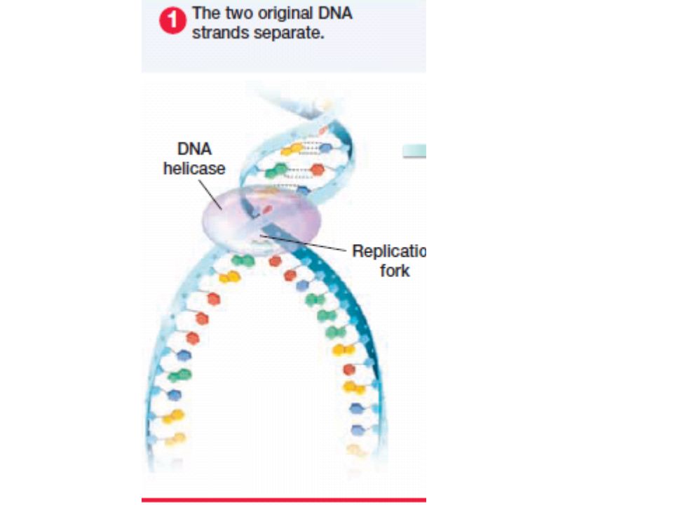
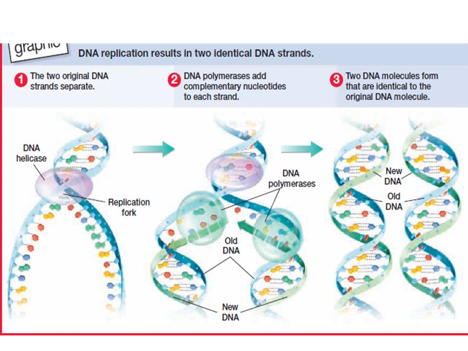
The Replication of DNA Before a cell divides, it copies its DNA by a process called DNA replication. ( s phase) In DNA replication, enzymes work to unwind and separate the double helix and add complementary nucleotides to the exposed strands.

130
The result of DNA replication is two exact copies of the cell’s original DNA.
Each new double helix is composed of one original DNA strand and one new DNA strand. .

131
DNA polymerase proofreads DNA during its replication so that very few errors occur

136
30. What is the direction of synthesis of the new strand?

137
5' to 3' direction

138
31. What are Okazaki fragments? How are they welded together?

139
Okazaki fragments are short segments of DNA synthesized away from the replication fork on a template strand during DNA replication. Many such segments are joined together by the enzyme DNA ligase to make up the lagging strand of newly synthesized DNA.

140
topoisomerase stabilizes the intact double helix
topoisomerase stabilizes the intact double helix. It also relieves strain in the DNA ahead of the replication fork helicase unwinds the double helix A short RNA primer is added by primase in the 5' to 3' direction

141
Helicase unwinds the parental double helix
Helicase unwinds the parental double helix. (Green) at the replication fork 2. Molecules of single-stranded binding protein stabilize the unwound template strands.( grey)
 at the replication fork. 2. Molecules of single-stranded binding protein stabilize the unwound template strands.( grey)")
142
3. The leading strand is synthesized continuously in the 5' to 3' direction by DNA polymerase III after being primed by primase. ( orange) 4. Primase begins synthesis of the RNA primer for the lagging strand. (pink) 5. DNA polymerase III synthesizes discontinuously the lagging strand in the 5' to 3' direction. 6. DNA polymerase I removes all the RNA primer sections and replaces them with DNA nucleotides ( yellow) 7.
 5. DNA polymerase III synthesizes discontinuously the lagging strand in the 5 to 3 direction. 6. DNA polymerase I removes all the RNA primer sections and replaces them with DNA nucleotides ( yellow) 7.")
143
6. DNA polymerase I removes all the RNA primer sections and replaces them with DNA nucleotides ( yellow) 7. The replacement of the primer with DNA leaves the new DNA nucleotides with a free 3' end. DNA ligase joins the free 3' end to its adjacent 5' end, forming a continuous and unbroken strand of DNA on both the leading and lagging strands.

144
33. Label the following figures
33. Label the following figures. Include 3' and 5' strands, RNA primer, primase, SSBP, topoisomerase, helicase, leading strand, lagging strand, DNA pol I, DNA pol III, DNA ligase, parental DNA, and new DNA On the second figure, also add arrows to indicate the direction of synthesis.

145
topoisomerase stabilizes the intact double helix
topoisomerase stabilizes the intact double helix. helicase unwinds the double helix A short RNA primer is added by primase in the 5' to 3' direction

148
34. Put it all together! Make a detailed list of the steps that occur in the synthesis of a new strand. 1. Helicase unwinds the parental double helix. 2. Molecules of single-stranded binding protein stabilize the unwound template strands. 3. The leading strand is synthesized continuously in the 5' 3' direction by DNA polymerase III after being primed by primase. 4. Primase begins synthesis of the RNA primer for the lagging strand. 5. DNA polymerase III synthesizes discontinuously the lagging strand in the 5' 3' direction. 6. DNA polymerase I removes all the RNA primer sections and replaces them with DNA nucleotides. 7. The replacement of the primer with DNA leaves the new DNA nucleotides with a free 3' end. DNA ligase joins the free 3' end to its adjacent 5' end, forming a continuous and unbroken strand of DNA on both the leading and lagging strands.

152
Enzymes for repair and proofreading

153
Prokaryotes have a single circular chromosome and eukaryotes have DNA packaged into several chromosomes that are found in the nucleus.

154
6. Describe how the DNA is packaged into a chromosome in a eukaryote and compare this to the prokaryotic chromosome._

155
6. Describe how the DNA is packaged into a chromosome in a eukaryote and compare this to the prokaryotic chromosome._ DNA is in chromosomes in eukaryotic cells which means it is wrapped around proteins called histones into units called nucleosomes to make chromosomes. In the prokaryotic cell the DNA is in a circular strand.

157
3. What enzyme first unwinds the DNA helix? _
4. What type of bond must be broken to “unzip the double helix ? 5. Leading and lagging strands are built using what enzyme? 6. The strands are bonded together to form 2 new strands each made up of one original and one new side. What is the name of the enzyme used to bond the strands together?

158
3. What enzyme first unwinds the DNA helix? _
DNA Helicase 4. What type of bond must be broken to “unzip the double helix ? hydrogen_ 5. Leading and lagging strands are built using what enzyme? DNA polymerase 6. The strands are bonded together to form 2 new strands each made up of one original and one new side. What is the name of the enzyme used to bond the strands together? DNA Ligase

159
Replication forks

160
7. How does this process make sure that the original information in the DNA is maintained in all new cells? 8. Why is this process important in terms of not making changes in the nitrogen base order? _ 9. How is the process of DNA replication in eukaryotes different from that of prokaryotes?

161
7. How does this process make sure that the original information in the DNA is maintained in all new cells? The original strands are used as templates to make the new copies, 8. Why is this process important in terms of not making changes in the nitrogen base order? _ If changes in the nitrogen bases are made the instructions are changes resulting in different proteins and this is called a mutation!__ 9. How is the process of DNA replication in eukaryotes different from that of prokaryotes? In eukaryotes, the process starts in many places and they then connect so that large amounts of DNA can be copies all at once. In prokaryotes it is a circular process starting in 2 places and then connecting in the middle.

162
36. What is a thymine dimer? How might it occur? How is it repaired?

163
A thymine dimer is the covalent linking of thymine bases that are adjacent on a DNA strand, causing the DNA to buckle and interfere with DNA replication. In order to repair this damage, a nuclease enzyme cuts the damaged DNA strand, and the damaged section is removed. DNA polymerase fills in the missing nucleotides, and DNA ligase seals the free end of the new DNA to the old DNA, making the strand complete

164
Thymine Dimer

165
37. Make a sketch of a chromosome and label the telomeres.

166
telomeres

167
38. Explain telomere erosion and the role of telomerase.

168
Telomeres provide their protective function by postponing the erosion of genes located near the ends of DNA molecules. Telomeres become shorter during every round of replication.

169
Telomeric DNA tends to be shorter in dividing somatic cells of older individuals and in
cultured cells that have divided many times. Importantly, some cell genomes (such as germ cells) must persist virtually unchanged from an organism to its offspring over many generations. In order to accomplish this, an enzyme called telomerase catalyzes the lengthening of telomeres in eukaryotic germ cells, thus restoring their original length and compensating for the shortening that occurs during DNA replication.
 must persist virtually unchanged from an organism to its offspring over many generations. In order to accomplish this, an enzyme called telomerase catalyzes the lengthening of telomeres in eukaryotic germ cells, thus restoring their original length and compensating for the shortening that occurs during DNA replication.")
170
39. Why are cancer cells immortal even though most body cells have a limited life span?

171
Researchers have found telomerase activity in cancerous somatic cells, suggesting that its ability to stabilize telomere length may allow these cancer cells to persist

172
40. On the following diagram, identify the following: 30-nm fiber, metaphase chromosome, double
helix, histone proteins, nucleosomes, protein scaffold, and looped domains (300-nm fiber). See pages in your text for the labeled figure.
. See pages in your text for the labeled figure.")
173
41. Distinguish between heterochromatin and euchromatin.

174
Heterochromatin is eukaryotic chromatin that remains highly compacted during interphase and is generally not transcribed. Euchromatin is a less condensed form of eukaryotic chromatin that is available for transcription. Heterochromatin is highly condensed, whereas euchromatin is less compact.

175
Protein production Vocabulary
1. mRNA 2. rRNA 3. tRNA 4. Transcription 5. RNA polymerase 6. Intron 7. Exon 8. Codon 9. Translation 10. Gene regulation 11. Operon 12. Mutation 13. Mutagen

176
links A very useful game to help comprehend DNA replication: A beneficial video about how DNA replication works: A quick video on DNA damage and repair:

Similar presentations


 Helicase Enzyme.>")


