Download presentation
Presentation is loading. Please wait.

1
The Circulatory System Chapter 37

2
Functions of the Circulatory System: Circulatory systems are used by large organisms that cannot rely on diffusion to exchange materials with the environment to transport –Oxygen –Carbon dioxide –Nutrients –Waste products –Hormones or cell products that are made in one place and used in another Helps to regulate body heat

3
Components of the Circulatory System: Heart Blood Blood vessels –Arteries –Arterioles –Capillaries –Venules –Veins

4
Parts of the Blood Blood is the only liquid tissue in the body It contains a liquid called plasma and 3 different cellular components –Red Blood Cells = RBC’s –White Blood Cells = WBC’s –Platelets

5
Plasma Is 46% to 63% of total blood volume Mostly water (92%) Transports dissolved materials such as –Plasma proteins –Hormones –Ions (charged atoms) –Nutrients –Wastes
 Transports dissolved materials such as –Plasma proteins –Hormones –Ions (charged atoms) –Nutrients –Wastes")
6
Red Blood Cells Also called erythrocytes Most numerous of all blood cells Produced in bone marrow Transport O 2 and CO 2 Contain HEMOGLOBIN, a protein that oxygen binds to No NUCLEUS Last 120 days

7
White Blood Cells Also called leukocytes Outnumbered by RBC’s 1000 to 1. Produced in bone marrow Contain a nucleus Guard against infection, fight parasites, and attack bacteria Can last days, weeks, or months.

8
Platelets Also called thrombocytes Cell fragments that function in blood clotting Certain cells in bone marrow break into pieces and the pieces are released into the blood stream When platelets come into contact with the edge of a broken blood vessel, they become very sticky. A cluster of platelets will form around the wound and begin to release chemicals called clotting factors, which start chemical reactions to clot blood.

9
Blood Vessels All mammals including humans have a closed circulatory system. This means that blood is always contained within the heart or a blood vessel There are 3 main types of blood vessels –Arteries –Veins –Capillaries

10
Arteries Large vessels that carry blood away from the heart Thick, muscular walls b/c they are exposed to high pressure All except pulmonary arteries carry oxygen rich blood The aorta is the largest blood vessel in the body

11
Veins Return blood to the heart MOST carry deoxygenated (or oxygen poor blood) Have muscular walls, but not nearly as thick as arteries. Have valves to prevent backflow of blood when it is being pumped from places below the heart

12
Capillaries Smallest of all blood vessels. Walls are only 1 cell thick. Nutrients, wastes, & gases are exchanged with cells through diffusion into and out of capillaries. So narrow, RBC’s must line up and go single file

13
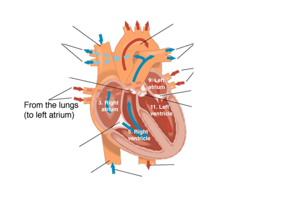
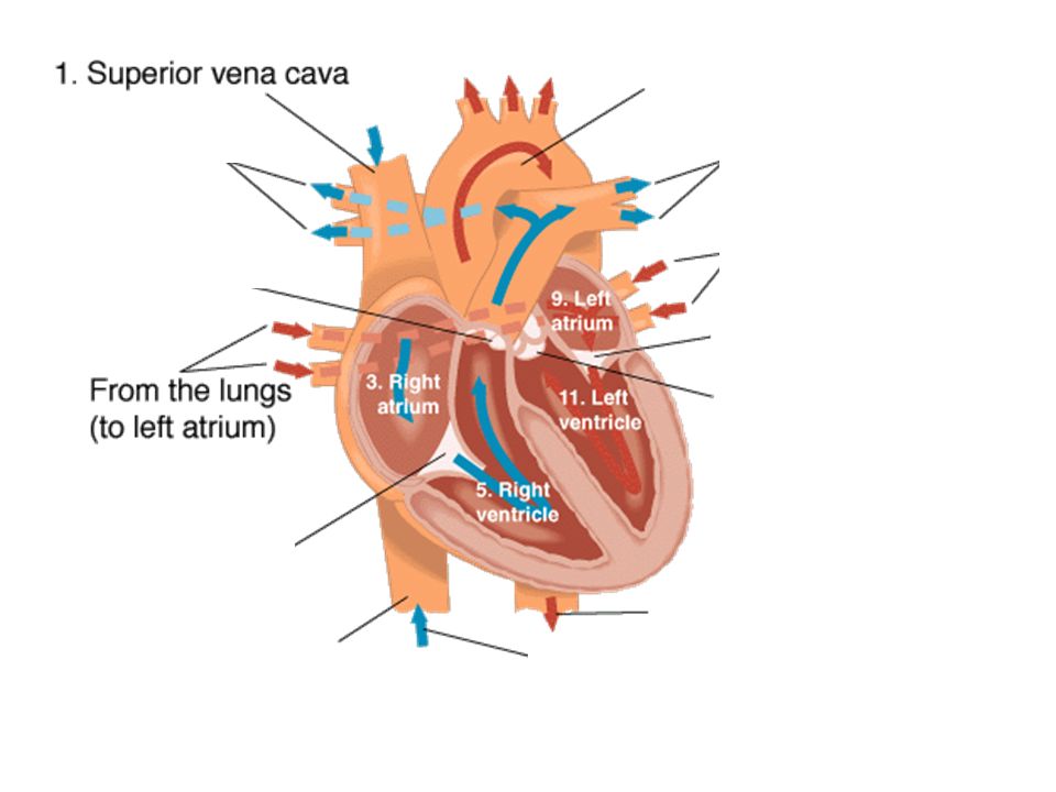
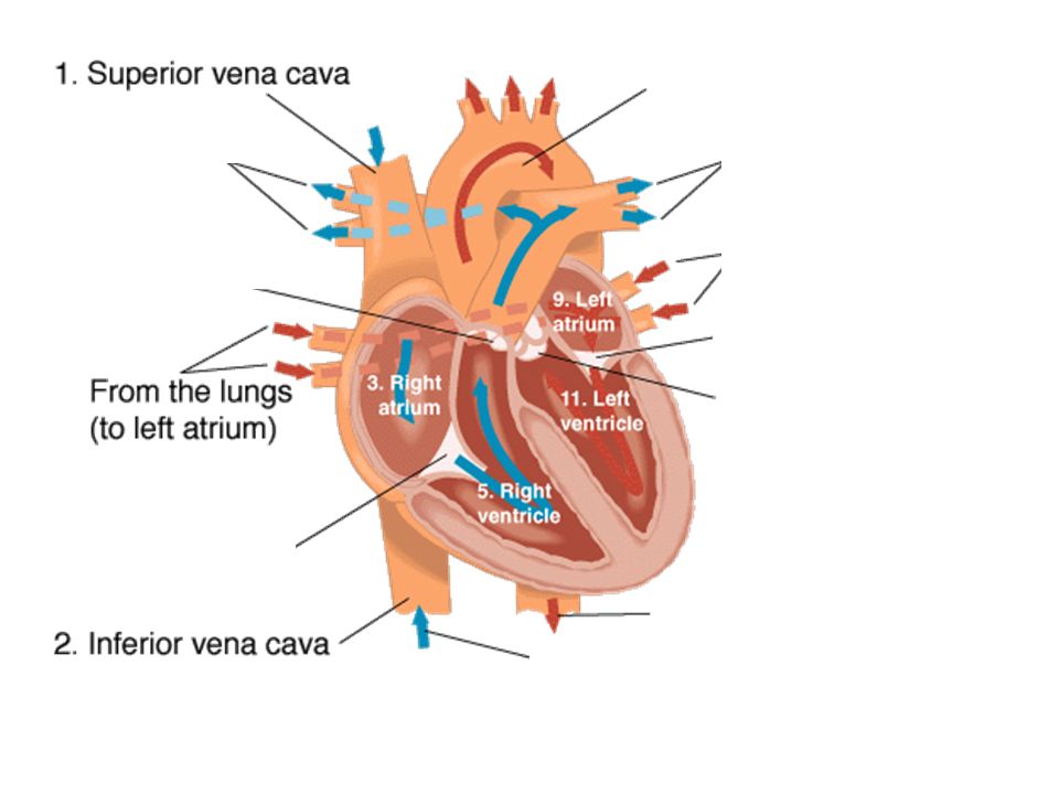
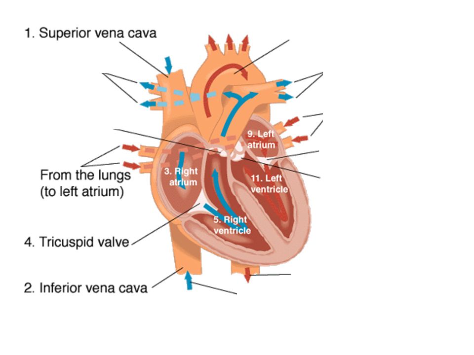
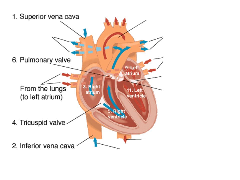
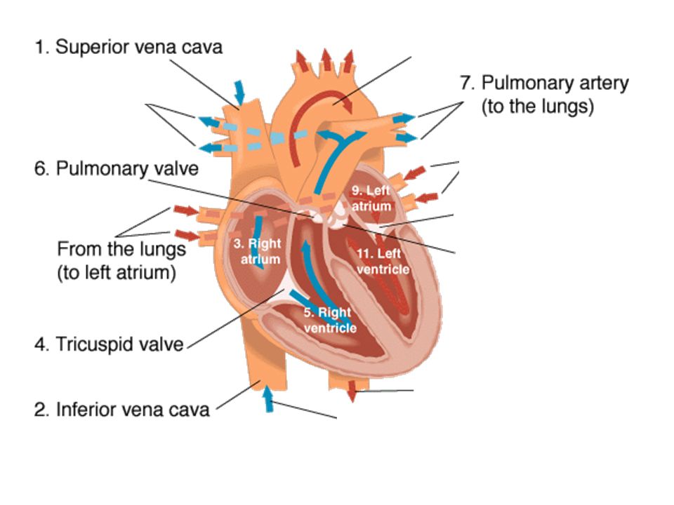
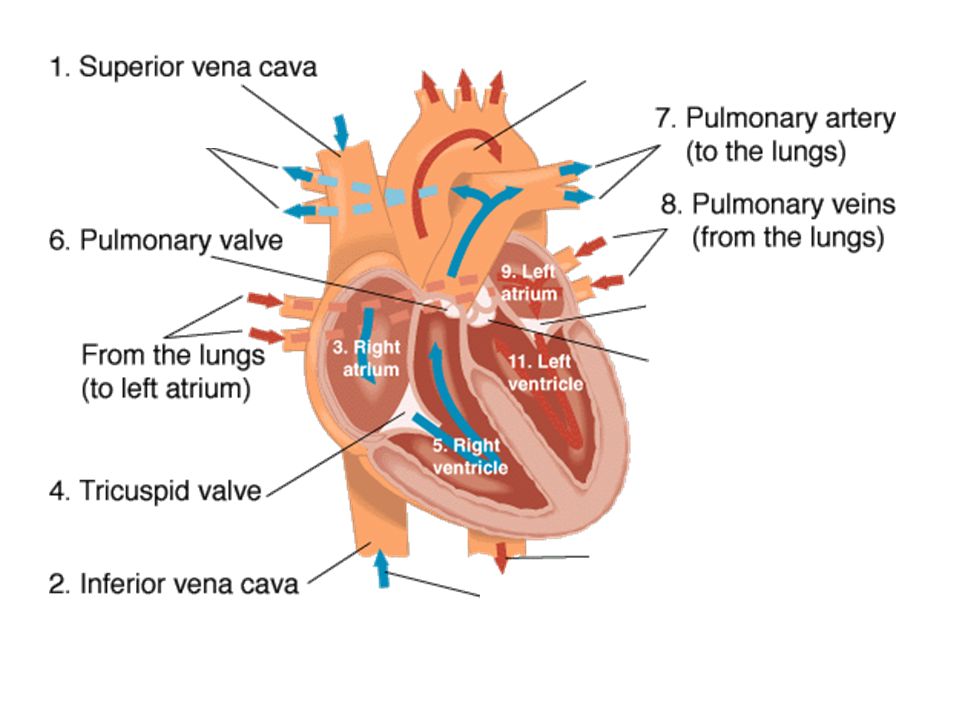
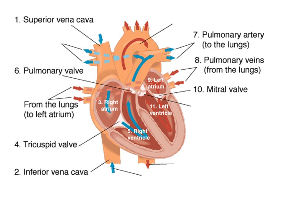
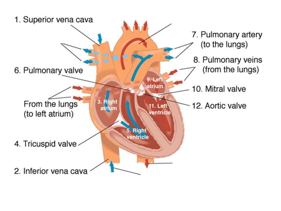
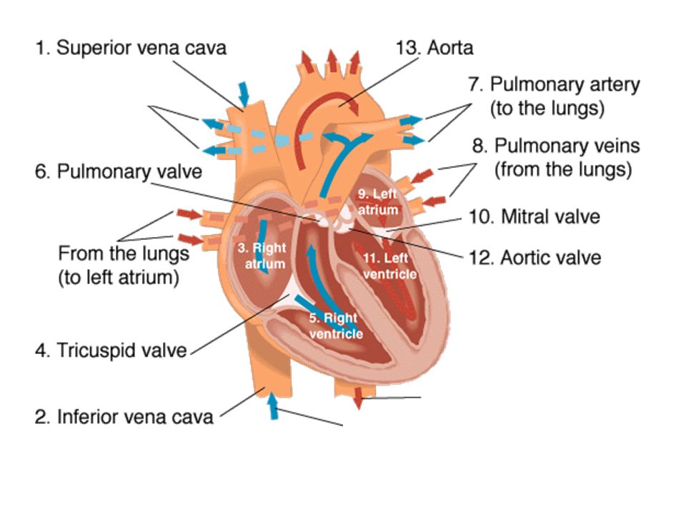
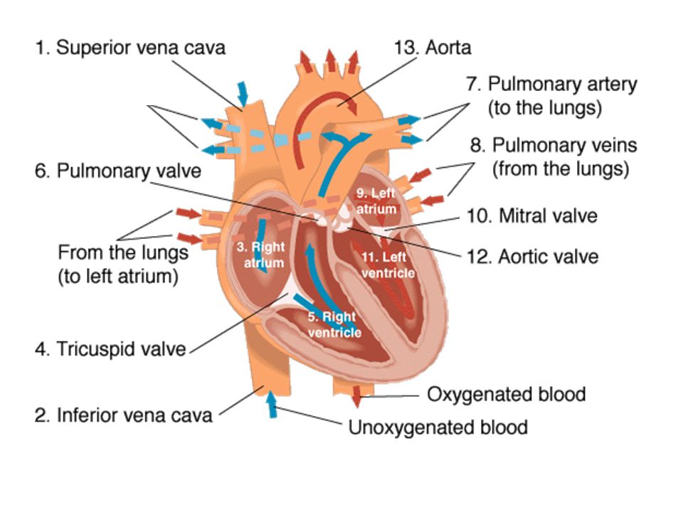
The Heart Mammals have a 4 chambered heart made of a type of muscle tissue called cardiac muscle. There are two atria and two ventricles. The atria are for collecting blood, the ventricles are for pumping blood. Oxygenated and deoxygenated DO NOT mix.

14
The Heart The walls of the ventricles are thicker than those of the atria. The walls of the left ventricle are the thickest of all because it must pump blood to the whole body, not just the lungs.

15
Pulmonary Circulation: –Blood returns from the body to the right atrium –It passes through the tricuspid valve to the right ventricle –It is pumped through the pulmonary arteries to the lungs. –Blood returns to the left atrium from the pulmonary veins

16
Systemic Circulation: –Blood returns to the heart from the lungs through the pulmonary veins, which empty into the left atrium. –Blood passes through the mitral valve, into the left ventricle. –Blood passes through the aortic valve and into the aorta. Blood passes through arteries, arterioles, capillaries, venules, and veins. –Blood returns to the heart through the vena cava, which empties into the right atrium.

17
The Heartbeat During the period of contraction (systole), the heart pumps blood out through the arteries; during the period of relaxation (diastole), the heart fills with blood. One complete sequence of filling and pumping blood is called a cardiac cycle, or heartbeat.

18
The pacemaker The heart's rhythm of contraction is controlled by the sinoatrial node (SA node), often called the pacemaker. This node is part of the heart's intrinsic conduction system, which is made up of specialized myocardial cells called nodal cells.

19
The activity of the conduction system, muscles, and valves of the heart are synchronized so that the heart can operate as a pump. The conduction system initiates and coordinates the muscular activity of the heart. Pressure differentials that result from muscle activity actuate the opening and closing of valves. The opening and closing of valves directs the flow of blood through the heart.

20
Flow of Blood

21
Path of blood You are a red blood cell in the superior vena cava. Trace your path through the heart and body using the following terms. Begin and end at the superior vena cava. –Right ventricle, right atrium, left ventricle, left atrium. –Tricuspid valve, mitral valve, aortic valve –Pulmonary arteries, pulmonary veins, lungs –Veins, venules, arteries, arterioles, capillaries, aorta

24
The Heart Mammalian hearts contain 4 chambers 2 atria (atrium singlular) for collecting blood 2 ventricles for pumping blood Oxygen rich and oxygen poor blood DO NOT mix Valves separate the atria and ventricles A wall called the septum separates right from left
 for collecting blood 2 ventricles for pumping blood Oxygen rich and oxygen poor blood DO NOT mix Valves separate the atria and ventricles A wall called the septum separates right from left")
25
The Heart

27
superior vena cava

28
aorta

29
superior vena cava aorta left pulmonary artery right pulmonary artery

30
superior vena cava aorta left pulmonary artery right pulmonary artery pulmonary veins

31
superior vena cava aorta left pulmonary artery right pulmonary artery pulmonary veins right atrium

32
superior vena cava aorta left pulmonary artery right pulmonary artery pulmonary veins right atrium left atrium

33
superior vena cava aorta left pulmonary artery right pulmonary artery pulmonary veins right atrium left atrium tricuspid valve

34
superior vena cava aorta left pulmonary artery right pulmonary artery pulmonary veins right atrium left atrium tricuspid valve bicuspid valve

35
superior vena cava aorta left pulmonary artery right pulmonary artery pulmonary veins right atrium left atrium tricuspid valve bicuspid valve right ventricle

36
superior vena cava aorta left pulmonary artery right pulmonary artery pulmonary veins right atrium left atrium tricuspid valve bicuspid valve right ventricle left ventricle

37
superior vena cava aorta left pulmonary artery right pulmonary artery pulmonary veins right atrium left atrium tricuspid valve bicuspid valve right ventricle left ventricle septum

38
superior vena cava aorta left pulmonary artery right pulmonary artery pulmonary veins right atrium left atrium tricuspid valve bicuspid valve right ventricle left ventricle septum semi-lunar valves

39
superior vena cava aorta left pulmonary artery right pulmonary artery pulmonary veins right atrium left atrium tricuspid valve bicuspid valve right ventricle left ventricle septum semi-lunar valves inferior vena cava

51
The heart Each side of the heart is completely separate and so deoxygenated blood and oxygenated blood do not mix

52
The heart The thickness of the walls of each chamber is related to the distance that it has to pump the blood. The atria just pump into the ventricles so are very thin

53
The heart The right ventricle has to pump the blood to the lungs and has a thinner wall than the left ventricle …because this has to pump blood all around the body

54
The heart The valves keep the blood flowing in one direction. The atrio-ventricular valves prevent the back flow of blood into the atria when the ventricles contract On the right side the tricuspid valve has three flaps, on the left the bicuspid has two flaps.

55
Structures of the Respiratory System pharynx larynx trachea bronchi bronchioles alveoli diaphragm

56
Respiratory System Function of the Respiratory System: To bring about the exchange of oxygen and carbon dioxide between the blood, the air, and the tissues.

58
Path of air to the lungs: Nose or mouth pharynx larynx trachea bronchi bronchioles alveloli

59
Path of air inside the lungs: Trachea bronchi bronchioles alveoli (air sacs)
")
60
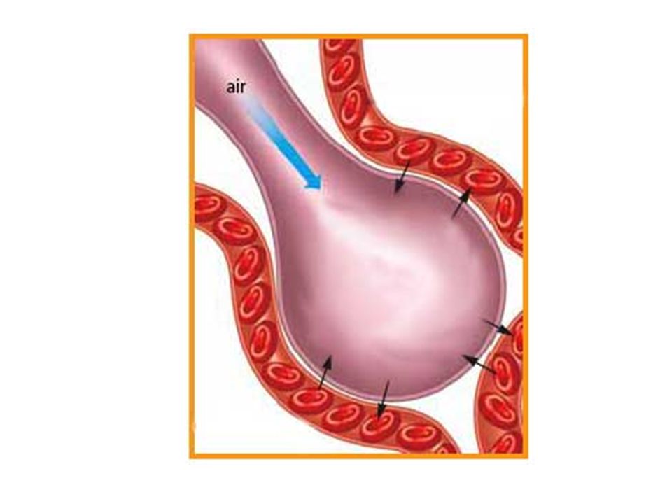
Gas Exchange: 150 million alveoli in each lung Oxygen dissolves in the moisture on the inner surface of the alveoli and then diffuses across the capillary wall into the blood Carbon dioxide in the blood moves from the capillaries to the alveoli and is then exhaled. Inhaled air = 21% oxygen and 0.04% carbon dioxide Exhaled air = 15% oxygen and 4% carbon dioxide

63
Breathing: Movement of air into and out of the lungs No muscles connected to the lungs. Air pressure forces air into the lungs The diaphragm is a muscle at the bottom of the body cavity. Inhaling: the diaphragm moves down the ribs move up and out the chest cavity volume increases creates a partial vacuum inside the body cavity air is forced in Exhaling : diaphragm moves up the ribs move down and in volume of chest cavity decreases internal pressure rises above air pressure outside the body air is forced out

64
Breathing

Similar presentations


, nutrient molecules and waste materials (from the digestive system) 2.Regulates.>")


