Download presentation
Presentation is loading. Please wait.

1
What do I mean by growth and development?

2
A secondary oocyte can be fertilized for about 24 hours after ovulation Sperm remain viable for up to 48 hours within the female reproductive tract This gives a three day “window” for intercourse to result in fertilization: two days before to one day after ovulation

3
Fertilization usually takes place in the outer one-third of the uterine tube, but can take place in the abdominal cavity Sperm swim up the female reproductive tract, aided by muscular contractions of the uterus stimulated by prostaglandins in the semen. The oocyte may also secrete a chemical that attracts sperm

4
Sperm undergo a functional change in the female tract – called capacitation During this process the membrane around the acrosome becomes fragile, and its enzymes are released. It requires the combined action of many sperm to allow one sperm to penetrate the oocyte.

6
When the first sperm enters the egg, the cell depolarizes causing the release of calcium ions inside the cell. This stimulates the release of granules that cause changes in the zona pellucida to prevent entry of other sperm. Secondary oocyte completes division, and nuclei of ovum and sperm unite to form a zygote.

7
Zygote undergoes rapid mitotic cell division, but these do not increase the size of the zygote – called cleavage divisions Cleavage produces a solid sphere of cells, still surrounded by zona pellucida – now called a morula. At 4.5 to 5 days, cells have developed into a hollow ball of cells – blastocyst. It is at this stage that it enters the uterus.

9
Blastocyst has an outer layer of cells called the trophoblast, an inner cell mass, and a fluid filled cavity called the blastocele. The trophoblast and part of the inner cell mass will form the membranes of the fetal portion of the placenta, the rest of the inner mass forms the embryo.

10
The blastocyst remains free in the uterus a short time, during which the zona pellucida disintegrates. Blastocyst nourished by glycogen from glands of the endometrium. At about 6 days after ovulation blastocyst implants – orients cell mass toward endometrium, and secretes enzymes which allow it to penetrate (digest) the endometrial wall. This nourishes the blastocyst for about a week after implantation.
 the endometrial wall. This nourishes the blastocyst for about a week after implantation..")
11
As early as 8 -12 days after fertilization, the blastocyst begins to secrete human chorionic gonadotropin or hCG. hCG keeps the corpus luteum active until the placenta can produce estrogens and progesterone. The presence of hCG is the basis for pregnancy tests.

12
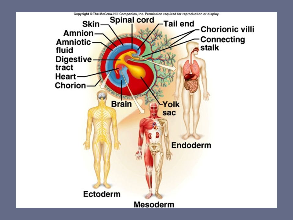
Inner cell mass forms two cavities: The yolk sac Amniotic cavity In humans the yolk sac produces blood cells and future sex cells The amniotic cavity becomes the cavity in which the embryo floats. Fluid is produced from fetal urine, and secretions from the skin, respiratory tract, and amniotic membranes.

15
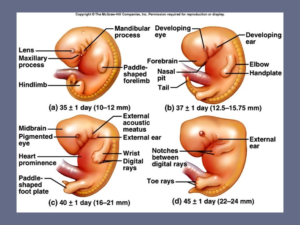
Divided into three trimesters. During first trimester individual starts out as a zygote, then morula, blastocyst, and after implantation, is called an embryo. Embryonic phase of development lasts from fertilization until the 8 th week of gestation, when it becomes a fetus. By day 35 the heart is beating, and eye and limb buds are present.

16
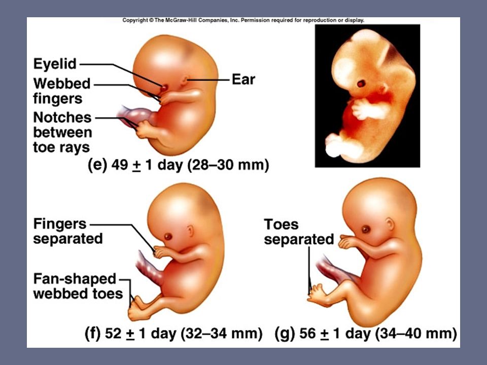
Week 4 - Curled upon itself, arm and leg buds, liver formed, heart present, primitive blood cells Weeks 6-8 – Rapid brain development, Fingers and toes, Eyes, ear and nose appear, GI tract, urogenital systems, Head very large because of brain Weeks 9-12 - Head still large, eye lids fused, nail beds, bile in intestines, sex traits, kidney secretion, respiratory-like movements, neck well defined

18
Weeks 13-19 - Fetus active, “quickening”, skeleton calcified, lanugo on head, placenta distinct, heart circulates blood, thyroid functions, total blood volume Weeks 17-20 - Fetal heart sounds with stethoscope, Scalp hair, eye brows present, fingernails and toe nails, some fat deposit Weeks 21-24 – Skin wrinkled, pink, eyebrows and eyelashes, external ear soft and shapeless, some breathing effort, lanugo on entire body

20
Weeks 25-28 – Skin wrinkled and covered with vernix caesosa, “Little old man” eyelids open, sub cutaneous fat, testes at internal inguinal ring Weeks 29-32 – Skin pink and smooth, areola of breast visible, testicles descend, female genitalia present (clitoris large), hair fine and wooly, can respond to noise outside body Weeks 33-36 – More rounded appearance, skin thicker and whiter, lanugo disappearing, sole creased, breast tissue beneath nipples
, hair fine and wooly, can respond to noise outside body Weeks – More rounded appearance, skin thicker and whiter, lanugo disappearing, sole creased, breast tissue beneath nipples")
22
The first movement of the fetus felt by the mother, usually occurring during the fourth or fifth month of pregnancy By month seven the fetus is quite active During the last month the fetus becomes less active (usually due to space considerations.)
")
23
Weeks 37-40 – After 38 weeks considered full term, body plump, lanugo gone from face, vernix leaving, testes in scrotum, labia majora and minora and clitoris developed, ear well defined, uniform color to eyes acquires antibodies from mother

25
At the end of pregnancy both the mother and the uterus become “irritable” The uterus undergoes Braxton-Hicks contractions: intermittent, painless contractions which can come 10 to 20 minutes apart. Become more frequent as gestation progresses, and can be mistaken for onset of labor Cervix begins to thin and dilate

26
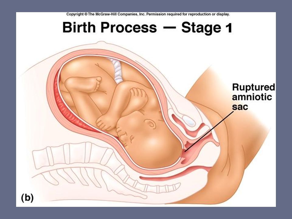
Stage one – the period from the onset of true labor contractions until the cervix is completely dilated at 10 cm. The uterine contractions cause the cervix to dilate, and the amniotic sac may rupture. Usually lasts 6 – 24 hours depending on the number of previous deliveries.

27
Stage one – the period from the onset of true labor contractions until the cervix is completely dilated at 10 cm. The uterine contractions cause the cervix to dilate, and the amniotic sac may rupture. Usually lasts 6 – 24 hours depending on the number of previous deliveries.

29
Inhered tied Prenatal period Three stages of labor

32
Period from maximal cervical dilation until the birth of the baby Lasts minutes to an hour Contractions become more intense and frequent.

34
The expulsion of the placenta Usually occurs within 15 minutes after the birth of the baby, but can range from 5 to 60 minutes.

36
Strictly speaking, most doctors define the age of viability as being about 24 weeks of gestation. In many hospitals, 24 weeks is the cutoff point for when doctors will use intensive medical intervention to attempt to save the life of a baby born prematurely. A baby born at 24 weeks would generally require a lot of intervention, potentially including mechanical ventilation and other invasive treatments followed by a lengthy stay in a neonatal intensive care unit (NICU). In the hands of experienced specialists, though, babies born slightly earlier may have a chance at survival. Babies born at 23 weeks may survive with these specialists in a state-of-the-art NICU, but the odds of survival are much lower. The earliest baby to have ever survived premature birth was born at 21 weeks and 6 days, and this was reported in the news as having been a miracle.
. In the hands of experienced specialists, though, babies born slightly earlier may have a chance at survival. Babies born at 23 weeks may survive with these specialists in a state-of-the-art NICU, but the odds of survival are much lower. The earliest baby to have ever survived premature birth was born at 21 weeks and 6 days, and this was reported in the news as having been a miracle..")
Similar presentations


>")


 – carrying of developing young within the female reproductive tract Fertilization to birth Humans = 266 days (38 weeks)>")