Download presentation
Presentation is loading. Please wait.

1
II- Globins (Histones): They are basic proteins rich in histidine amino acid. They are present in : a - combined with DNA b - combined with heme to form hemoglobin of RBCs. III- Fibrous proteins: also called Scleroproteins : They are structural proteins, not digested. include: keratin, collagen and elastin. a- α-keratin: protein found in hair, nails, enamel of teeth and outer layer of skin. It is α-helical polypeptide chain, rich in cysteine and hydrophobic (non polar) amino acids so it is water insoluble.
 amino acids so it is water insoluble..")
2
Keratin Straightening Treatments Are you tired of flat ironing your hair every morning? Is your hair wavy or curly and you want super straight silky healthy hair? Get a Keratin treatment.

3
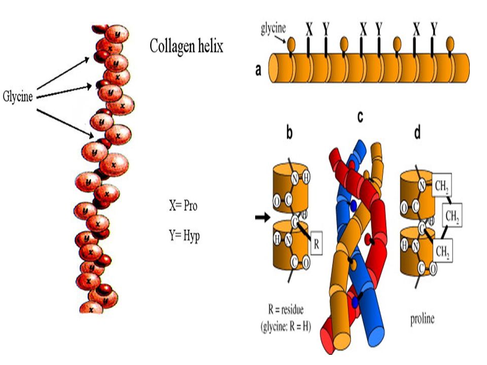
b- collagens: protein of connective tissues found in bone, teeth, cartilage, tendons, skin and blood vessels. Collagen may be present as gel e.g. in extracellular matrix or in vitreous humor of the eye. Collagens are the most important protein in mammals. They form about 30% of total body proteins. There are more than 20 types of collagens, the most common type is collagen I which constitutes about 90% of cell collagens. Structure of collagen: three helical polypeptide chains (trimer) twisted around each other forming triplet-helix molecule. ⅓ of structure is glycine, 10% proline, 10% hydroxyproline and 1% hydroxylysine. Glycine is found in every third position of the chain. The repeating sequence –Gly-X-Y-, where X is frequently proline and Y is often hydroxyproline and can be hydroxylysine.
 twisted around each other forming triplet-helix molecule. ⅓ of structure is glycine, 10% proline, 10% hydroxyproline and 1% hydroxylysine. Glycine is found in every third position of the chain. The repeating sequence –Gly-X-Y-, where X is frequently proline and Y is often hydroxyproline and can be hydroxylysine..")
5
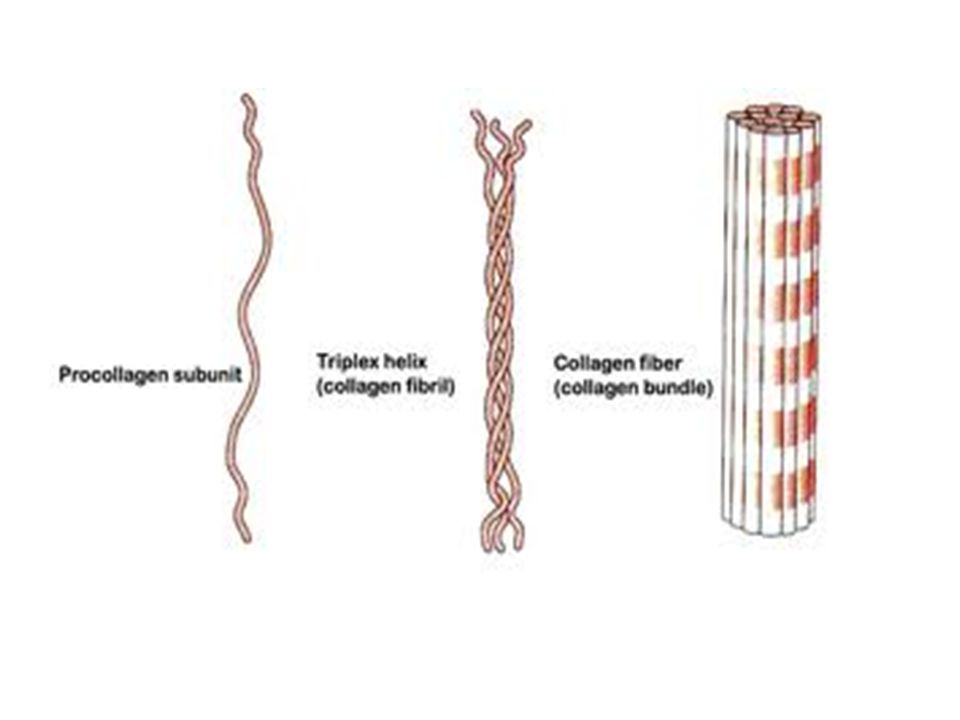
Solubility: collagen is insoluble in all solvents and not digested. When collagen is heated with water or dil. HCl it will be converted into gelatin which is soluble, digestible and used as diet (as jelly). Gelatin is classified as derived protein. Steps in Biosynthesis of Collagen: 1.Translation on ribosomes to form 3 polypeptide chains called α chains. This is the preprocollagen 2.Hydroxylation of some of proline and lysine into hydroxyproline and hydroxylysine. This step needs enzymes lysyl hydroxylase and prolyl hydroxylase in the presence of vitamin C. 3.Release of α chains into from ribosome to endoplasmic reticulum and addition of glucose and galactost to hydroxy group of some hytdroxylysine forming procollagen. 4.Combination of the 3 chains and formation of triple helix (also is a procollagen) 5. Release into extracellular space and removal of segments from N- and C- terminal to form collagen or called tropocollagen 6. Cross linkage of collagen molecules to form mature collagen fibres (or collagen network). This cross linkage is essential for achieving strength necessary for proper function of connective tissues.
. Gelatin is classified as derived protein. Steps in Biosynthesis of Collagen: 1.Translation on ribosomes to form 3 polypeptide chains called α chains. This is the preprocollagen 2.Hydroxylation of some of proline and lysine into hydroxyproline and hydroxylysine. This step needs enzymes lysyl hydroxylase and prolyl hydroxylase in the presence of vitamin C. 3.Release of α chains into from ribosome to endoplasmic reticulum and addition of glucose and galactost to hydroxy group of some hytdroxylysine forming procollagen. 4.Combination of the 3 chains and formation of triple helix (also is a procollagen) 5. Release into extracellular space and removal of segments from N- and C- terminal to form collagen or called tropocollagen 6. Cross linkage of collagen molecules to form mature collagen fibres (or collagen network). This cross linkage is essential for achieving strength necessary for proper function of connective tissues..")
7
Note that, Hydroxylation reaction to form hydroxy proline and hydroxylysine are important for the following: a- attachment of sugars (glucose and galactose) to OH groups of hydroxylysine and hydroxyproline b- hydroxylation is very important to organize the three chains in the form a triple helix. The three chains are linked together by hydrogen bonding between OH groups in the three chains. c- OH groups of hydroxylysine is needed to permit the cross-linking of the triple helices into the fibers and networks of the tissues.

8
cross-linking which links hydroxylysine and lysine residues. Multiple collagen fibrils form into collagen fibers.

9
Collagen biosynthesis (Fig. 2)
")
10
Collagen biosynthesis (Fig. 3)
")
12
Some collagen diseases: 1- Scurvy: disease due to deficiency of vitamin C which is important coenzyme for conversion of proline into hydroxyproline and lysine into hydroxylysine. Thus, synthesis of collagen is decreased leading to abnormal bone development, bleeding, loosing of teeth and swollen gum. 2- Osteogenesis Imperfecta (OI): Inherited disease resulting from genetic deficiency or mutation in gene that synthesizes collagen type I leading to abnormal bone formation in babies and frequent bone fracture in children. It may be lethal. What is the type of mutation that causes this disease
: Inherited disease resulting from genetic deficiency or mutation in gene that synthesizes collagen type I leading to abnormal bone formation in babies and frequent bone fracture in children. It may be lethal. What is the type of mutation that causes this disease.")
13
OI Type I OI type I is the most common type of OI. People with OI type I have an average of 20 to 40 fractures before puberty. Fewer fractures occur after puberty. Prenatal diagnosis often is possible. People with OI type I have one or more of the following features: -fragile bones -- hearing loss beginning in the teens, twenties, or thirties -curvature of the spine -thin, smooth skin In other severe types of OI: Person have 20 or more fractures during the first three years of life and more than 100 fractures by puberty. Loose joints and poor muscle development are common. Characteristic features include: - soft bones that not only break but also bend easily -misshapen arms and legs. severe curvature of the spine -severely misshapen chest that may cause problems with breathing - poor development of the teeth, causing teeth to be discolored and to break easily - short stature (some people may only grow to be three feet tall).
..")
15
IV-Conjugated proteins i.e. On hydrolysis, give protein part and non protein part and subclassified into: 1- Phosphoproteins: These are proteins conjugated with phosphate group. Phosphorus is attached to OH group of serine or threonine. e.g. Casein of milk and vitellin of yolk. 2- Lipoproteins: These are proteins conjugated with lipids. Functions: a- help lipids to transport in blood b- Enter in cell membrane structure helping lipid soluble substances to pass through cell membranes.

16
3- Glycoproteins: proteins conjugated with sugar (carbohydrate) e.g. - Some hormones such as erythropoeitin, LH, FSH - present in cell membrane structure - blood groups that are present on the surface of RBCs ( A, B, O) - 4- Nucleoproteins: These are basic proteins ( e.g. histones) conjugated with nucleic acid (DNA or RNA). e.g. a- chromosomes: are proteins conjugated with DNA b- Ribosomes: are proteins conjugated with RNA
 - 4- Nucleoproteins: These are basic proteins ( e.g. histones) conjugated with nucleic acid (DNA or RNA). e.g. a- chromosomes: are proteins conjugated with DNA b- Ribosomes: are proteins conjugated with RNA.")
17
5- Metalloproteins: These are proteins conjugated with metal like iron, copper, zinc, …… a- Iron-containing proteins: - hemoglobin (Hb), - myoglobin (protein of skeletal muscles and cardiac muscle), NB: Hb and myoglobin are hemeproteins that bind O 2 - Ferritin: Main store of iron in the body in non toxic form, because free iron is toxic and oxidize cells (form reactive oxygen species). Ferritin is present in liver, spleen and bone marrow. The amount of ferritin stored reflects the amount of iron stored. Ferritin releases iron to areas where it is required. - Hemosidrin: another iron store. (report hemosidrosis) -Transferrin: is the iron carrier protein in plasma.
 -Transferrin: is the iron carrier protein in plasma..")
18
Typically in the healthy individual, about 65% of the iron in the body is in hemoglobin (in red blood cells) and about 4% in myoglobin (protein in skeletal muscle). About 30% of the iron in the body is stored (as ferritin or hemosiderin) in liver, bone marrow, and the spleen. A small percentage of the body's iron is in transport between various compartments of the body (in association with transferrin) or is a component of enzymes in cells throughout the body. Ferritin is a globular protein complex consisting of 24 subunits and is the primary intracellular iron-storage protein keeping iron in a soluble and non-toxic form. Ferritin that is not combined with iron is called apoferritin.
 in liver, bone marrow, and the spleen. A small percentage of the body s iron is in transport between various compartments of the body (in association with transferrin) or is a component of enzymes in cells throughout the body. Ferritin is a globular protein complex consisting of 24 subunits and is the primary intracellular iron-storage protein keeping iron in a soluble and non-toxic form. Ferritin that is not combined with iron is called apoferritin..")
19
Ferritin: The Iron-Storage Protein Most of us are able to maintain appropriate levels of available iron in the body (enough available iron to ensure an adequate supply of hemoglobin, but not so much as to produce toxic effects), Ferritin (Figure) is the key to this important control of the amount of iron available to the body. Ferritin is a protein that stores iron and releases it in a controlled fashion. Hence, the body has a "buffer" against iron deficiency (if the blood has too little iron, ferritin can release more) and, to a lesser extent, iron overload (if the blood and tissues of the body have too much iron, ferritin can help to store the excess iron).
 and, to a lesser extent, iron overload (if the blood and tissues of the body have too much iron, ferritin can help to store the excess iron)..")
20
hemoglobin Ferritin

21
b- Copper containing proteins: e.g. - Ceruloplasmin which oxidizes ferrous ions into ferric ions. c- Mg containing proteins: e.g. Kinases and phosphatases. Kinases are enzymes that add phosphate to a molecule. Phoshatases are enzymes that remove phosphate from a molecule.

Similar presentations


 Hb is found in RBCs its main function is to transport O2 to tissues. Structure: 2 parts : heme + globin Globin: four globin chains (2 α.>")

 keratin, myosin, fibrinogen - mainly alpha helix.>")


: They are basic proteins rich in histidine amino acid. They are present in : a - combined with DNA b - combined with heme to form.>")