Download presentation
Presentation is loading. Please wait.

1
Jenna Scholnick, MD January 21, 2011

2
Goals Identify common benign skin conditions Describe skin disorders characterized by papules and pustules Recognize vascular, hyperpigmented, and hypopigmented birthmarks Identify and treat various types of diaper rashes

4
Cutis Marmorata Clinical Features: When infants become cold, capillaries in the extremities constrict, causing characteristic violaceous, reticulated mottling of the skin. Treatment: No treatment needed. Resolves spontaneously with warming. May persist in patients with Down syndrome, trisomy 18, or Cornelia de Lange syndrome. When to Refer: Localized or exaggerated mottling may represent a related disorder referred to as cutis marmorata telangiectatica congenita

5
Harlequin color change Occurs when newborn lies on side Erythema of dependent side of body with simultaneous blanching of contralateral side. Develops suddenly and persists for 30 sec to 20 minutes Resolves with increased muscle activity or crying Affects up to 10% of full term infants but often goes unnoticed due to bundling. 2 nd to 5 th day until three weeks Thought to be caused by immatturity of hypothalamic center

7
Erythema Toxicum Neonatorum Cause: Inflammatory disorder of unknown etiology. Clinical Features: Blotchy, erythematous macules and patches are present and often have a central papule, vesicle, or pustule. Fluid from vesicles contains eosinophils. Lesions appear within 24 to 48 hours of birth. Occurs in up to 50% of term infants (all races) and is uncommon in premature infants. Treatment: No treatment is required. Lesions typically resolve within 4 to 5 days, although the process may begin later or persist longer in some infants.
 and is uncommon in premature infants. Treatment: No treatment is required. Lesions typically resolve within 4 to 5 days, although the process may begin later or persist longer in some infants..")
10
Milia Cause: Epidermal inclusion cysts. Clinical Features: Tiny (1- to 2-mm) white papules most commonly located on the forehead, nose, and cheeks. The lesions may be present at birth or develop during the first few months of life. Treatment: No therapy is necessary. Milia generally resolve spontaneously over several weeks to months
 white papules most commonly located on the forehead, nose, and cheeks. The lesions may be present at birth or develop during the first few months of life. Treatment: No therapy is necessary. Milia generally resolve spontaneously over several weeks to months.")
12
Miliaria Cause: Miliaria is caused by obstruction of the eccrine ducts, which may be superficial (resulting in miliaria crystallina) or deep (resulting in miliaria rubra or pustulosa) Miliaria occurs primarily during hot, humid weather or febrile illness. It may also occur if an infant is dressed too warmly.

13
Miliaria rubra (“prickly heat”) Clinical Features: Most common form of miliaria Obstruction of the eccrine ducts leads to dilation and rupture, producing swelling and inflammation. Characterized by tiny erythematous macules or papules often concentrated on areas of skin covered by clothing. Treatment: Best management is prevention, avoiding excessive environmental heat, overdressing, and application of thick emollients (which may occlude eccrine ducts). Infants with established miliaria may benefit from a low- humidity air-conditioned environment. Cool baths or sponge baths may be helpful.
. Infants with established miliaria may benefit from a low- humidity air-conditioned environment. Cool baths or sponge baths may be helpful..")
15
Miliaria Crystallina Clinical Features: Characterized by clear, thin-walled vesicles without surrounding erythema. The vesicles rupture easily. Treatment: Management and prevention are as described for miliaria rubra.

17
Mongolian spots Cause: Arrested migration of melanocytes from the neural crest to the skin. Clinical Features: Brown to gray-blue macules and patches most commonly distributed over the lumbosacral spine and buttocks but may also occur on the trunk or extremities. Differential Diagnosis: Mongolian spots are sometimes confused with melanocytic nevi, vascular lesions, and bruises. Treatment: No treatment is necessary, as there is no potential for malignant change, and the lesions usually fade spontaneously by adolescence.

19
Neonatal acne Cause: Obstruction and inflammation within pilosebaceous follicles. Attributed to androgens. Clinical Features: occurs in approximately 20% of all neonates during first few weeks of life and consists of papules, pustules, and closed comedones located primarily on the face. Rarely, larger papulonodular or scarring lesions may occur. Treatment: Treatment usually unnecessary. As androgen levels decline during the first months of life, the condition resolves. When to Refer: Infants with persistent or unusually severe acne should be evaluated for signs of androgen excess (eg, virilization).
..")
21
Neonatal cephalic pustulosis Cause: A recently described condition that may be result of an inflammatory response to yeasts of the Malassezia species. May represent disorder in the spectrum of neonatal acne. Clinical Features: Characterized by an erythematous papular and pustular eruption that usually affects the face, although scalp and upper part of chest may be involved. Lesions begin to appear at 2 to 3 weeks of age. Closely resembles neonatal acne but does not produce comedones (blackheads and whiteheads) and often involves the scalp. Treatment: Resolves spontaneously over several weeks. Treatment with an imidazole cream twice daily may hasten resolution
 and often involves the scalp. Treatment: Resolves spontaneously over several weeks. Treatment with an imidazole cream twice daily may hasten resolution.")
23
Sebaceous gland hyperplasia Cause: Hypertrophy of sebaceous glands caused by elevated androgen levels present during the first months of life. Clinical Features: Pinpoint, yellow macules and papules (enlarged sebaceous glands) are commonly located on the nose, forehead, cheeks, and upper lip. The condition is often present in term newborns. Treatment: No therapy is necessary. The condition resolves as androgen levels decline
 are commonly located on the nose, forehead, cheeks, and upper lip. The condition is often present in term newborns. Treatment: No therapy is necessary. The condition resolves as androgen levels decline.")
25
Postdates desquamation Cause: Thought to be the result of loss of vernix and the transition from the aqueous intrauterine environment to the dry extrauterine environment. Clinical Features: Desquamation or cracking of the skin is most pronounced in acral locations (eg, the wrists, hands, ankles, and feet) but may be generalized. The degree of desquamation increases proportionately with the degree of postmaturity. Treatment: No treatment is required, but an emollient may be applied. The condition usually resolves during the first weeks of life.
 but may be generalized. The degree of desquamation increases proportionately with the degree of postmaturity. Treatment: No treatment is required, but an emollient may be applied. The condition usually resolves during the first weeks of life..")
28
Sucking blister Cause: Vigorous sucking in utero. Clinical Features: Present at birth in 0.5% of newborns. Appear as shallow erosions or intact bullae, most commonly on the fingers, wrists, lips, or forearms. Differential Diagnosis: Occasionally mimic other blistering disorders (eg, epidermolysis bullosa) and infections (eg, bullous impetigo and herpes simplex virus infection). However, their characteristic appearance and location suggest the diagnosis. Treatment: The condition is self-limited, and treatment is supportive. A topical antibiotic and dressing may be applied as needed.
 and infections (eg, bullous impetigo and herpes simplex virus infection). However, their characteristic appearance and location suggest the diagnosis. Treatment: The condition is self-limited, and treatment is supportive. A topical antibiotic and dressing may be applied as needed..")
30
Pseudo ingrown nails Cause: Hypoplasia and soft consistency of the infantile nail plate. Clinical Features: Most commonly affects the great toenails and presents as swelling or redness of the lateral nail fold, simulating an ingrown nail. Treatment: Intervention is rarely required. With age, the nail plates grow and assume the appearance of mature nails

32
Seborrheic dermatitis Cause: Unknown but possibly related to increased sebaceous gland function due to elevated androgen levels during the first months of life. Some believe that it may be the result of an inflammatory response to the yeast Pityrosporum ovale. Clinical Features: Characterized by scaling and/or erythematous patches concentrated in areas where sebaceous glands are most numerous, such as the scalp, face, and postauricular and intertriginous areas. The umbilicus is frequently involved. Treatment: The condition usually resolves spontaneously by 8 to 12 months of age. For scaling of the scalp ("cradle cap”): small amount of mineral oil applied to affected area followed by gentle brushing with a soft brush may be effective. Alternatively, an antiseborrheic shampoo may be used. For lesions on the skin: a low-potency topical corticosteroid (eg, hydrocortisone 1%) may be applied twice daily as needed.
: small amount of mineral oil applied to affected area followed by gentle brushing with a soft brush may be effective. Alternatively, an antiseborrheic shampoo may be used. For lesions on the skin: a low-potency topical corticosteroid (eg, hydrocortisone 1%) may be applied twice daily as needed..")
34
Transient neonatal pustular melanosis Cause: Benign pustular disorder of unknown etiology. Clinical Features: Present at birth and occurs in approximately 5% of African-American newborns; it is rare in other races. Pustules and vesiculopustules that rupture easily (often before birth), leaving small hyperpigmented macules, each with a rim (collarette) of scale. Polymorphonuclear leukocytes are present in the fluid from pustules. The lesions are concentrated on the forehead, chin, neck, and trunk. The palms and soles also may be affected. Treatment: No treatment is required. The pustules resolve within a few days, and the hyperpigmented macules fade completely by 3 to 4 months of age.
, leaving small hyperpigmented macules, each with a rim (collarette) of scale. Polymorphonuclear leukocytes are present in the fluid from pustules. The lesions are concentrated on the forehead, chin, neck, and trunk. The palms and soles also may be affected. Treatment: No treatment is required. The pustules resolve within a few days, and the hyperpigmented macules fade completely by 3 to 4 months of age..")
38
Acropustulosis of infancy Cause: An uncommon disorder of unknown etiology, although some believe it may represent a postscabies hypersensitivity phenomenon. Clinical Features: May be present at birth but more often presents in the first months of life. Characterized by a rash composed of tense, pruritic vesiculopustules concentrated on the hands and feet, including the palms and soles. In some patients, a few lesions may be present on the trunk, proximal extremities, or scalp. The lesions persist for 5 to 10 days, but crops of new lesions appear every 2 to 4 weeks..

39
Acropustulosis of infancy Differential Diagnosis: Most often confused with scabies. However, scabies does not recur with regularity, and a scraping of lesions will reveal mites, eggs, or fecal material. Dyshidrotic eczema may cause vesicles on the hands or feet but rarely has its onset in infancy. Treatment: The condition resolves spontaneously over 1 to 2 years. Flares may be managed with a potent topical corticosteroid used cautiously or an oral antihistamine

41
Scabies Cause: Infestation with the mite, Sarcoptes scabiei, which rarely occurs during the first month of life. Clinical Features: Unlike in older children, in whom lesions are concentrated in skin flexures, in infants, scabies causes a generalized rash composed of erythematous papules, vesicles, or pustules. The rash may involve the palms, soles, and scalp. Diagnosis: The diagnosis may be confirmed by microscopic examination of scrapings from several papules placed in immersion oil on a glass slide. Mites, eggs, or fecal material may be observed.

42
Scabies Differential Diagnosis: When vesicles are present, scabies may be confused with acropustulosis of infancy. However, the latter condition can be differentiated by its recurrence every 2 to 4 weeks and the concentration of lesions on the hands and feet. Treatment: Treatment is topical permethrin 5%, applied to the infant and to all close contacts and rinsed 8-14 hours later. Treatment is often repeated in 1 week for infested individuals.

43
Infant birthmarks Vascular birthmarks are the most common of all birthmarks and include hemangiomas, port-wine stains, and nevus simplex. Hyperpigmented birthmarks include congenital melanocytic nevi (increased risk of malignant transformation), and café au lait spots (macules). Hypopigmented birthmarks are common in infancy. Do not have malignant potential, but may represent a cutaneous marker of systemic disease. Examples include ash-leaf spots and nevus depigmentosus
, and café au lait spots (macules). Hypopigmented birthmarks are common in infancy. Do not have malignant potential, but may represent a cutaneous marker of systemic disease. Examples include ash-leaf spots and nevus depigmentosus.")
45
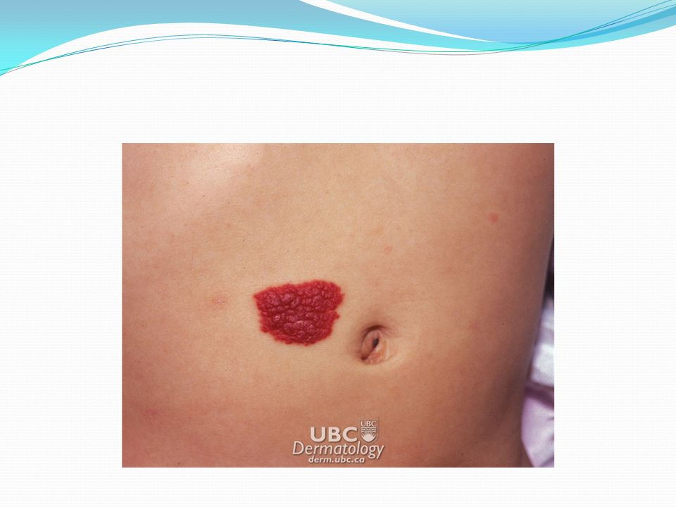
Hemangiomas Cause: Benign, self-limited vascular tumors of unknown etiology. Development may be related to placental factors. Risk factors include prematurity and advanced maternal age and, possibly, pregnancy-induced hypertension and placenta previa. Clinical Features: Typically appear at 2 to 4 weeks of age and affect 2.6% of newborns and 10% to 12% of 1-year-old infants. More common in females and preterm infants. Appear as enlarging erythematous papules, nodules, or plaques, most commonly on the head or neck. They can be superficial, deep, or mixed. Deeper lesions have a blue color. Grow rapidly in the first 4 to 6 months of age. Between 6 and 12 months of age, growth slows and involution begins (appearance of areas of gray mottling on the surface of the lesion)
.")
46
Hemangiomas Treatment: Half of all hemangiomas resolve by age 5 years, and 90% resolve by age 9 years. If small and does not threaten a vital structure, no treatment is required. If redundant skin or telangiectasias remain after involution, patients may be referred to a pediatric dermatologist or plastic surgeon for pulsed dye laser treatment. Patients should be referred to a pediatric dermatologist if the hemangioma compromises a vital structure (eg, the eye or airway); ulcerates; is located on the tip of the nose, lip, or midline of the back; or is large, particularly if it involves the face or head.
; ulcerates; is located on the tip of the nose, lip, or midline of the back; or is large, particularly if it involves the face or head..")
47
Hemangiomas Hemangiomas of the eyelids or periorbital skin may obstruct the visual axis (leading to amblyopia) or may compress the globe (resulting in glaucoma or astigmatism) Any hemangioma that obstructs the visual axis or causes ptosis should be evaluated immediately by a dermatologist and an ophthalmologist. May extend into the orbit, imaging (eg, magnetic resonance imaging [MRI]) should be considered Hemangiomas located on the tip of the nose or lip often cause permanent distortion. Early referral is indicated. These lesions may be treated with intralesional or oral corticosteroids or propranolol. Life- threatening or treatment-resistant hemangiomas may be treated with vincristine, interferon alfa, or other agents
 should be considered Hemangiomas located on the tip of the nose or lip often cause permanent distortion. Early referral is indicated. These lesions may be treated with intralesional or oral corticosteroids or propranolol. Life- threatening or treatment-resistant hemangiomas may be treated with vincristine, interferon alfa, or other agents.")
50
Nevus simplex Cause: A nevus simplex (“salmon patch”) represents an area of superficially dilated capillaries. Clinical Features: These erythematous lesions occur in 30% to 40% of newborns and are commonly located at the glabella, on the eyelids, or at the nape of the neck. Treatment: Most lesions on the face resolve during the first 2 years of life. Those at the nape of the neck may persist.

52
Port wine stain Cause: Vascular malformation of unknown etiology. Clinical Features: These erythematous or violaceous patches are present in 0.5% of newborns. They tend to be permanent, although some may fade slightly with time. Many of these lesions become darker and thicker with time, and they may develop vascular nodules that bleed. Most port-wine stains are not associated with underlying disease. However, a facial port-wine stain may be associated with Sturge-Weber syndrome (SWS); those involving the extremities or trunk rarely are associated with Klippel-Trénaunay or Proteus syndromes. Treatment: Early treatment with pulsed dye laser improves the appearance of facial port-wine stains and may prevent later local complications.
; those involving the extremities or trunk rarely are associated with Klippel-Trénaunay or Proteus syndromes. Treatment: Early treatment with pulsed dye laser improves the appearance of facial port-wine stains and may prevent later local complications..")
54
Congenital melanocytic nevi Cause: Unknown. Clinical Features: Congenital melanocytic nevi (CMN) are hyperpigmented macules or thin plaques present at birth. The lesion may exhibit hypertrichosis. All CMN have an increased risk of malignant transformation; the larger the nevus, the greater the risk. Risk of transformation is 2% to 3% in small lesions ( 20 cm). The presence of hair does not imply a poor prognosis (ie, risk for malignancy). The risk of malignant transformation in African-American patients is much lower. Treatment: Management of CMN remains controversial. Small lesions may be observed or electively excised at puberty; malignant change before puberty is extraordinarily rare.
 are hyperpigmented macules or thin plaques present at birth. The lesion may exhibit hypertrichosis. All CMN have an increased risk of malignant transformation; the larger the nevus, the greater the risk. Risk of transformation is 2% to 3% in small lesions ( 20 cm). The presence of hair does not imply a poor prognosis (ie, risk for malignancy). The risk of malignant transformation in African-American patients is much lower. Treatment: Management of CMN remains controversial. Small lesions may be observed or electively excised at puberty; malignant change before puberty is extraordinarily rare..")
57
Acrodermatitis enteropathica Cause: Rare autosomal recessive disorder Defective gastrointestinal transport protein results in impaired absorption of zinc. Because human milk contains a protein that facilitates zinc absorption, clinical manifestations often appear after an infant is weaned from human milk to formula or cow’s milk. An acrodermatitis enteropathica-like presentation may occur with zinc deficiency occurring in full-term, breast- fed infants, in which case it has been ascribed to defects in maternal mammary zinc secretion. In these patients, zinc replacement is only required until the time of weaning.

58
Acrodermatitis enteropathica Clinical Features: In addition to a scaling erythematous eruption located around the mouth and in the diaper area, infants may have sparse hair, diarrhea, or failure to gain weight. Diagnosis: The diagnosis is suggested by a low serum zinc concentration. Treatment: Treatment is zinc supplementation. While awaiting improvement from supplementation, affected areas may be treated with a low-potency topical corticosteroid (eg, hydrocortisone 1%) twice daily.
 twice daily..")
60
Candidal diaper dermatitis Cause: C albicans infection of the diaper area may be primary or, more often, secondary to an existing irritant diaper dermatitis. Clinical Features: Characterized by erythematous patches involving the convex skin surfaces as well as skin creases. Satellite papules and pustules are present, as is scaling at the margin of the involved area. Treatment: Treatment consists of application of a topical antifungal preparation (eg, nystatin, clotrimazole, or miconazole). Severe cases occasionally require treatment with an oral antifungal agent (eg, fluconazole) for 2 to 4 weeks.
. Severe cases occasionally require treatment with an oral antifungal agent (eg, fluconazole) for 2 to 4 weeks..")
61
Irritant diaper dermatitis Cause: Multiple factors may contribute to irritant diaper dermatitis, including moisture (which reduces the skin's ability to withstand frictional forces), friction (applied by the diaper), and enzymes in stool (which act as irritants). Clinical Features: The rash consists of erythematous patches involving the lower abdomen, buttocks, and thighs. Convex skin surfaces are involved, whereas the inguinal folds are often spared (suggesting that contact with the diaper is partly responsible). Treatment: Initial management includes frequent diaper changes and the application of a barrier cream or ointment that protects the skin from moisture and further trauma. Low-potency topical corticosteroids (eg, hydrocortisone 1% or alclometasone 0.05% ointment) may be used adjunctively to reduce inflammation
. Treatment: Initial management includes frequent diaper changes and the application of a barrier cream or ointment that protects the skin from moisture and further trauma. Low-potency topical corticosteroids (eg, hydrocortisone 1% or alclometasone 0.05% ointment) may be used adjunctively to reduce inflammation.")
62
Psoriasis Cause: unknown Clinical Features: In infant is characterized by erythematous scaling papules or plaques. Scaling of the scalp and umbilicus may be present May manifest as diaper dermatitis, although lesions in the diaper area often lack the scale characteristic of psoriatic lesions located elsewhere. Lesions in the diaper area often are the result of the Koebner phenomenon (eg, the appearance of lesions at sites of trauma Treatment: emollients and low potency corticosteroid twice daily When to Refer: Referral is useful to confirm the diagnosis (skin biopsy may be helpful) and to design a treatment plan for infants with recalcitrant disease.
 and to design a treatment plan for infants with recalcitrant disease..")
63
You are asked by a nursery nurse to check a 3 day old newborn with a rash. The infant was delivered by c- section, has been feeding well, and has had normal vital signs. The rash is scattered on the trunk and consists of individual white, 1-2 mm papules on blanching erythematous base. Six to eight such lesions are evident. Your diagnosis would likely be: A. Milia B. Impetigo C. Herpes simplex virus infection D. Erythema toxicum neonatorum

64
References AAP Pedialink Pediatric Dermatology-Newborn Skin Pediatric Dermatology, Mancini http://newborns.stanford.edu/PhotoGallery American Academy of Family Physicians

Similar presentations