Download presentation
Presentation is loading. Please wait.

1
Chapter 3 Cell Structure and Function

2
Eukaryotic Cell Structure

3
Chemical Components of Cells
Most cells are composed of 4 elements Carbon Hydrogen Oxygen Nitrogen Cells are about 60% water

4
Anatomy of a Generalized Cell
Cells have 4 main regions (parts) Nucleus Cytoplasm Plasma Membrane
 Nucleus. Cytoplasm. Plasma Membrane.")
5
Nucleus Control center Cell reproduction DNA --Visible Chromosomes
Nuclear envelope Double membrane Pores Nucleoli rRNA and tRNA assembly

6
Cell Membrane-fluid mosaic
Controls movement into and out of the cell Composed of lipid and protein bilayer Cholesterol Glycolipids Glycoproteins

7
Components of Cell Membrane (Fluid Mosaic Model)
Phospholipids (bilayer) Phospholipids Hydrophillic (water loving) Head: Phosphate and glycerol Hydrophobic Tails (water hating): impermeable to most water soluble molelcules Cholesterol – membrane fluidity Proteins: Receptors, enzymes, transport channels or carriers Receptors: Glycoproteins and Glycolipids Blood type, organ transplant rejection
 Phospholipids. Hydrophillic (water loving) Head: Phosphate and glycerol. Hydrophobic Tails (water hating): impermeable to most water soluble molelcules. Cholesterol – membrane fluidity. Proteins: Receptors, enzymes, transport channels or carriers. Receptors: Glycoproteins and Glycolipids. Blood type, organ transplant rejection.")
8
ORGANELLES Specialized cellular compartments Many membrane bound

9
Cytoplasm (Cytosol) Semi-fluid material suspends other elements
Contains enzymes

10
Mitochondrion Double membrane Cellular Respiration!!! Energy for cell
Internal folds Cellular Respiration!!! Energy for cell Contains its own DNA and RNA

11
Ribosomes Site of Protein synthesis Found free in cytoplasm
As a part of the Rough ER

12
Endoplasmic Reticulum
Fluid Filled Tubules Rough ER Contains Ribosomes Moves proteins within cell Smooth ER No Ribosomes Protein modification Lipid metabolism

13
Rough ER

14
Golgi Apparatus Flattened sacs
Modifies, Sorts, and packages proteins arriving from ER for delivery

15
Golgi Animation Materials are transported from Rough ER to Golgi to the cell membrane by VESICLES

16
3 types of packages

17
Lysosomes Intracellular digestion (enzymes)
Membranous “bags” from golgi apparatus Fuse with vesicles Ingested food Damaged organelles Tay-Sachs disease-missing or inactive lysosomal enzymes

18
Cytoskeleton Protein network made of… Cell Shape Internal Organization
Microfilaments intermediate filaments Microtubules Cell Shape Internal Organization Organelle Movement!

19
(b) Intermediate filaments (c) Microtubules
Figure 3.7 Cytoskeletal elements support the cell and help to generate movement. (a) Microfilaments (b) Intermediate filaments (c) Microtubules Tubulin subunits Fibrous subunits Actin subunit 10 nm 25 nm 7 nm Microfilaments form the blue batlike network. Intermediate filaments form the purple network surrounding the pink nucleus. Microtubules appear as gold networks surrounding the cells’ pink nuclei.
 Microfilaments. (b) Intermediate filaments. (c) Microtubules. Tubulin subunits. Fibrous subunits. Actin subunit. 10 nm. 25 nm. 7 nm. Microfilaments form the blue batlike network. Intermediate filaments form the purple network surrounding the pink nucleus. Microtubules appear as gold networks surrounding the cells’ pink nuclei.")
20
Centrioles Rod shaped made of Microtubules
Before mitosis-pairs duplicate + separate Produces Mitotic Spindles

21
Cilia and Flagella Cell movement Movement of materials along surface
Sperm cells-flagella Movement of materials along surface Respiratory tract-cilia Microvilli – fingerlike extensions Increase surface area for absorption

22
Cilia Moving Away Dust Particles from the Lungs Respiratory System

23
Membrane Transport Two basic methods
Passive Transport (no energy required) Active Transport (energy required ATP)
 Active Transport (energy required ATP)")
24
Passive Transport Diffusion Simple: lipid soluble or small
Osmosis: water moves thru aquaporins Facilitated: use carriers Filtration

25
Passive Transport: Filtration
Water and solutes are forced through a membrane because of a pressure gradient Through capillary walls Movement of water or small solutes Kidneys-blood filtration

26
Active Transport Solute pumping Requires protein carriers ATP used
Examples: sodium/potassium pump

27
Active Transport Endocytosis: into the cell
Phagocytosis: engulfing large particles Pinocytosis: cell drinking Exocytosis: movement out of the cell

28
Figure 3.12b Exocytosis. (b) Electron micrograph of a secretory vesicle in exocytosis (190,000×)
 Electron micrograph of a secretory vesicle in exocytosis (190,000×)")
29
Figure 3.13b Events and types of endocytosis.
Cytoplasm Extracellular fluid Bacterium or other particle Pseudopod (b)
")
30
Figure 3.13a Events and types of endocytosis.
Slide 4 Extracellular fluid Cytosol Plasma membrane Vesicle Lysosome 1 Vesicle fusing with lysosome for digestion Release of contents to cytosol 2 Transport to plasma membrane and exocytosis of vesicle contents Detached vesicle Ingested substance Membranes and receptors (if present) recycled to plasma membrane 3 Pit (a)
 recycled to plasma membrane. 3. Pit. (a)")
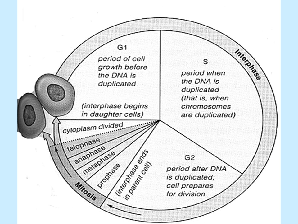
31
Cell Life Cycle INTERPHASE CELL DIVISION Cell growth
Carries on regular cell activities CELL DIVISION Cell replicates itself to produce more cells for growth and repair

32
Interphase G1: “growth” S: “synthesis” phase G2:
protein synthesis, organelles double S: “synthesis” phase DNA replication/duplicated chromosomes G2: Protein synthesis, chromatin condenses, chromosomes visible, final preparation to divide

34
Cell Division Mitosis – division of the nucleus
Result: 2 daughter nuclei Cytokinesis – division of the cytoplasm Result: 2 daughter cells

35
Figure 3.15 Stages of mitosis.
Slide 1 Centrioles Chromatin Centrioles Spindle microtubules Centromere Forming mitotic spindle Centromere Plasma membrane Nuclear envelope Chromosome, consisting of two sister chromatids Fragments of nuclear envelope Spindle pole Nucleolus Interphase Early prophase Late prophase Metaphase plate Nucleolus forming Cleavage furrow Nuclear envelope forming Spindle Sister chromatids Daughter chromosomes Metaphase Anaphase Telophase and cytokinesis

36
Cytokinesis Division of cytoplasm Cell pinched into 2 daughter cells

37
Protein Synthesis (into) (into) DNA mRNA Protein
transcription translation

38
Figure 3.16 Protein synthesis.
Slide 1 Nucleus (site of transcription) DNA Cytoplasm (site of translation) 1 mRNA specifying one polypeptide is made on DNA template. 2 mRNA leaves nucleus and attaches to ribosome, and translation begins. Amino acids mRNA Nuclear pore Nuclear membrane Correct amino acid attached to each species of tRNA by an enzyme Synthetase enzyme 4 As the ribosome moves along the mRNA, a new amino acid is added to the growing protein chain. Growing polypeptide chain Met 3 Incoming tRNA recognizes a complementary mRNA codon calling for its amino acid by binding via its anticodon to the codon. Gly Ser Phe Ala Peptide bond 5 Released tRNA reenters the cytoplasmic pool, ready to be recharged with a new amino acid. tRNA “head” bearing anticodon Large ribosomal subunit Codon Direction of ribosome advance; ribosome moves the mRNA strand along sequentially as each codon is read. Portion of mRNA already translated Small ribosomal subunit
 DNA. Cytoplasm (site of translation) 1. mRNA specifying one polypeptide is made on DNA template. 2. mRNA leaves nucleus and attaches to ribosome, and translation begins. Amino acids. mRNA. Nuclear pore. Nuclear membrane. Correct amino acid attached to each species of tRNA by an enzyme. Synthetase enzyme. 4. As the ribosome moves along the mRNA, a new amino acid is added to the growing protein chain. Growing polypeptide chain. Met. 3. Incoming tRNA recognizes a complementary mRNA codon calling for its amino acid by binding via its anticodon to the codon. Gly. Ser. Phe. Ala. Peptide bond. 5. Released tRNA reenters the cytoplasmic pool, ready to be recharged with a new amino acid. tRNA head bearing anticodon. Large ribosomal subunit. Codon. Direction of ribosome advance; ribosome moves the mRNA strand along sequentially as each codon is read. Portion of mRNA already translated. Small ribosomal subunit.")
Similar presentations


 Relationship.>")


